
Acl ligamnet anatomy
ACL anterior cruciate ligament anatomy bundles attachments
Anterior Cruciate Ligament (ACL) Anatomy
Overview
- Length: 22-41 mm (variable)
- Width: 7-12 mm (consistently narrowest at the midsubstance)
Two-Bundle Structure
| Feature | Anteromedial (AM) Bundle | Posterolateral (PL) Bundle |
|---|---|---|
| Length | ~38 mm | ~20 mm |
| Femoral origin | Superior and anterior portion of the lateral femoral condyle attachment (9:30-11:30 o'clock) | Posterior and inferior portion (8:30-10 o'clock) |
| Tibial insertion | Anteromedial part of the intercondylar fossa | Posterolateral part of the intercondylar fossa |
| Tension in extension | Relatively lax | Taut |
| Tension in flexion | Taut (especially beyond 45°) | Becomes lax |
| Primary function | Resists anterior tibial translation during flexion | Resists anterior translation in extension + rotational control |
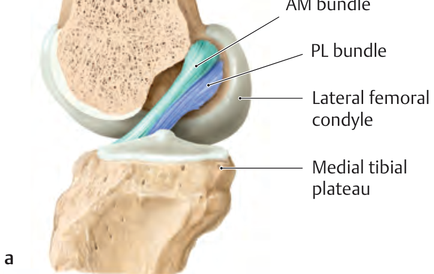
Femoral Attachment
- Located on the posteromedial edge of the lateral femoral condyle, posterior to the lateral intercondylar ridge (also called the Resident's Ridge)
- The attachment is oval in shape
- The AM bundle arises from the superior and anterior aspect
- The PL bundle arises from the posterior and inferior aspect
- AM bundle: 9:30 to 11:30
- PL bundle: 8:30 to 10:00
- Distance between the two bundle centers: 8-10 mm
Tibial Attachment
- Located on the tibial plateau in the anterior intercondylar fossa, between the medial and lateral tibial spines
- Positioned anterolateral to the medial tibial spine
- The tibial footprint is ~120% larger than the femoral footprint
- Some fibers pass deep to the transverse meniscal ligament
- Some fibers merge with the anterior horn of the lateral meniscus

Course and Orientation
Blood Supply and Innervation
- Primary blood supply comes from the middle genicular artery (a branch of the popliteal artery), which penetrates the posterior capsule and travels within the synovial sheath
- Nerve supply includes fibers from the posterior articular nerve (branch of the tibial nerve), carrying both pain fibers and mechanoreceptors (Ruffini endings, Pacinian corpuscles) important for proprioception
Functional Summary
| Function | Bundle Responsible |
|---|---|
| Primary resistance to anterior tibial translation | Both (AM dominant in flexion, PL in extension) |
| Rotational stability / screw-home mechanism | Primarily PL bundle |
| Hyperextension prevention | PL bundle |
| Stability beyond 45° flexion | AM bundle (dominant) |
Create a visual poster showing ACL anatomy with bundle locations
Medical education poster titled "ACL Anatomy" showing the anterior cruciate ligament of the knee. The poster includes: (1) a large central anatomical diagram of the right knee joint opened anteriorly showing the two ACL bundles - the Anteromedial (AM) bundle colored in teal/blue and the Posterolateral (PL) bundle colored in orange/gold, both running from the lateral femoral condyle down to the tibial plateau; (2) a top inset showing the femoral attachment with clock-face positions labeled - AM bundle at 9:30-11:30, PL bundle at 8:30-10:00, with the lateral intercondylar ridge (Resident's Ridge) and bifurcate ridge labeled; (3) a bottom inset showing the tibial footprint on the tibial plateau with AM and PL bundle insertion zones labeled, noting the tibial footprint is 120% larger than the femoral; (4) a side panel with a comparison table showing AM vs PL bundle properties: length (38mm vs 20mm), tension in flexion vs extension, and function; (5) labels pointing to key structures: lateral femoral condyle, medial tibial spine, lateral tibial spine, transverse meniscal ligament, lateral meniscus, medial meniscus; (6) clean white background, professional medical illustration style with clear labels, bold title, color-coded legend for AM (teal) and PL (orange) bundles
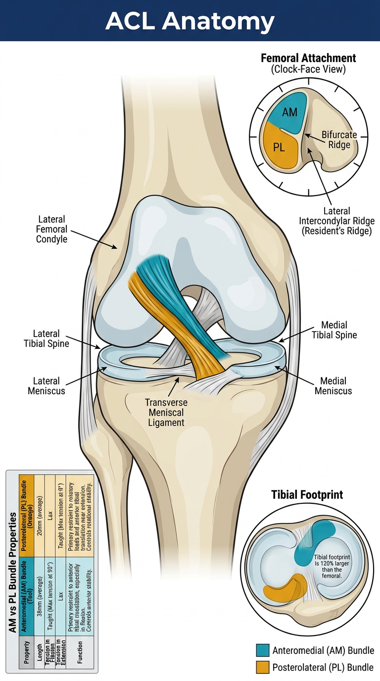
- Central knee diagram with the AM bundle (teal) and PL bundle (orange) clearly distinguished
- Femoral attachment inset with clock-face positions (AM: 9:30-11:30, PL: 8:30-10:00) and the Resident's Ridge / bifurcate ridge landmarks
- Tibial footprint inset showing the larger tibial insertion zone in the anterior intercondylar fossa
- AM vs PL comparison with length, tension behavior, and functional roles
- Key surrounding structures labeled (menisci, tibial spines, transverse meniscal ligament)