
What is brain scar? How does it work And how does it effect persons life Lets take case of girl stronge and lively got hot on her head in a fight and She discovered at age of 17 she had a scar
brain scar gliosis after head injury symptoms effects on life
brain gliosis glial scar after traumatic head injury MRI
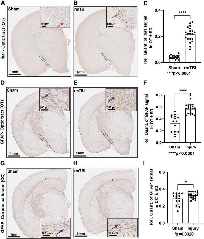
This composite diagnostic image presents immunohistochemical staining of murine coronal brain sections used to evaluate gliosis following repetitive mild traumatic brain injury (rmTBI). Panels A-C focus on microglial activation in the optic tract (OT) using Iba1 staining. Compared to the Sham group (A), the rmTBI group (B) displays a significant increase in Iba1 immunoreactivity, characterized by a higher density of Iba1-positive microglia with amoeboid morphology (red arrowheads in insets). Panels D-I illustrate astrogliosis using Glial Fibrillary Acidic Protein (GFAP) staining in the optic tract (D-E) and corpus callosum (G-H). The rmTBI group shows increased GFAP intensity and astrocyte hypertrophy (blue arrowheads) in both white matter tracts compared to Sham controls. Accompanying bar graphs (C, F, I) provide relative quantification of the signals, demonstrating statistically significant increases in inflammatory markers (p<0.0001 for OT Iba1 and GFAP; p=0.0320 for CC GFAP). Scale bars indicate 1 mm for whole brain views and 100 m for magnified insets. These findings highlight the localized neuroinflammatory response and microstructural disruption characteristic of repetitive head injuries.
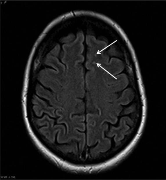
This diagnostic image is an axial Fluid-Attenuated Inversion Recovery (FLAIR) Magnetic Resonance Imaging (MRI) scan of the human head at the level of the superior cerebral hemispheres. The scan displays a cross-sectional view of the brain tissue, clearly showing the convoluted surface anatomy of the gyri and sulci. The cerebral cortex and white matter appear generally homogenous; however, the image highlights focal pathological changes. Two white arrows point to small, hyperintense (bright) punctate spots located in the subcortical white matter of the left frontal lobe. These findings represent nonspecific gliosis spots, which are areas of reactive glial cell changes resulting from previous neural injury. In a clinical context, such bihemispheric white matter lesions can be associated with various inflammatory or infectious conditions, including neuroborreliosis (Lyme disease). The image serves as an educational example of how systemic infections can manifest with subtle, multifocal white matter abnormalities on neuroimaging.
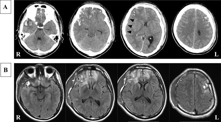
This medical comparison image displays diagnostic neuroimaging of a traumatic brain injury across two modalities and time points. Panel A presents a non-contrast axial head CT scan acquired on the day of injury. It demonstrates multiple hemorrhagic brain contusions, characterized by a mixed-density 'salt and pepper' appearance, primarily involving the right frontal and temporal lobes and the left frontal lobe. A hyperdense, crescentic collection along the right cerebral hemisphere (indicated by black arrowheads) signifies an acute subdural hematoma, associated with a mild midline shift. Panel B displays follow-up axial MRI using Fluid-Attenuated Inversion Recovery (FLAIR) sequences 23 days post-injury. The MRI reveals persistent hyperintense signals in the previously noted contusion sites within the frontal and temporal lobes. Additionally, diffuse high-intensity signals are visible in the surrounding white matter, representing perilesional vasogenic edema and gliosis. The comparison illustrates the evolution of traumatic lesions from acute hemorrhage and density changes on CT to late-stage edema and parenchymal signal alterations on MRI, essential for monitoring post-traumatic neuro-regeneration and complications.
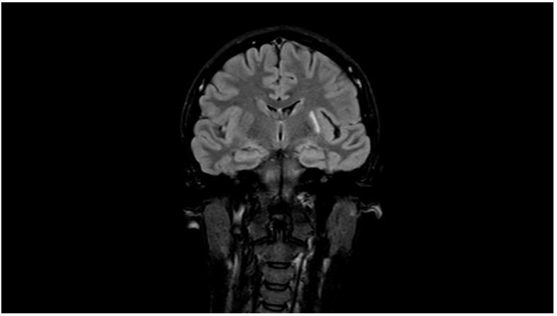
This diagnostic image is a coronal Fluid-Attenuated Inversion Recovery (FLAIR) MRI of the human head, demonstrating chronic neuroanatomical changes following a migrainous infarction. The primary findings are localized in the left parietal subcortical region. Visible pathology includes hyperintense signals indicative of gliosis and leukoaraiosis (white matter changes) surrounding well-defined hypointense cystic cavities, representing encephalomalacia. The image illustrates the long-term sequelae of ischemic injury in the context of migraine with aura, highlighting the evolution from an acute infarct to permanent parenchymal loss and surrounding glial scarring. These features are clinically significant for neuroradiological follow-up of stroke and the assessment of cerebral tissue viability. The modality effectively suppresses cerebrospinal fluid (CSF) signal to enhance the visibility of perilesional hyperintensities against the brain parenchyma.
Brain Scar (Glial Scar / Gliosis) - A Complete Guide
What Is a Brain Scar?
- Robbins & Kumar Basic Pathology explains it clearly: "Astrocytes are the principal cells responsible for repair and scar formation in the brain, a process termed gliosis. In response to injury, astrocytes undergo both hypertrophy and hyperplasia... the cellular processes become more tightly interwoven (fibrillary astrocytes)."
How Does It Form? (The Biology, Step by Step)
| Stage | What Happens | Time Frame |
|---|---|---|
| Immediate injury | Neurons and blood vessels are damaged; blood-brain barrier breaks down; brain swells | Minutes to hours |
| Microglial activation | The brain's immune cells (microglia) rush to the site, engulf debris, release inflammatory signals | Hours to days |
| Astrocyte activation | Nearby astrocytes enlarge, multiply, and grow more processes (arms); fill with GFAP protein filaments | Days to weeks |
| Scar formation | The astrocytic processes pack densely together, forming a permanent fibrous boundary | Weeks to months |
| Chronic scar (plaque) | The scar shrinks slightly, becomes tightly woven, permanent | 6+ months onward |
Now - The Story of the Girl
Why Was It Not Caught Earlier?
- May cause no obvious symptoms for years
- Is invisible without MRI (regular X-rays and CT scans often miss it)
- Only becomes apparent when it starts causing problems (like seizures)
- Or may be found incidentally on a scan done for another reason
What Could She Experience?
| Location of Scar | Possible Effects |
|---|---|
| Frontal lobe | Personality changes, impulsivity, poor planning, mood swings |
| Temporal lobe | Memory problems, difficulty finding words, seizures with strange smells/sounds |
| Motor cortex | Weakness or twitching on one side of the body |
| Occipital lobe | Visual disturbances |
| General/diffuse | Headaches, fatigue, slower thinking |
The Biggest Risk: Post-Traumatic Epilepsy
"True post traumatic epilepsy due to the scar tissue formation in the brain or between the brain and the membranes will take no less than 6 months to develop. Post traumatic epilepsy is mainly Jacksonian type - uncontrollable twitching may affect the thumb or the hand in the beginning. Gradually the muscles of the arm, shoulder or even face will be affected. The convulsion may become generalized."
How Does This Affect Her Life?
Physical Effects
- Seizures (most common and serious) - can occur without warning
- Persistent headaches
- Fatigue
- In some cases, weakness or sensory changes on one side of the body
Cognitive Effects
- Difficulty concentrating in school
- Slower information processing
- Memory gaps (especially short-term memory)
- Trouble multitasking
Emotional and Behavioral Effects
- Mood swings - she might feel irritable or sad without a clear reason
- Anxiety (especially about having a seizure in public)
- Depression - her sense of identity ("strong and lively") may be challenged
- She may feel misunderstood because she "looks fine" but struggles internally
Social and Practical Effects
- Driving restrictions if she has seizures
- Possible limitations in certain sports or physical activities
- May need academic accommodations at school
- Sleep disruption (seizures are more common during sleep and on waking)
Key Point: The Scar Itself vs. Its Consequences
- Anti-epileptic drugs (AEDs) if seizures develop - medications like levetiracetam, lamotrigine, or valproate can control seizures in most patients
- Neuropsychological support - therapy and cognitive rehabilitation
- Regular MRI/EEG monitoring to track electrical activity
- In some cases, neurosurgery (resecting the scar focus) if seizures are drug-resistant
The Emotional Truth for Someone Like Her
- Robbins & Kumar Basic Pathology - Astrocyte injury and gliosis
- Histology: A Text and Atlas (Ross & Pawlina) - Reactive gliosis and scar formation in the CNS
- S Das - A Manual on Clinical Surgery - Post-traumatic epilepsy and head injury complications
- Frontiers in Cellular Neuroscience (2024) - Reactive gliosis in TBI: comprehensive review
- Orlando Health - Effects of Brain Injuries
Sometime like occasionally after heavy work she get mild headaches If the load was went beyond her limit Her head start to explode with pain randomly and sometimes faint
brain scar post-traumatic severe exertional headache fainting syncope causes mechanisms
thunderclap headache exertional severe sudden onset mechanism diagram
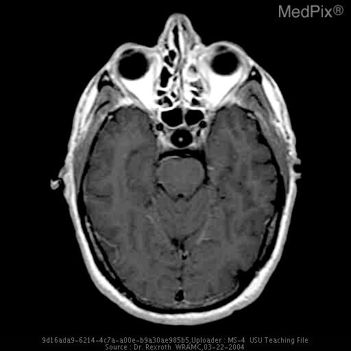
**Imaging Modality:** Magnetic Resonance Imaging (MRI), T1-weighted axial sequence. **Anatomical Region:** Axial section of the neurocranium at the level of the sella turcica, midbrain (pons), and orbits. **Observed Pathology:** Pituitary Apoplexy. The image demonstrates a significantly enlarged pituitary gland within an expanded sella turcica. **Characteristic Visual Features:** - **Sellar Mass:** There is a well-circumscribed, hyperintense lesion occupying the sella turcica. - **Signal Intensity:** The T1-weighted hyperintensity is highly suggestive of acute to subacute hemorrhage or high protein content within a pituitary adenoma. - **Mass Effect:** The enlarged gland shows evidence of superior extension (suprasellar extension), potentially compromising the optic chiasm. - **Surrounding Structures:** The sphenoid sinus is visible anterior to the sella, and the cavernous sinuses border the lesion laterally. The brainstem and temporal lobes appear unremarkable at this level. **Clinical Context:** In a patient presenting with sudden-onset severe headache ("thunderclap" presentation), these radiologic findings are pathognomonic for pituitary apoplexy, resulting from hemorrhage or infarction of a preexisting pituitary macroadenoma. **Key Diagnostic Features:** T1-hyperintense sellar mass with expansion of the bony sella and suprasellar protrusion.
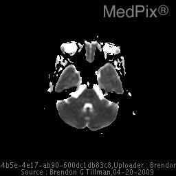
**Imaging Modality:** Magnetic Resonance Imaging (MRI), Apparent Diffusion Coefficient (ADC) map. **Anatomical Region:** Axial section of the brain at the level of the pons, cerebellum, and temporal lobes. **Observed Pathology:** The image demonstrates areas of altered signal intensity. In the context of the provided clinical history of a postpartum patient with a sudden onset "thunderclap" headache, these findings are highly suspicious for Reversible Posterior Leukoencephalopathy Syndrome (PRES) or Reversible Cerebral Vasoconstriction Syndrome (RCVS). **Characteristic Visual Features:** - The ADC map is used to differentiate between cytotoxic edema (low ADC/dark) and vasogenic edema (high ADC/bright). - There is evidence of hyperintensity in the posterior regions, suggesting vasogenic edema rather than acute infarction. - Symmetrical or asymmetrical involvement of the subcortical white matter may be present, though the resolution of this specific map primarily highlights the posterior fossa and temporal regions. **Key Diagnostic Features:** The absence of restricted diffusion (which would appear dark on ADC) helps exclude acute ischemic stroke, pointing instead toward a reversible encephalopathy or vasoconstriction syndrome often associated with the peripartum period.
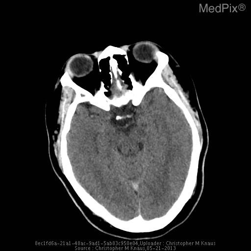
**Imaging Modality:** Non-contrast Computed Tomography (NCCT) of the head. **Anatomical Region:** Axial section at the level of the suprasellar cistern, midbrain, and skull base. **Observed Pathology:** There is a hyperdense signal located within the suprasellar cistern, consistent with acute subarachnoid hemorrhage (SAH). The hyperdensity appears concentrated in the region of the circle of Willis, suggesting potential aneurysmal rupture. **Characteristic Visual Features:** * **Hyperdensity:** High-attenuation (bright) material is visible in the basal cisterns, specifically the suprasellar cistern and extending slightly into the ambient cisterns. * **Vascular Structures:** A focal hyperdense area is noted near the expected location of the anterior communicating artery or internal carotid artery terminus, often referred to as a "bright artery sign" or sentinel clot. * **Ventricular System:** The visible portions of the temporal horns of the lateral ventricles and the fourth ventricle do not show immediate evidence of gross hydrocephalus or intraventricular extension in this single slice. **Clinical Context:** Findings are highly indicative of acute nontraumatic subarachnoid hemorrhage, correlating with the clinical presentation of a sudden-onset "thunderclap" headache. **Key Diagnostic Features:** Presence of extravasated blood in the subarachnoid space (cisterns) on non-contrast CT.
What's Happening to Her: Exertional Headaches + Fainting with a Brain Scar
The Two-Level Pattern You Described
| Situation | What She Gets | Medical Term |
|---|---|---|
| Normal daily activity | No symptoms | Compensated state |
| Heavy/physical work | Mild headache | Post-traumatic exertional headache |
| Beyond her limit | Explosive, sudden, severe head pain | Possible thunderclap / pressure headache |
| Beyond her limit | Fainting/loss of consciousness | Exertional syncope |
Why Does Physical Effort Trigger This?
1. Blood Pressure and Pressure Inside the Skull Rise Together
2. The Scar Disrupts Neurovascular Coupling
3. The "Exploding" Sensation - What Is It?
Why Does She Faint?
Mechanism A: Vasovagal/Reflex Syncope
Mechanism B: Direct Pressure Effect
The "Limit" Concept - Her Brain Is Actually Telling Her Something
Light work → no pain → SAFE ZONE
Heavy work → mild headache → WARNING ZONE
Beyond limit → explosive pain + faint → DANGER ZONE
What Are the Possible Causes to Rule Out?
| Condition | Why Relevant |
|---|---|
| Post-traumatic cerebral vascular damage | The old blow may have weakened a blood vessel near the scar |
| Cerebral arteriovenous malformation (AVM) | Tangles of abnormal vessels, more common after trauma, bleed under pressure |
| Cortical spreading depression | Wave of electrical suppression spreading from the scar, similar to migraine aura |
| Scar-triggered seizure equivalent | Brief electrical discharge causing loss of consciousness without full convulsion |
| Reversible Cerebral Vasoconstriction Syndrome (RCVS) | Exertion-triggered spasm of brain arteries causing thunderclap headache |
| Hydrocephalus (CSF pressure buildup) | Scar tissue blocking cerebrospinal fluid drainage |
What This Looks Like in Her Real Life
- Hide it because she doesn't want to seem weak
- Push through the warning signs which makes it worse
- Start avoiding physical activity out of fear - which leads to deconditioning and isolation
- Develop anxiety around any physical effort, always wondering "will today be the day?"
- Lose confidence in her own body, which for an active girl is a significant identity blow
What She Should Do - Practically
- See a neurologist urgently - describe the exertion threshold clearly
- MRI with MRA (angiography) - to look at the blood vessels around and near the scar for any abnormality
- EEG - to check if the fainting episodes are actually brief seizures (ictal syncope)
- Exercise stress test under monitoring - to document exactly what happens to her brain activity and blood pressure when she crosses the threshold
- No unmonitored heavy exertion until evaluated - particularly no activities where fainting would be dangerous (swimming alone, heights, driving)
What Management Might Look Like
- If vascular cause found: possible intervention or medication to protect vessels
- If seizure related: anti-epileptic medication
- If pressure-related: medication to reduce intracranial pressure
- Activity modification with clear, defined safe limits - she does not have to stop being active, she needs to know her actual boundary
- Cognitive behavioral therapy to process the anxiety and identity disruption this causes
The Key Message
Brain scar symptoms
brain lesion scar location symptoms cortex regions functions diagram
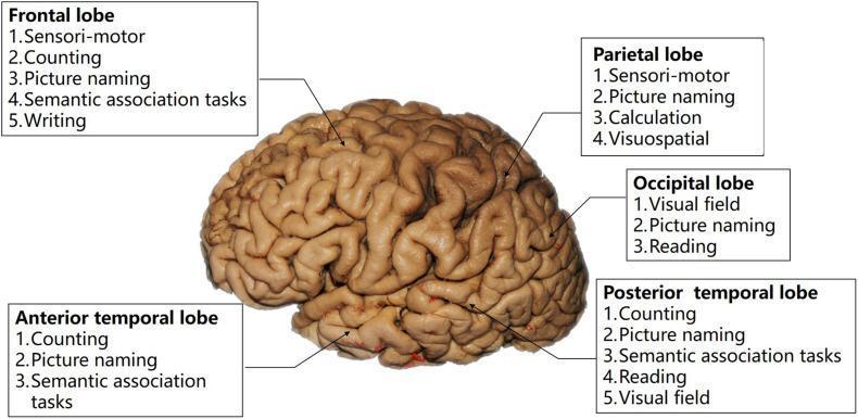
Anatomical diagram and flowchart presenting a lateral view of the human brain cortex, specifically mapped for intraoperative cognitive tasks used during neurosurgical mapping. The illustration identifies five major regions: Frontal lobe, Parietal lobe, Occipital lobe, Anterior temporal lobe, and Posterior temporal lobe. Each region is annotated with recommended clinical tasks to preserve functional integrity during lesion resection. Key tasks listed include: 1) Frontal lobe: sensori-motor, counting, picture naming, semantic association, and writing; 2) Parietal lobe: sensori-motor, picture naming, calculation, and visuospatial tasks; 3) Occipital lobe: visual field, picture naming, and reading; 4) Temporal regions: counting, picture naming, and semantic association, with the posterior temporal lobe specifically adding reading and visual field assessments. This diagram serves as a clinical guide for selecting direct electrical stimulation (DES) tasks based on tumor location to protect eloquent cortical areas involved in language, motor, and cognitive processing.
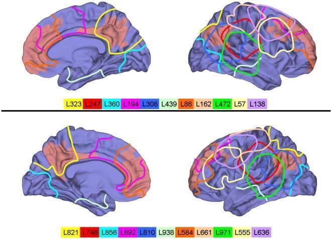
Anatomical diagram presenting four renderings of a standard human cortical surface used to map simulated focal brain lesions. The brain is shown in lateral and medial views across two rows. The cortical surface is rendered in blue, with regions of interest (ROIs) belonging to the default mode network (DMN) highlighted in light red; these include the precuneus, posterior cingulate, medial prefrontal cortex, and lateral parietal cortex. Superimposed on the cortex are various colored outlines representing 22 distinct lesion locations (labeled L323 through L636). Each lesion encompasses 50 ROIs, approximately 5% of the cortical surface. The diagram illustrates lesion distribution across major functional zones, including the frontal lobe, temporo-parietal junction, and cortical midline. A color-coded legend at the bottom of each row correlates specific colors with lesion identifiers, serving as a reference for neuroanatomical location and clinical significance in studies of functional connectivity and network disruption (diaschisis).
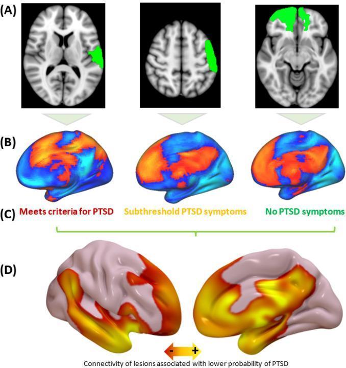
Educational diagram illustrating Lesion Network Mapping (LNM) in the context of Post-Traumatic Stress Disorder (PTSD) pathophysiology. (A) Representative axial brain slices (T1-weighted MRI template) show localized focal brain lesions highlighted in green across three different subjects, demonstrating anatomical heterogeneity. (B) Functional connectivity maps (normative connectomes) for these subjects, with orange-red areas indicating high connectivity to the respective lesion sites. (C) Clinical categorization of subjects based on symptoms: 'Meets criteria for PTSD', 'Subthreshold PTSD symptoms', and 'No PTSD symptoms'. (D) A group-level statistical brain map (medial and lateral views) showing the 'PTSD circuit.' Regions in yellow-orange represent areas where high lesion connectivity is associated with a lower probability of developing PTSD. These regions prominently include the medial prefrontal cortex (mPFC), amygdala, and medial temporal lobe. The infographic explains how lesion locations and their functional networks can influence psychiatric clinical outcomes and potentially inform future neuromodulation (TMS) targets.
brain scar glial scar symptoms complete list location-based effects
Brain Scar Symptoms - Complete Guide
The Golden Rule: Location Determines Symptoms

Full Symptom List by Category
🔴 PHYSICAL SYMPTOMS
| Symptom | How It Feels |
|---|---|
| Headaches | The most common symptom. Dull, pressure-like, or pounding. Often worse with effort, stress, or lack of sleep |
| Exertional headaches | Triggered specifically by physical activity - the brain's pressure system is less flexible around the scar |
| Explosive / thunderclap headache | Sudden severe "head explosion" during heavy work or strain - a red-flag that needs urgent attention |
| Seizures | From brief twitching or blank stares to full convulsions. The scar acts as an electrical short-circuit |
| Fainting / loss of consciousness | Can happen with severe pain surges or brief seizure equivalents |
| Weakness on one side | If scar is near the motor cortex - arm, leg, or face may feel weak or clumsy |
| Numbness / tingling | If scar is near sensory areas - one side of body may feel odd or numb |
| Balance problems | Dizziness, unsteadiness, or coordination issues - especially if near cerebellum |
| Vision problems | Blurred vision, visual disturbances, or blind spots - if near occipital (back) lobe |
| Fatigue | Profound tiredness even after normal activity - the brain works harder to compensate |
| Sleep disturbances | Insomnia, broken sleep, or sleeping too much |
| Nausea | Often accompanies severe headaches or raised pressure episodes |
| Sensitivity | Heightened sensitivity to light (photophobia), loud sounds, or strong smells |
🟡 THINKING & MEMORY SYMPTOMS
| Symptom | How It Shows Up |
|---|---|
| Short-term memory problems | Forgetting recent conversations, losing items, forgetting what she was about to do |
| Concentration difficulty | Mind drifts off, hard to stay focused on reading or study for long |
| Slow processing speed | Takes longer to respond, think through problems, or complete tasks |
| Word-finding problems | Knows what she wants to say but can't find the word - called anomia |
| Poor multitasking | Easily overwhelmed when doing more than one thing at once |
| Learning difficulties | New information takes longer to absorb and stick |
| Mental fog | Brain feels cloudy, especially after exertion, poor sleep, or stress |
| Calculation or planning problems | If frontal lobe involved - difficulty organizing tasks, managing time |
🟠 EMOTIONAL & BEHAVIORAL SYMPTOMS
| Symptom | What It Looks Like |
|---|---|
| Irritability | Snapping at people unexpectedly, low frustration tolerance |
| Mood swings | Emotions that shift quickly without obvious reason |
| Anxiety | Constant low-level worry, or fear of having a headache/blackout in public |
| Depression | Persistent low mood, loss of interest in things she used to enjoy |
| Impulsivity | Acting without thinking, especially with frontal lobe scars |
| Emotional blunting | Feeling less emotionally reactive - "flat" or disconnected |
| Social withdrawal | Pulling away from friends and activities due to symptoms or fear |
| Low self-esteem | Identity disruption - "I used to be strong, what happened to me?" |
🟢 SYMPTOMS SPECIFIC TO SCAR LOCATION
| Scar Location | What She May Experience |
|---|---|
| Frontal lobe | Personality changes, poor planning, impulsivity, low motivation, emotional blunting |
| Temporal lobe | Memory loss, déjà vu, hearing strange sounds, unexplained fear or emotion, word-finding problems |
| Parietal lobe | Numbness, clumsiness on one side, difficulty with spatial tasks |
| Occipital lobe | Visual disturbances, flashing lights, blind spots |
| Motor cortex | Weakness or twitching on the opposite side of the body |
| Cerebellum | Balance problems, uncoordinated movements, dizziness |
| Near blood vessels | Pressure headaches, exertional pain, risk of vascular events |
Symptoms That Are RED FLAGS - Seek Help Immediately
- ❗ Sudden "worst headache of life" - especially with exertion
- ❗ Fainting or loss of consciousness
- ❗ Seizure (shaking, blank staring, brief blackout)
- ❗ Sudden weakness or numbness on one side of the body
- ❗ Sudden confusion or speech difficulty
- ❗ Vision loss
- ❗ Vomiting with severe headache
- ❗ Headache that wakes her from sleep
Invisible vs. Visible Symptoms
- Other people may not take her symptoms seriously
- She may push herself too hard because she "looks fine"
- Teachers, employers, friends may not understand why she struggles sometimes
- She may internalize it as weakness rather than recognizing it as a real, structural issue
One Key Insight
If dr see Someone collapsed And his symptoms was Head exploded randomly out of no where Seizure and then collapsed
sudden explosive headache then seizure then collapse emergency differential diagnosis doctor response
subarachnoid hemorrhage CT scan emergency sudden headache seizure
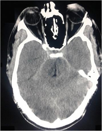
This diagnostic image is a non-contrast computed tomography (CT) scan of the head in an axial section at the level of the midbrain and suprasellar region. The primary finding is the presence of linear, hyperdense (bright white) signals within the subarachnoid spaces, specifically involving the suprasellar and ambient cisterns. These findings are highly characteristic of acute subarachnoid hemorrhage (SAH). The brain parenchyma shows relatively preserved gray-white matter differentiation at this level, though there is a subtle mass effect on the basal cisterns. Bony structures of the skull vault and the sphenoid bone demonstrate normal high attenuation. The orbits and paranasal sinuses are partially visualized in the anterior field. This image serves as a clinical demonstration of the sensitivity of non-contrast CT in detecting acute intracranial bleeding within the CSF-filled spaces of the basal cisterns, a critical step in the emergency diagnostic pathway for patients presenting with sudden-onset severe headache.
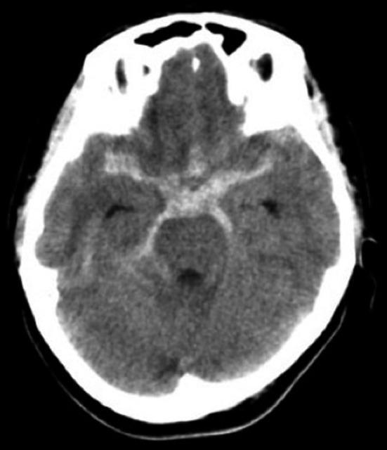
This diagnostic image is an axial non-contrast computed tomography (CT) scan of the brain at the level of the basal cisterns. The primary finding is an acute subarachnoid hemorrhage (SAH), characterized by high-attenuation (hyperdense) material filling the normally hypodense cerebrospinal fluid (CSF) spaces. The hyperdensity is diffusely distributed throughout the suprasellar cistern, interpeduncular cistern, and the ambient cisterns. The distribution is notably prominent on the right side, extending into the right sylvian fissure and outlining the contours of the midbrain. This visual evidence of 'blood in the cisterns' creates a characteristic pentagonal or star-shaped appearance in the center of the scan. The presence of these findings in a 42-year-old patient with sudden severe headache is consistent with an aneurysmal rupture, specifically a blood blister-like aneurysm of the internal carotid artery as suggested by the clinical context. The image serves as a critical educational example of intracranial hemorrhage recognition and anatomical localization for neuroradiology and emergency medicine training.
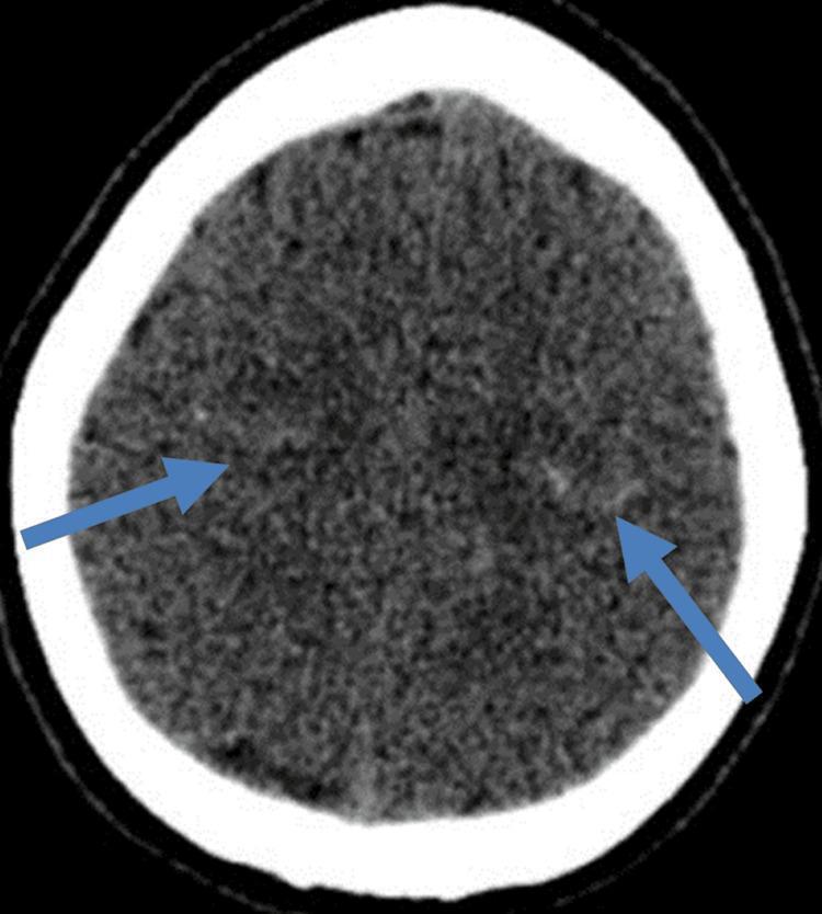
This unenhanced axial computerized tomography (CT) scan of the head demonstrates the superior cerebral hemispheres. The primary finding is the presence of focal hyperdensity within the cortical sulci bilaterally, specifically localized to the precentral sulci as indicated by blue arrows. These hyperdense signals are consistent with acute subarachnoid hemorrhage (SAH) confined to the sulcal spaces, often referred to as convexal subarachnoid hemorrhage (cSAH). The surrounding brain parenchyma shows preserved gray-white matter differentiation without evidence of large-scale edema, mass effect, or midline shift. The skull remains intact with normal bony density and no signs of fracture. The extracerebral space (subarachnoid space) elsewhere appears typically hypodense. This diagnostic image is clinically significant for identifying non-aneurysmal or focal SAH in patients presenting with sudden severe headache (thunderclap headache) or focal neurological deficits, serving as a critical teaching tool for neuroradiology and emergency medicine.
What a Doctor Sees: Explosive Headache → Seizure → Collapse
The Doctor's First Thought: "Is This a Bleed?"
"Subarachnoid hemorrhage typically presents with a sudden and severe headache, often accompanied by nausea, vomiting, loss of consciousness, and nuchal rigidity."
"Onset of thunderclap headache during periods of exertion raises suspicion for subarachnoid hemorrhage or arterial dissection... Headaches associated with loss of consciousness, seizure, diplopia... should raise concern."
What the Doctor Thinks Is Happening (Differential Diagnosis)
🔴 MOST DANGEROUS - Must Rule Out First
| Condition | What It Is | Why This Pattern Fits |
|---|---|---|
| Subarachnoid Hemorrhage (SAH) | Bleeding into the space around the brain, usually from a burst aneurysm | Classic: thunderclap headache + seizure + collapse. #1 killer in this scenario |
| Intracerebral Hemorrhage | Bleeding inside the brain tissue itself | Sudden headache + rapid neurological collapse |
| Ruptured Brain AVM | Tangled abnormal blood vessels burst | Especially in young people with prior head trauma; causes exactly this triad |
| Hypertensive Crisis | Blood pressure catastrophically high, vessels burst | Explosive headache, seizure, LOC |
🟠 SERIOUS - Also Considered
| Condition | What It Is |
|---|---|
| Scar-triggered Seizure (especially in this girl's case) | The glial scar fires an electrical storm - seizure causes collapse |
| Reversible Cerebral Vasoconstriction Syndrome (RCVS) | Arteries in the brain suddenly spasm - thunderclap headache, can cause seizures |
| Cerebral Venous Thrombosis | Clot in the brain's veins - raised pressure, seizure, collapse |
| Posterior Reversible Encephalopathy Syndrome (PRES) | Pressure overload disrupts brain - seizure, altered consciousness |
| Pituitary Apoplexy | Bleeding into the pituitary gland - sudden severe headache + collapse |
What the Doctor Does - Step by Step
Step 1: Immediate ABC Assessment (First 60 seconds)
- A - Airway: Is she breathing? Is the airway clear after the seizure?
- B - Breathing: Oxygen saturation - brain needs oxygen NOW
- C - Circulation: Pulse, blood pressure - is she in shock?
- Call for backup. This is not a one-doctor job.
Step 2: Rapid Neurological Check (Next 2 minutes)
- GCS score (Glasgow Coma Scale) - how conscious is she?
- Pupils - are they equal and reactive to light? Unequal pupils = brain herniation, life-threatening
- Any focal weakness - one side not moving = stroke territory
- Neck stiffness (meningism) - classic sign of blood in the CSF around the brain
Step 3: Emergency CT Scan - No Delays
Step 4: If CT is Negative - Lumbar Puncture
"Clinical presentation suggests subarachnoid hemorrhage even if CT is normal: sudden, severe headache, often with loss of consciousness at onset"
Step 5: CT Angiography (CTA)
If It's a Scar-Triggered Seizure (No Active Bleed)
Scar fires electrical storm
        ↓
Seizure begins (brain in chaos)
        ↓
Blood pressure spikes dramatically during seizure
        ↓
Explosive headache felt
        ↓
Post-ictal collapse (brain exhaustion after seizure)
        ↓
She falls, unconscious for minutes
        ↓
Wakes up confused, head pounding
The Hunt and Hess Scale - How Doctors Grade Severity
| Grade | Condition | Survival |
|---|---|---|
| 1 | Mild headache, alert | ~70% |
| 2 | Severe headache, alert, maybe neck stiffness | ~60% |
| 3 | Drowsy/confused | ~50% |
| 4 | Stuporous, semi-conscious | ~20% |
| 5 | Deep coma, near death | ~10% |
Treatment if Bleed Confirmed
| Priority | Action |
|---|---|
| Stop the bleeding source | Surgery (clipping the aneurysm shut) or endovascular coiling (filling it from inside with coils) |
| Control brain swelling | Medications to reduce intracranial pressure |
| Prevent vasospasm | Blood vessel spasm after SAH kills brain tissue - nimodipine drug given to prevent this |
| Seizure control | Anti-epileptic medications IV |
| ICU monitoring | 24-hour intensive care, repeated CT scans |
The Critical Sentence Every Emergency Doctor Knows
"One quarter of patients with SAH die before reaching the hospital. Median mortality in the United States is about one-third, with approximately one-third of survivors having functional and cognitive deficits." - Rosen's Emergency Medicine
Connecting Back to the Girl
- Seizure-induced collapse (most likely cause in her case)
- Abnormal vessels growing around old scar tissue (increased AVM/aneurysm risk)
- Scar-adjacent vessel fragility
What symptoms will lead dr to brain scar
how doctors diagnose brain scar gliosis symptoms that lead to MRI investigation
MRI FLAIR T2 brain gliosis scar hyperintense white spot diagnosis
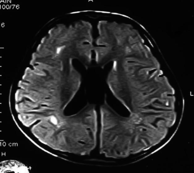
This diagnostic image is an axial Fluid-Attenuated Inversion Recovery (FLAIR) MRI of the human brain, focusing on the supratentorial region. The image demonstrates multiple focal and confluent T2-hyperintense lesions located within the deep and periventricular white matter. These hyperintensities are most prominent in the parieto-occipital areas, characterized by patchy signal changes adjacent to the lateral ventricles. The ventricular system maintains a relatively normal morphology and size, though the surrounding white matter disturbances suggest focal gliosis or leukoencephalopathy. The cortical ribbon appears intact with darker signal intensity compared to the underlying white matter pathology. This radiological finding is clinically significant for evaluating demyelinating diseases, chronic small vessel ischemic changes, or metabolic white matter disturbances in neurology and neuroradiology education.
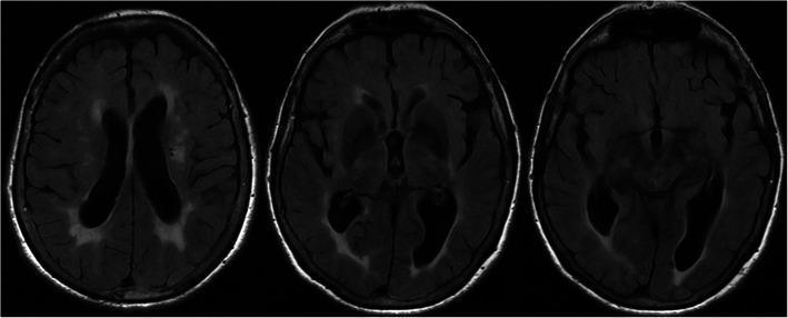
Diagnostic axial MRI of the brain using T2-Fluid Attenuated Inversion Recovery (FLAIR) sequences, shown in three representative slices at different neuroanatomical levels. The images demonstrate extensive, confluent periventricular white matter hyperintensities (WMH) involving the frontal and occipital horns of the lateral ventricles, characteristic of chronic small vessel ischemic disease. In the leftmost slice, a focal area of encephalomalacia and gliosis is visible in the left anterior region, appearing as a central hypointense signal surrounded by a hyperintense rim, consistent with sequelae of a prior left anterior choroidal artery territory stroke. The ventricular system appears mildly dilated, likely representing ex-vacuo changes secondary to global brain volume loss. These findings are clinically relevant for assessing chronic cerebrovascular burden and correlating imaging with neurological symptoms such as cognitive decline, motor fluctuations, or previous ischemic events in elderly patients.
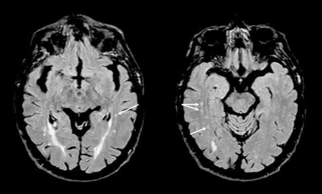
This diagnostic image consists of two axial T2-weighted fluid-attenuated inversion recovery (FLAIR) MRI scans of the human brain at the level of the temporal lobes and midbrain. The primary findings are small, punctate, hyperintense foci located within the deep white matter of both the left and right temporal lobes, indicated by white arrows. These hyperintensities represent gliotic changes or white matter hyperintensities (WMHs). The cortical gray matter, sulcal patterns, and ventricular systems appear otherwise unremarkable for the age-matched clinical context. These images are used to demonstrate neuroanatomical correlates in chronic pain conditions, such as burning mouth syndrome, emphasizing the presence of small vessel ischemic changes or gliosis in brain regions associated with mood and pain modulation. The target audience includes neurology and radiology students or clinicians studying white matter disease progression and its clinical associations.
How a Doctor Gets from Symptoms to "Brain Scar"
The Key Starting Point: Gliosis Is Often Silent
The Symptom Trail - What Raises the Doctor's Suspicion
🔑 Clue 1: History of Head Trauma
For the girl in our story: she was hit on the head → that history alone should have put every doctor she ever saw on alert.
🔑 Clue 2: First Seizure (Most Common Route to Diagnosis)
| Seizure Type | What It Looks Like | Why It Points to a Scar |
|---|---|---|
| Focal (Jacksonian) seizure | Twitching starts in one finger, spreads up the arm to the shoulder | Classic for a localized irritating scar near the motor cortex |
| Temporal lobe seizure | Strange smell/taste, déjà vu, fear, staring blankly, lip smacking | Points to scar in temporal lobe - very common post-trauma |
| Tonic-clonic (grand mal) | Full body convulsion, loss of consciousness | Can result from scar anywhere, especially if electrical activity spreads |
| Absence-like episode | Brief blank stare, unresponsive for seconds | Subtle - often missed for months before diagnosis |
🔑 Clue 3: Chronic Headaches with a Specific Pattern
- Headaches that are consistently on the same side of the head
- Headaches that worsen with physical effort or straining
- Headaches that have been present for months to years after a head injury
- Headaches that don't respond to normal painkillers
- Headaches accompanied by any neurological symptom (visual changes, weakness, tingling)
🔑 Clue 4: New Focal Neurological Symptoms
| Symptom | What it Suggests |
|---|---|
| Arm or leg suddenly weaker on one side | Motor cortex or pathway affected by scar |
| Trouble finding words, speech hesitation | Language area (Broca's/Wernicke's) scarred |
| Memory gaps, especially recent memory | Temporal lobe / hippocampus scarring |
| Personality change noticed by family | Frontal lobe scar |
| Strange sensations on one side of body | Sensory cortex scar |
| Vision problems in one field | Occipital or parietal scar |
🔑 Clue 5: Behavioral or Psychiatric Changes After Head Injury
- Sudden personality change after a head injury (more aggressive, more impulsive, more emotional)
- Depression or anxiety that started after a blow to the head
- Memory problems in a young person with no other explanation
- Cognitive decline in someone who was previously sharp
🔑 Clue 6: Fainting + Headache Combination
The Doctor's Decision Tree
Patient presents with symptoms
           ↓
Doctor asks: "Any history of head injury?"
           ↓
     YES ──────────────────────────────────────────────────┐
           │                                               │
     + New seizure        + Chronic one-sided headaches   + Focal neuro symptom
     + Fainting           + Personality change            + Memory problems
           │                                               │
           └───────────────────┬───────────────────────────┘
                               ↓
                    ORDER BRAIN MRI
                    (with FLAIR sequence)
                               ↓
                    Radiologist reports:
               "Focal T2/FLAIR hyperintensity"
               "Areas of encephalomalacia"
               "Gliotic changes"
                               ↓
                    Doctor connects to history:
               "This is post-traumatic gliosis"
                    = BRAIN SCAR CONFIRMED
What the MRI Actually Shows

- T2/FLAIR = bright white spot where the scar is
- T1 = slightly dark in the same area
- No contrast enhancement (unlike tumors, which light up with contrast dye)
- No mass effect (the scar doesn't push brain tissue aside like a tumor does)
How the Doctor Confirms It Is a Scar (Not Something Else)
| Feature | Glial Scar | Tumor | Active Inflammation (MS) |
|---|---|---|---|
| History of trauma | ✅ Yes | ❌ No | ❌ No |
| Changes over time on repeat MRI | ❌ Stable, no change | ✅ Grows | ✅ May change |
| Contrast enhancement | ❌ None | ✅ Usually | ✅ Sometimes |
| Location matches old injury | ✅ Yes | ❌ Random | ❌ Periventricular |
| EEG abnormality nearby | ✅ Often | Sometimes | Rarely |
The Tests the Doctor Orders
| Test | What It Finds |
|---|---|
| MRI Brain (FLAIR + T2 + T1) | Shows the scar directly - the gold standard |
| EEG (brain electrical recording) | Shows abnormal electrical activity coming FROM the scar zone - confirms it as a seizure source |
| MRI with contrast | Rules out tumor (scar does NOT enhance; tumor does) |
| MRA (MR Angiography) | Checks blood vessels near the scar for damage |
| Neuropsychological testing | Maps which cognitive functions are affected - tells doctor which brain areas are impaired |
| Full history interview | The trauma history is often the most important diagnostic tool of all |
Summary: The Symptom Checklist That Points a Doctor to Brain Scar
- ✅ Past head injury (even years ago, even minor)
- ✅ New seizure - especially focal (one side, or strange sensations)
- ✅ Chronic headaches on the same side, worse with effort
- ✅ Fainting with severe headache
- ✅ Memory problems in a young person
- ✅ Personality change after a head injury
- ✅ Weakness, numbness, or clumsiness on one side
- ✅ Unexplained psychiatric symptoms after head trauma
Can mri can say how old a scar is?
can MRI tell how old a brain scar gliosis is dating age of lesion radiology
MRI brain stages acute subacute chronic lesion signal change T1 T2 evolution timeline
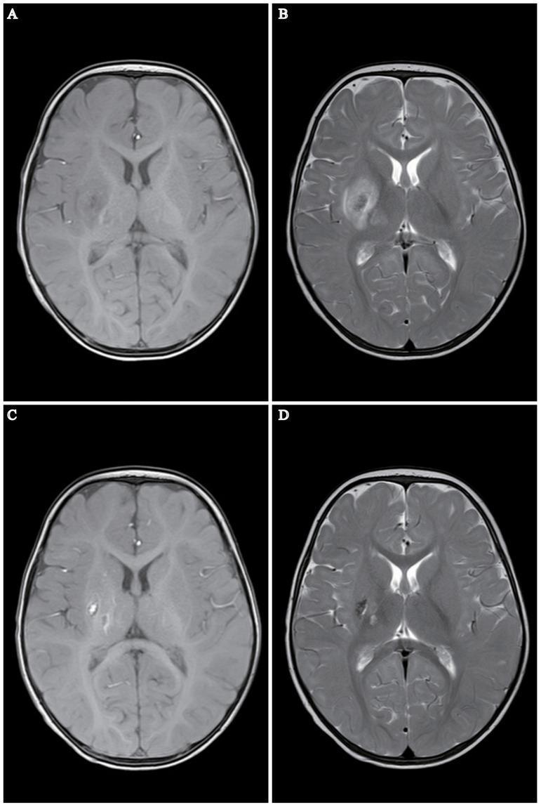
A multi-panel axial brain MRI series from a pediatric patient (7-month-old) demonstrating the evolution of a cerebral infarction. Panels A and B represent initial imaging, showing a patchy, heterogeneous lesion involving the right basal ganglia (putamen and globus pallidus) and the right thalamus. The lesion displays characteristic signal intensities for acute-to-subacute infarction, including long T1 and long T2 signals, with associated hyperintensity on DWI and FLAIR sequences indicating vasogenic and cytotoxic edema. Panels C and D show a one-month follow-up MRI. The previously identified lesion in the right basal ganglia and thalamus has significantly reduced in size. The signal has evolved into areas of encephalomalacia and gliosis, characterized by cystic-like changes and focal contraction of the brain tissue. There is a shift toward shorter T1 and T2 signals within parts of the lesion, suggesting the resolution of acute edema and the transition into a chronic post-ischemic state.
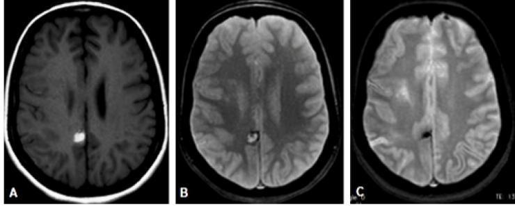
This diagnostic imaging composite displays three axial MRI brain slices (A, B, and C) illustrating the evolution of an intraparenchymal hematoma. Image A is a T1-weighted axial sequence showing a focal, rounded hyperintense (bright) lesion located in the posterior cingulate gyrus, indicative of acute to subacute hemorrhage. Image B is a T2*-weighted (gradient echo/susceptibility-weighted) sequence of the same lesion, appearing hyperintense with a distinct peripheral hypointense (dark) halo, representing early blood product breakdown. Image C shows a follow-up T2*-weighted axial scan during the chronic stage, demonstrating a significant reduction in the lesion's size and a transition to marked, uniform hypointensity. This dark signal in the chronic phase is characteristic of hemosiderin deposition. The comparison effectively demonstrates the radiological progression of intracranial hemorrhage and the utility of T2* sequences in detecting susceptibility effects from hemoglobin degradation products over time.
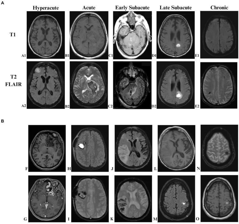
This composite educational graphic details the MRI characteristics of intracranial hemorrhage (ICH) through axial brain slices. Part A displays the evolution of blood signal intensity across five stages: hyperacute (oxyhemoglobin), acute (deoxyhemoglobin), early subacute (intracellular methemoglobin), late subacute (extracellular methemoglobin), and chronic (hemosiderin). Modalities include T1-weighted, T2 FLAIR, and Gradient Echo (GRE) sequences, illustrating the progression from isointense to bright T1 signals, and the eventual development of a hypointense 'blooming' rim on GRE indicative of chronic iron deposition. Part B serves as a diagnostic comparison chart for different etiologies of ICH. It features arteriovenous malformation (AVM) showing flow voids and post-contrast enhancement (F, G); cavernous hemangioma with a 'popcorn' core on T2 FLAIR and blooming artifact on GRE (H, I); hemorrhagic conversion of ischemic stroke with typical parenchymal distribution (J, K, L); and hemorrhagic metastasis displaying multifocal hyperintensities and significant perilesional edema (M, N, O). The image provides a comprehensive overview of neuroradiological signs essential for staging and diagnosing stroke-related and vascular pathologies.
https://globalrph.com/2025/10/can-mri-detect-old-strokes-new…
Can MRI Tell How Old a Brain Scar Is?
How the Brain "Ages" on MRI - The Science
The 5 Stages (Using Hemorrhage as the Clearest Example)
| Stage | Time After Injury | What's in the Tissue | T1 Signal | T2 Signal |
|---|---|---|---|---|
| Hyperacute | 0-6 hours | Fresh oxyhaemoglobin | Isointense (grey) | Bright |
| Acute | 6 hours - 3 days | Deoxyhaemoglobin | Dark | Very dark |
| Early subacute | 3-7 days | Intracellular methaemoglobin | Bright | Dark |
| Late subacute | 1-4 weeks | Extracellular methaemoglobin | Bright | Bright |
| Chronic | Months to years | Hemosiderin ring + gliosis | Dark rim | Dark rim |
"Acute subdural hematomas are initially isointense on T1 and hyperintense on T2, but as deoxyhemoglobin appears, the signal on T2-weighted images becomes hypointense. In the subacute phase, the signal is hyperintense on T1 and hypointense on T2..."
For a Pure Gliotic Scar (No Bleed) - The Clues Are Different
| What the Radiologist Sees | What It Means About Age |
|---|---|
| Swelling + bright DWI signal | Very recent injury - hours to days |
| Swelling still present + T2 bright | Subacute - days to 2 weeks |
| Swelling gone, FLAIR bright, tissue shrinking | Weeks to months old |
| Cavity (dark hole) + bright FLAIR rim around it | Months to years old - fully chronic |
| Shrunken tissue, ventricle pulled toward the scar, no swelling | Old scar - years old |
| Brain shrinkage (atrophy) in the region | Long-standing - years old |
"Chronic infarcts typically appear hypointense on T1 and hyperintense on T2/FLAIR images. These signal changes reflect tissue loss, gliosis, and encephalomalacia secondary to previous injury."
Visual Guide: Fresh vs. Old Scar
What the MRI Report Actually Says
- "Focal area of encephalomalacia and gliosis in the left frontal lobe, consistent with old traumatic injury"
- "Hemosiderin deposition noted, suggesting remote hemorrhagic contusion"
- "Ex vacuo dilation of the left lateral ventricle adjacent to area of gliosis, indicating chronic (long-standing) injury"
- "No restricted diffusion or contrast enhancement - findings consistent with chronic, not acute pathology"
The Limits - What MRI Cannot Tell You
| Limitation | Why |
|---|---|
| Cannot give an exact date | Biology varies between people - one person's 6-month scar can look like another's 2-year scar |
| Cannot distinguish 2 years from 10 years | Once fully chronic, all old scars look broadly similar |
| Confounding factors | Age of patient, medications (blood thinners change bleeding patterns), repeated injuries |
| No bleed = harder to date | Pure traumatic gliosis without hemorrhage has no hemoglobin clock to read |
| Individual variation | Some people scar faster or slower biologically |
Connecting Back to the Girl
- No swelling around the scar = not recent
- No restricted diffusion = not acute
- Hemosiderin rim (if there was any bleeding at impact) = consistent with years-old injury
- Shrunken/gliotic tissue with normal surrounding brain = chronic, established scar
- Tissue volume loss in the affected area = long-standing
Summary
| Question | MRI Answer |
|---|---|
| Is the injury recent or old? | ✅ Yes, reliably |
| Is it days vs. weeks? | ✅ Yes, using DWI + swelling |
| Is it months vs. acute? | ✅ Yes, very clearly |
| Is it 2 years vs. 10 years? | ⚠️ Rough estimate only |
| Exact date? | ❌ No - not possible |