
Veins histology
Reading File
Reading File
Reading File
Searching the Web
veins histology tunica intima media adventitia venous valves histology
Finding Sources
Searching Images
histology of vein cross section tunica intima thin media thick adventitia venous valve
Searching PubMed
veins histology
Reading File
Histology of veins
Veins are thin-walled, low-pressure vessels that return blood to the heart. Like arteries, they have three tunics, but their wall is less muscular and their lumen is usually wider and more irregular.
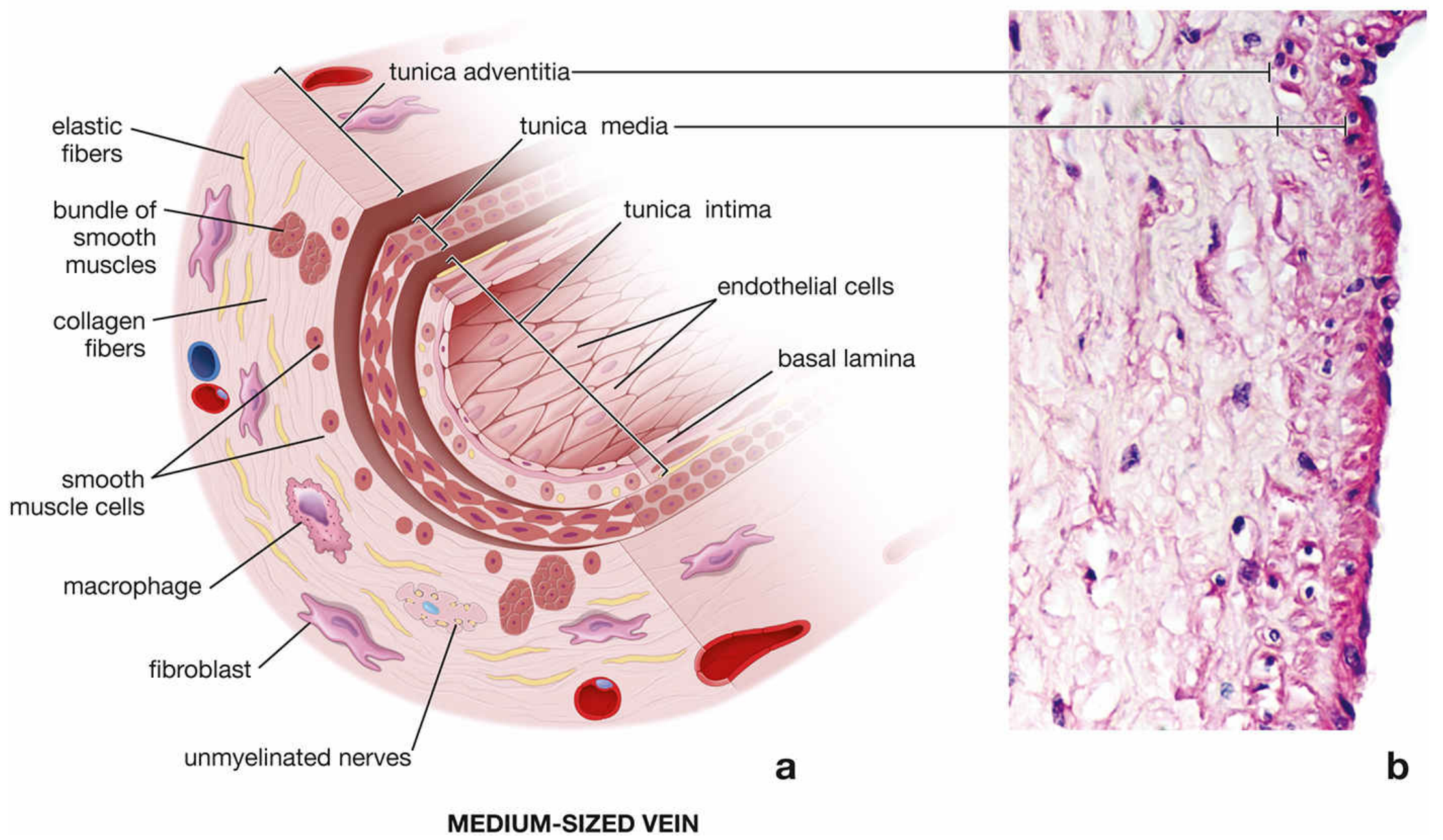
Layers of a vein
| Layer | Histological features |
|---|---|
| Tunica intima | Simple squamous endothelium on a basal lamina; very thin subendothelial connective tissue. The internal elastic lamina is absent or inconspicuous. |
| Tunica media | Much thinner than in an accompanying artery. Has few circular or spiral smooth-muscle layers, with collagen and elastic fibers. |
| Tunica adventitia | Usually the thickest layer of a medium or large vein. It contains abundant collagen, elastic fibers, fibroblasts, nerves, and in large veins, vasa vasorum. |
Types
-
Venules
- Postcapillary venules consist mainly of endothelium with pericytes.
- They are the principal site of leukocyte migration from blood into tissues.
- Muscular venules acquire 1-2 layers of smooth muscle.
-
Medium-sized veins
- Typical examples: radial, tibial, and popliteal veins.
- The three tunics are most easily identified.
- The adventitia is thicker than the media.
- Venous valves are folds of the tunica intima that prevent retrograde flow, particularly in the lower limbs.
-
Large veins
- Examples: vena cava and major pulmonary veins.
- Thin media; very thick adventitia.
- The adventitia may contain bundles of longitudinal smooth muscle and vasa vasorum.
- Large veins close to the heart can have myocardial muscle sleeves in the adventitia.
How to distinguish a vein from an artery on a slide
- Vein: wide, irregular or collapsed lumen; thin wall; media poorly developed; adventitia relatively thick.
- Artery: smaller, more circular lumen; thick muscular media; prominent elastic lamina in many arteries.
The medium-vein wall has endothelium and a thin subendothelial layer in its intima, a few smooth-muscle layers in its media, and a collagen-rich adventitia that is the thickest layer. Histology: A Text and Atlas with Correlated Cell and Molecular Biology, p. 1113.
This is a shared conversation. Sign in to Orris to start your own chat.