
Heme synthesis
I'll look up heme synthesis from the medical library first.
Reading File
Now I'll search for heme synthesis content in the library - running parallel searches for broad coverage.
Finding Sources
Finding Sources
Excellent results. Now I'll read the key sections in parallel - Lippincott Biochemistry (most detailed) and Basic Medical Biochemistry.
Reading File
Reading File
Good content. Let me now read more of the Lippincott section (steps 2-8 of heme biosynthesis) and fetch the key pathway diagram.
Reading File
Loading Image
Great. Let me fetch the protoporphyrin IX diagram and also read the rest of the biosynthesis steps (heme formation + regulation + porphyrias).
Loading Image
Reading File
I have the full pathway diagram showing PBG → Protoporphyrin IX steps. Let me now fetch the final heme formation image and read the porphyrias + regulation content.
Loading Image
Reading File
I now have all the information needed for a thorough answer. Let me compile the comprehensive response.
Heme Synthesis
Heme is an iron-porphyrin complex that serves as the prosthetic group of hemoglobin, myoglobin, cytochromes (including CYP450), catalase, and peroxidases. Its biosynthesis is one of the most important pathways in human biochemistry.
Structure of Heme
Heme consists of protoporphyrin IX (a type III porphyrin) coordinated with Fe²⁺ at its center. The porphyrin ring is formed by four pyrrole rings joined by methenyl (-CH=) bridges, with eight side chains (methyl, vinyl, propionate) in the specific order M-V-V-M-M-P-P-M around the ring.
Sites of Synthesis
- >85% of heme is synthesized in erythroid precursor cells of the bone marrow
- The liver is the second major site (produces CYP450 proteins)
- Mature RBCs cannot synthesize heme (lack mitochondria)
The pathway is split between mitochondria (first and last 3 steps) and the cytosol (intermediate steps).
The 8-Step Pathway
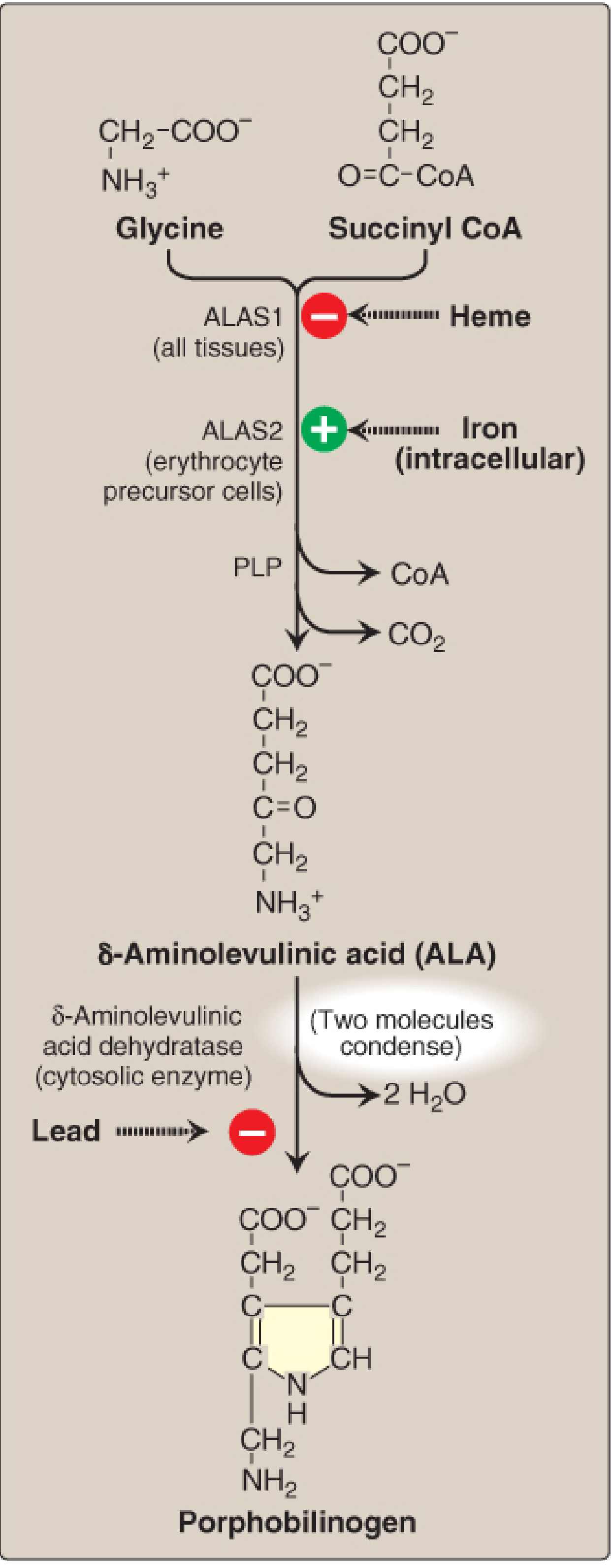
Step 1 - ALA Formation (Mitochondria) - RATE-LIMITING STEP
Glycine + Succinyl CoA → δ-Aminolevulinic acid (ALA)
- Enzyme: ALA synthase (ALAS); cofactor: pyridoxal phosphate (PLP, vitamin B6)
- This is the committed, rate-limiting step
- Two isoforms:
- ALAS1: ubiquitous (all tissues); regulated by heme (negative feedback - heme inhibits)
- ALAS2: erythroid-specific; stimulated by intracellular iron (positive regulation)
- Loss-of-function mutations in ALAS2 cause X-linked sideroblastic anemia
Step 2 - Porphobilinogen (PBG) Formation (Cytosol)
2 ALA → Porphobilinogen (PBG) + 2 H₂O
- Enzyme: ALA dehydratase (also called PBG synthase); zinc-containing
- Highly sensitive to inhibition by heavy metals (especially lead), which displace zinc
- This is one of the two steps inhibited in lead poisoning (causes elevated ALA)
Step 3 - Hydroxymethylbilane Formation (Cytosol)
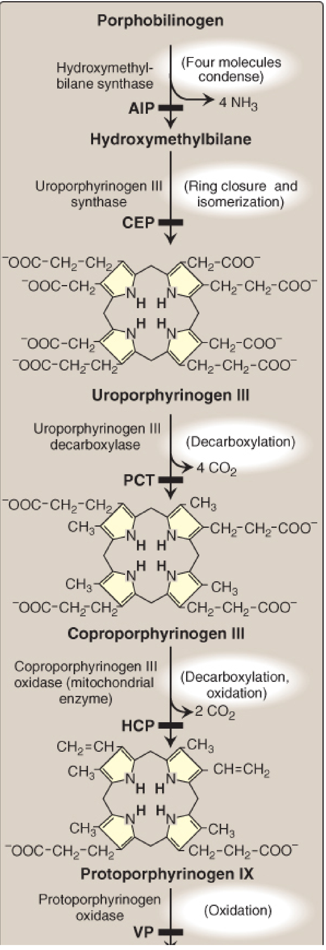
4 PBG → Hydroxymethylbilane + 4 NH₃
- Enzyme: Hydroxymethylbilane synthase (also called PBG deaminase or uroporphyrinogen I synthase)
- Deficiency: Acute Intermittent Porphyria (AIP) - most common acute hepatic porphyria
Step 4 - Uroporphyrinogen III Formation (Cytosol)
Hydroxymethylbilane → Uroporphyrinogen III
- Enzyme: Uroporphyrinogen III synthase (ring closure + isomerization)
- Creates the asymmetric type III porphyrin (physiologically active series)
- Deficiency: Congenital Erythropoietic Porphyria (CEP)
Step 5 - Coproporphyrinogen III Formation (Cytosol)
Uroporphyrinogen III → Coproporphyrinogen III + 4 CO₂
- Enzyme: Uroporphyrinogen III decarboxylase (UROD) - decarboxylates all 4 acetate groups to methyl groups
- Deficiency: Porphyria Cutanea Tarda (PCT) - most common porphyria overall
Step 6 - Protoporphyrinogen IX Formation (Mitochondria)
Coproporphyrinogen III → Protoporphyrinogen IX + 2 CO₂
- Enzyme: Coproporphyrinogen III oxidase (mitochondrial)
- Deficiency: Hereditary Coproporphyria (HCP)
Step 7 - Protoporphyrin IX Formation (Mitochondria)
Protoporphyrinogen IX → Protoporphyrin IX
- Enzyme: Protoporphyrinogen oxidase (6-electron oxidation)
- Deficiency: Variegate Porphyria (VP)
Step 8 - Heme Formation (Mitochondria) - FINAL STEP
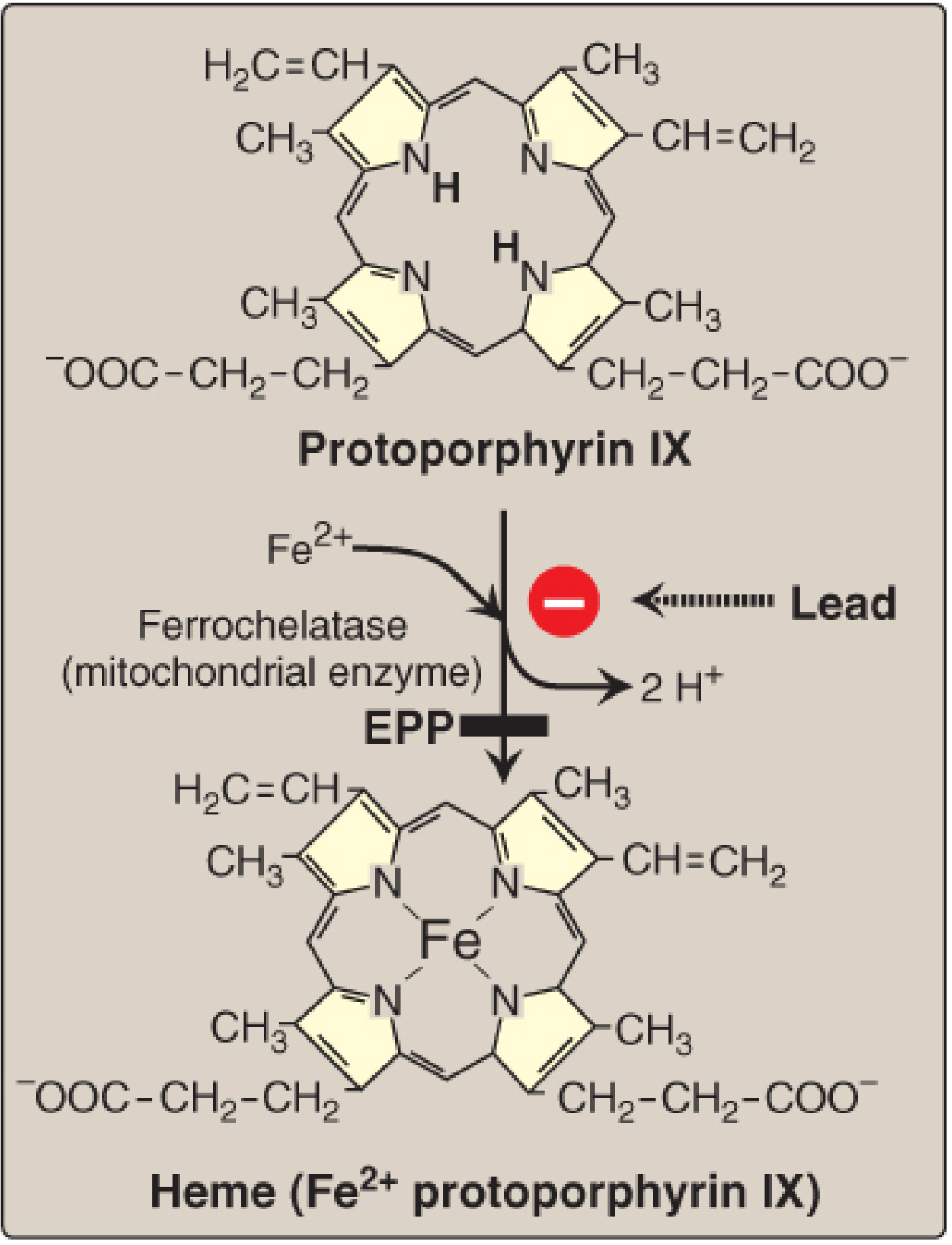
Protoporphyrin IX + Fe²⁺ → Heme + 2 H⁺
- Enzyme: Ferrochelatase (also called heme synthase; mitochondrial)
- Requires ferrous iron (Fe²⁺) - not ferric
- Inhibited by lead (second lead-sensitive step)
- Deficiency: Erythropoietic Protoporphyria (EPP)
Summary Table
| Step | Substrate → Product | Enzyme | Compartment | Disease if Deficient |
|---|---|---|---|---|
| 1 | Glycine + Succinyl CoA → ALA | ALAS (PLP) | Mitochondria | X-linked sideroblastic anemia (ALAS2) |
| 2 | 2 ALA → PBG | ALA dehydratase (Zn²⁺) | Cytosol | ALA dehydratase deficiency porphyria |
| 3 | 4 PBG → Hydroxymethylbilane | HMB synthase | Cytosol | AIP |
| 4 | HMB → Uroporphyrinogen III | Uroporphyrinogen III synthase | Cytosol | CEP |
| 5 | Uroporphyrinogen III → Coproporphyrinogen III | UROD | Cytosol | PCT (most common) |
| 6 | Coproporphyrinogen III → Protoporphyrinogen IX | Coproporphyrinogen oxidase | Mitochondria | HCP |
| 7 | Protoporphyrinogen IX → Protoporphyrin IX | Protoporphyrinogen oxidase | Mitochondria | VP |
| 8 | Protoporphyrin IX + Fe²⁺ → Heme | Ferrochelatase | Mitochondria | EPP |
Net inputs: 8 glycine + 8 succinyl CoA → 1 heme molecule
Regulation
In the Liver (ALAS1 regulation)
- Heme (hemin) acts as the key negative regulator - it represses ALAS1 gene transcription via the Erg-1 aporepressor/NAB corepressor system, and also promotes ALAS1 mRNA degradation before it enters the mitochondria
- When heme is depleted (e.g., by drugs that induce CYP450 synthesis), ALAS1 is upregulated, increasing ALA production
- Drugs (barbiturates, rifampin, alcohol, estrogens, griseofulvin) that induce CYP450 consume heme, thereby de-repressing ALAS1
In Erythroid Cells (ALAS2 regulation)
- Iron positively regulates ALAS2 - when iron is plentiful, ALAS2 mRNA translation is enhanced (via iron-responsive element/iron-regulatory protein system)
- Heme synthesis is matched to globin synthesis to ensure balanced hemoglobin production
Clinical Correlates
Lead Poisoning
Lead inhibits two enzymes: ALA dehydratase (step 2) and ferrochelatase (step 8). This causes:
- Elevated urinary ALA and coproporphyrin III
- Accumulation of zinc protoporphyrin (zinc substitutes for Fe²⁺ as ferrochelatase substrate)
- Microcytic anemia
- Neurotoxicity (ALA is toxic to neurons; lead crosses the blood-brain barrier)
- Treatment: chelation with succimer (DMSA) or EDTA for severe poisoning (>45 μg/dL)
Porphyrias - Clinical Classification
| Type | Disease | Enzyme Deficient | Manifestations |
|---|---|---|---|
| Acute hepatic | AIP | HMB synthase | Abdominal pain, neuropsychiatric, NO photosensitivity |
| Chronic hepatic | PCT | UROD | Photosensitivity, skin blistering, dark urine |
| Acute hepatic | HCP | Coproporphyrinogen oxidase | Abdominal + neuropsychiatric + photosensitivity |
| Acute hepatic | VP | Protoporphyrinogen oxidase | Abdominal + neuropsychiatric + photosensitivity |
| Erythropoietic | CEP | Uroporphyrinogen III synthase | Photosensitivity, hemolytic anemia (childhood onset) |
| Erythropoietic | EPP | Ferrochelatase | Photosensitivity (childhood onset) |
Key principle: Enzyme defects early in the pathway (before tetrapyrroles form) → neurovisceral/abdominal symptoms (from ALA/PBG accumulation). Defects later in the pathway → photosensitivity (from porphyrin accumulation).
Triggers for acute hepatic porphyrias: barbiturates, sulfonamides, rifampin, alcohol, estrogens, fasting, infections - all increase demand for CYP450, depleting heme and upregulating ALAS1.
Treatment of acute attacks: IV glucose (suppresses ALAS1), IV hemin (represses ALAS1 directly), supportive care. Givosiran (RNAi therapy targeting ALAS1 mRNA) is approved for recurrent acute hepatic porphyria.
Sources: Biochemistry, 8th ed - Lippincott Illustrated Reviews, pp. 783-792; Basic Medical Biochemistry - A Clinical Approach 6e, pp. 1520-1521
This is a shared conversation. Sign in to Orris to start your own chat.