
Management and diagnosis of paralysis
"paralysis" AND "diagnosis" AND "management"
upper motor neuron vs lower motor neuron lesion diagram paralysis
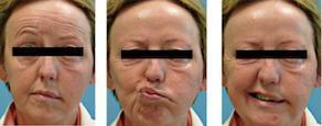
A series of three clinical photographs showing a female patient with complete left-sided peripheral facial nerve paralysis (House-Brackmann Grade VI). The images demonstrate classic clinical signs of a lower motor neuron lesion affecting the Cranial Nerve VII. There is significant facial asymmetry characterized by: (1) Total loss of forehead wrinkling on the left side during attempted elevation, (2) widened palpebral fissure on the left with potential lagophthalmos, and (3) effacement of the left nasolabial fold. During dynamic movements (middle and right frames), such as puckering or smiling, the mouth deviates markedly toward the unaffected right side due to the flaccid paralysis of the left circumoral muscles and unopposed contraction of the right-sided musculature. The cheek on the affected side appears flattened. This presentation is clinically relevant for diagnosing peripheral facial nerve injury, such as Bell's palsy or, as in this case context, secondary infiltration by a malignant process like lymphoma.
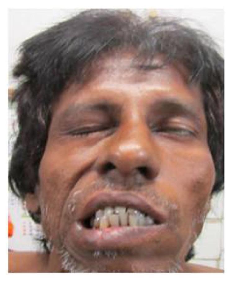
A clinical photograph of an adult male patient demonstrating classic signs of left-sided facial nerve paralysis (Bell's palsy), specifically a lower motor neuron (LMN) type lesion. The patient is shown attempting a facial expression, which highlights significant facial asymmetry. On the left side of the face (anatomical left), there is noticeable flattening of the nasolabial fold and inability to fully close the left eyelid (lagophthalmos), suggesting weakness of the orbicularis oculi muscle. The mouth exhibits marked deviation toward the unaffected right side due to the unopposed action of the contralateral facial muscles, indicating paralysis of the left buccinator, zygomaticus major, and orbicularis oris muscles. The absence of forehead wrinkling on the left side is consistent with an LMN pattern of cranial nerve VII palsy. This image serves as an educational example for neurological examination and the clinical presentation of House-Brackmann grade 5 facial dysfunction.
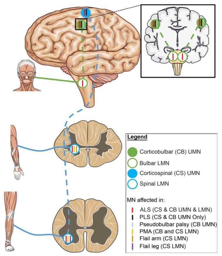
A medical schematic diagram illustrating the neuroanatomical pathways of upper motor neurons (UMN) and lower motor neurons (LMN) and their selective involvement in Amyotrophic Lateral Sclerosis (ALS) variants. The diagram features a lateral view of the human brain, a coronal section of the brain, and transverse sections of the spinal cord (cervical and lumbar levels). Two primary tracts are highlighted: the corticobulbar (CB) tract, represented by green solid circles (UMN) and open circles (LMN), and the corticospinal (CS) tract, represented by blue solid circles (UMN) and open circles (LMN). Solid circles represent UMN cell bodies in the motor cortex, while open circles denote LMNs in the brainstem (bulbar) and spinal cord. Colored vertical tick marks inside these circles indicate specific disease phenotypes: red for classic ALS (CS & CB UMN and LMN), black for Primary Lateral Sclerosis (PLS, UMN only), and yellow for Progressive Muscular Atrophy (PMA, LMN only). Other variants shown include Pseudobulbar palsy (CB UMN), Flail arm, and Flail leg (specific CS LMN segments). This illustration serves as an educational tool for differentiating motor neuron disease phenotypes based on the anatomical site of degeneration.
Paralysis: Diagnosis and Management
1. Definition and Classification
- Monoplegia - one limb
- Hemiplegia - one side of the body (arm + leg)
- Paraplegia - both legs (usually thoracic/lumbar SCI)
- Quadriplegia/Tetraplegia - all four limbs (usually cervical SCI)
- Diplegia - bilateral symmetric involvement
- Flaccid (hypotonic) - lower motor neuron (LMN) lesion
- Spastic (hypertonic) - upper motor neuron (UMN) lesion
2. UMN vs. LMN - The Key Diagnostic Distinction
| Feature | UMN Lesion | LMN Lesion |
|---|---|---|
| Tone | Increased (spastic) | Decreased (flaccid) |
| Deep tendon reflexes | Increased/hyperreflexia | Absent/diminished |
| Plantar response | Extensor (Babinski +) | Flexor |
| Muscle wasting | Minimal/late | Prominent/early |
| Fasciculations | Absent | Present |
| Location of lesion | Cortex, internal capsule, brainstem, spinal cord | Anterior horn cell, nerve root, peripheral nerve, NMJ |
3. Common Causes by Level
Cortical / Subcortical (UMN)
- Stroke (most common cause of acute hemiplegia)
- Brain tumors, abscesses
- Multiple sclerosis
- Traumatic brain injury
Spinal Cord (UMN or Mixed)
- Traumatic spinal cord injury (SCI)
- Cord compression (disc herniation, epidural hematoma, tumor)
- Transverse myelitis
- Vascular (anterior spinal artery syndrome)
Nerve Root / Peripheral Nerve (LMN)
- Guillain-Barre syndrome (ascending flaccid paralysis)
- Cauda equina syndrome
- Radiculopathy
- Mononeuropathy (e.g., common peroneal nerve palsy)
Neuromuscular Junction
- Myasthenia gravis
- Botulism (descending flaccid paralysis in conscious patient)
- Organophosphate poisoning
- Envenomation (Elapidae - kraits, cobras, mambas) causing descending flaccid paralysis from ptosis → ophthalmoplegia → facial paralysis → respiratory failure - Pye's Surgical Handicraft, p. 430
Muscle (Myopathic)
- Muscular dystrophies
- Inflammatory myopathies (dermatomyositis, polymyositis)
- Periodic paralysis (hypo/hyperkalemic)
4. Diagnostic Approach
History
- Onset (sudden = vascular/traumatic; subacute = inflammatory/infective; gradual = neoplastic/degenerative)
- Pattern of weakness, progression direction (ascending vs. descending)
- Associated symptoms: sensory loss, bladder/bowel dysfunction, pain, fever, recent infection
- Trauma, toxin exposure, tick bite (tick paralysis), prior episodes
Physical Examination
| Level | Loss of Function |
|---|---|
| C4 | Spontaneous breathing (diaphragm) |
| C5 | Shoulder shrug |
| C6 | Elbow flexion |
| C7 | Elbow extension |
| C8-T1 | Finger flexion |
| L1-L2 | Hip flexion |
| L4-L5 | Dorsiflexion of foot |
| S1-S2 | Plantar flexion |
| S2-S4 | Rectal sphincter tone |
- Rosen's Emergency Medicine, p. 437
- C6: biceps; C7: triceps; L4: patellar; S1: Achilles
Investigations
- MRI spine/brain - imaging of choice for cord/brain lesions; gadolinium-enhanced for tumors and Bell palsy workup
- CT scan - faster for trauma (fractures, hemorrhage); CT for thoracolumbar fractures to differentiate burst from wedge fracture (20-23% misdiagnosis rate with plain X-ray alone)
- EMG/Nerve conduction studies - localizes LMN vs. myopathic lesion
- Lumbar puncture - CSF analysis for Guillain-Barre, MS, infection
- Laboratory: CBC, metabolic panel, CK (rhabdomyolysis), acetylcholine receptor antibodies (myasthenia), toxicology
5. Management
A. Acute Traumatic Spinal Cord Injury
- C3-C5 injuries may require ventilatory support; C2 and above require immediate ventilation
- Phrenic nerve (C3-C5) supplies the diaphragm; injuries below C5 can still require ventilatory support depending on comorbidities
- Bradley and Daroff's Neurology, p. 558-560
- Transfer to ICU (preferably dedicated SCI unit) per 2013 AANS/CNS guidelines
- Maintain MAP >85 mmHg for 7 days to limit secondary cord ischemia
- Fluid resuscitation first; then vasopressors: norepinephrine (combined α/β agonist, preferred), dopamine, phenylephrine
- Neurogenic shock (bradycardia + hypotension) must be distinguished from hemorrhagic shock
- Bradley and Daroff's Neurology, p. 551-555
- Remains controversial and should NOT be given routinely in acute blunt SCI
- NASCIS II and III trials studied 30 mg/kg bolus then 5.4 mg/kg/hr for 23 hours
- The U.S. FDA has not approved corticosteroids for acute SCI; major neurosurgery societies advise against routine use
- "A treatment option at best and certainly not 'standard of care'" - Miller's Orthopaedics; Tintinalli's Emergency Medicine
- Indicated for progressive deficit, cord compression, unstable fractures, penetrating injuries
- Reduction of fracture-dislocation may not always be sufficient for decompression
B. Stroke (Hemiplegia)
- Ischemic stroke: IV tPA within 4.5 hours of symptom onset; mechanical thrombectomy for large vessel occlusion within 6-24 hours depending on imaging
- Hemorrhagic stroke: blood pressure control, neurosurgical consultation for eligible patients
- Early rehabilitation (physiotherapy, speech therapy) initiated as soon as medically stable
C. Bell Palsy (Peripheral Facial Nerve Paralysis)
- Corticosteroids (prednisolone) - strong evidence for benefit in preventing unsatisfactory recovery
- Antivirals (acyclovir or valaciclovir) added to steroids: combination is more effective than steroids alone; antivirals alone are insufficient
- Eye protection (lubricating drops, taping) to prevent exposure keratopathy
- Electroneuronography (ENoG) used to select patients for surgical vs. non-surgical management
- Differential diagnosis must exclude malignancy, herpes zoster (Ramsay Hunt), cholesteatoma, parotid tumors
- Bradley and Daroff's Neurology; Kanski's Ophthalmology; Tintinalli's Emergency Medicine
D. Guillain-Barre Syndrome
- IV Immunoglobulin (IVIg) or plasmapheresis - both equally effective, not combined
- Respiratory monitoring - forced vital capacity (FVC) <20 mL/kg or negative inspiratory force <-30 cmH2O indicates impending respiratory failure
- Supportive: DVT prophylaxis, pain management, autonomic monitoring
E. Myasthenia Gravis
- Acetylcholinesterase inhibitors (pyridostigmine) - symptomatic
- Immunosuppressants: prednisone, azathioprine, mycophenolate
- Thymectomy (especially with thymoma)
- Myasthenic crisis: IVIg or plasmapheresis, ICU admission
F. Envenomation-Induced Paralysis (Neurotoxic Snakebite)
- Anti-venom (specific to species)
- Mechanical ventilation for respiratory paralysis (can develop within 15-30 min after large elapid bites, though usually over several hours)
- Anticholinesterases (neostigmine) may partially reverse paralysis from some postsynaptic neurotoxins
- Pye's Surgical Handicraft, p. 430
6. Rehabilitation
- Physiotherapy: passive ROM exercises to prevent contractures, progressive strengthening, gait training, transfer training
- Occupational therapy: ADL training, adaptive equipment, splinting to prevent deformity
- Spasticity management: baclofen (oral or intrathecal), tizanidine, botulinum toxin injections, phenol nerve blocks
- Bladder/bowel program: intermittent catheterization, bowel regimen
- Pressure ulcer prevention: repositioning, specialized mattresses
- Psychological support: depression is extremely common post-paralysis
- Assistive devices: wheelchairs, AFOs (ankle-foot orthoses), FES (functional electrical stimulation)
7. UMN/LMN Pathway Diagram
Key Summary Points
- Localize first - cortex, spinal cord, nerve root, NMJ, or muscle. The UMN vs. LMN distinction (tone, reflexes, Babinski) drives workup and management.
- MRI is the gold standard for cord/brain imaging; CT for acute trauma (fractures).
- Steroids in SCI are no longer standard of care - do not give routinely.
- Bell palsy - treat with prednisolone + acyclovir/valaciclovir within 72 hours for best outcomes.
- Stroke - time is brain; act on IV tPA/thrombectomy windows immediately.
- GBS - watch respiratory function closely; IVIg or plasmapheresis.
- Rehabilitation is not an afterthought - early initiation improves functional outcomes significantly.