
Dermatome and mayoyomes of upperlimb explain
dermatome upper limb diagram sensory nerve distribution
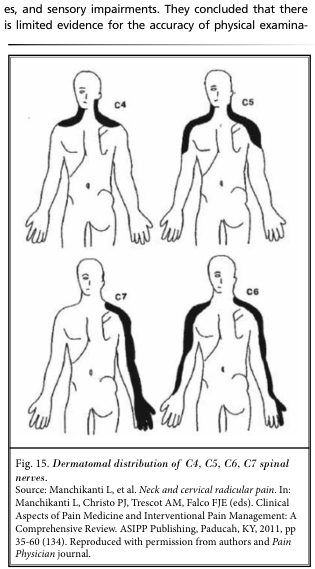
Summary : This figure illustrates the dermatomal distribution patterns of the C4, C5, C6, and C7 spinal nerves on the human body, showing the specific skin areas innervated by each nerve root. illustration: # Overview : • Four schematic diagrams of the human upper body (front and back views) are shown, each labeled with a different cervical spinal nerve (C4, C5, C6, C7). • Each diagram highlights a distinct region of the skin corresponding to the sensory distribution (dermatome) of the respective nerve. # Dermatome Regions : • C4: – Shaded area covers the lower neck, upper shoulder, and extends over the clavicle region. • C5: – Shaded area includes the lateral (outer) aspect of the upper arm and shoulder, extending down to just above the elbow. • C6: – Shaded area runs from the lateral neck and shoulder, down the lateral arm and forearm, to include the thumb. • C7: – Shaded area extends from the posterior neck and shoulder, down the posterior arm and forearm, to include the middle finger. # Design Encodings : • Black shading is used to indicate the dermatome for each nerve root on a simplified human outline. • Each diagram is labeled in the upper left or right with the corresponding nerve root (C4, C5, C6, C7). # Spatial Relationships : • The diagrams are arranged in a 2x2 grid, with C4 and C5 on the top row, C7 and C6 on the bottom row. • Each dermatome is shown on one side of the body for clarity. # Analysis : • The figure visually distinguishes the unique, non-overlapping cutaneous territories supplied by each cervical nerve root. • The C4 dermatome is most superior and medial, while C7 is more posterior and extends to the middle finger. • The C6 dermatome uniquely includes the thumb, and C5 is limited to the upper lateral arm. • This layout aids in clinical localization of nerve root pathology based on sensory symptoms.
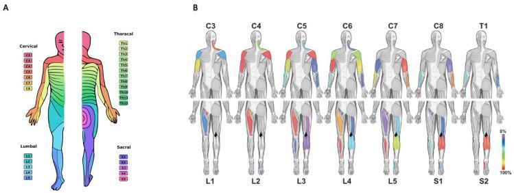
This medical illustration displays the sensory and motor innervation patterns of the human spinal nerves. Panel A is a dermatome map showing the horizontal and longitudinal sensory distribution of 31 pairs of spinal nerves (Cervical C2-C8, Thoracic Th1-Th12, Lumbar L1-L5, and Sacral S1-S5) across both anterior and posterior views of the human body. Panel B illustrates myotomal distributions through 14 distinct anatomical figures. The top row tracks cervical and first thoracic (C3-T1) root contributions to upper limb musculature, while the bottom row tracks lumbosacral (L1-S2) root contributions to the lower limbs. Specific muscle groups are highlighted with a color-coded heat map (0-100%) to represent response rates during intraoperative nerve root stimulation. This educational graphic serves as a clinical reference for mapping peripheral neurological deficits to their corresponding spinal levels, useful in neurology, physiatry, and neurosurgery for diagnosing nerve root entrapment or spinal cord injuries.
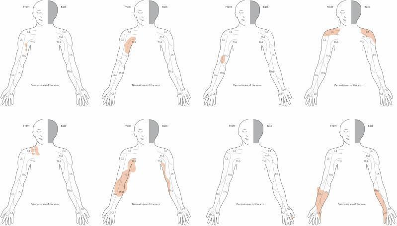
This medical illustration consists of eight schematic drawings depicting dermatomal distribution of the upper limb and hand, used to map the extent of plexiform neurofibromas (PNF). Each figure shows a frontal and dorsal (back) outline of a human torso and right arm. The dermatomes are labeled C4 through C8 and Th1 through Th3, illustrating the sensory innervation pathways from the shoulder to the fingertips. Areas of tumor involvement—representing either diffuse cutaneous neurofibroma or subcutaneous nodular PNF—are shaded in light orange/brown. The diagrams demonstrate the variable presentation of Neurofibromatosis type 1 (NF1), ranging from small, localized lesions restricted to a single dermatome (e.g., C5 or Th2) to large, confluent masses that cross multiple dermatomal boundaries and alter the anatomical contour of the limb. This visual tool aids in the clinical assessment and surgical planning of PNF by correlating physical tumor topography with spinal nerve segments, highlighting that tumor expansion often occurs independently of strict anatomical dermatomal borders.
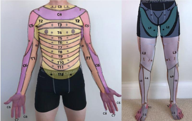
This clinical dermatome map, presented as an anatomical diagram overlaid on a human subject, illustrates the cutaneous sensory distribution of spinal nerves. The anterior trunk displays a sequential horizontal banding pattern representing thoracic dermatomes T2 through T12, with T4 roughly at the nipple line and T10 at the umbilicus. The upper extremities show cervical and upper thoracic distribution: C5 (lateral upper arm), C6 (lateral forearm and thumb), C7 (middle finger), C8 (medial hand and little finger), and T1 (medial forearm and arm). The lower extremity map details the lumbar and sacral levels: L1 (inguinal region), L2 (anterior mid-thigh), L3 (distal thigh and knee), L4 (medial lower leg), L5 (lateral lower leg and dorsal foot), and S1 (lateral foot and small toe). This resource is designed for primary care musculoskeletal medicine to assist in the neurological examination of sensory deficits, radiculopathy, or spinal cord injury level localization.
myotome upper limb muscle nerve root innervation
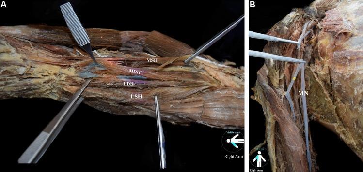
Anatomical dissection of a right upper limb demonstrating a four-headed variant of the brachialis muscle and associated neural supply. Panel A shows the gross morphology of the brachialis muscle, subdivided into four distinct bellies: the main superficial head (MSH), main deep head (MDH), lateral superficial head (LSH), and lateral deep head (LDH). The bellies exhibit a light tan color with clear longitudinal striations and are retracted with surgical instruments to show their relative deep-to-superficial layering. Panel B illustrates the innervation pattern in the absence of the musculocutaneous nerve. The median nerve (MN), depicted as a white, linear trunk, is shown providing atypical muscular branches (marked with an asterisk) to the anterior compartment of the arm. This clinical image highlights rare anatomical variations in upper limb myology and neurovascular architecture, emphasizing the surgical significance of identifying accessory muscle heads and variant motor innervation from the median nerve during orthopedic or neurosurgical procedures.
This medical illustration displays the sensory and motor innervation patterns of the human spinal nerves. Panel A is a dermatome map showing the horizontal and longitudinal sensory distribution of 31 pairs of spinal nerves (Cervical C2-C8, Thoracic Th1-Th12, Lumbar L1-L5, and Sacral S1-S5) across both anterior and posterior views of the human body. Panel B illustrates myotomal distributions through 14 distinct anatomical figures. The top row tracks cervical and first thoracic (C3-T1) root contributions to upper limb musculature, while the bottom row tracks lumbosacral (L1-S2) root contributions to the lower limbs. Specific muscle groups are highlighted with a color-coded heat map (0-100%) to represent response rates during intraoperative nerve root stimulation. This educational graphic serves as a clinical reference for mapping peripheral neurological deficits to their corresponding spinal levels, useful in neurology, physiatry, and neurosurgery for diagnosing nerve root entrapment or spinal cord injuries.
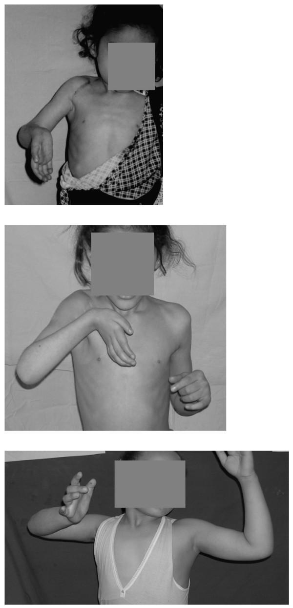
This sequence of three clinical photographs documents the longitudinal outcomes of reconstructive nerve surgery (neurotization) for late-presentation brachial plexus injuries in pediatric patients. The images illustrate the progression of upper limb function and joint alignment over a four-year period. Initial frames show a patient with classic signs of C5-T1 nerve root involvement, including a severe supination deformity of the forearm, wrist extension deformity, and shoulder weakness characterized by biceps co-contraction during attempted abduction. Middle and final frames demonstrate varying degrees of recovery in the deltoid and infraspinatus muscles, leading to improved active shoulder abduction and some restoration of forearm pronation. The visual data highlights the clinical significance of nerve augmentation for improving muscle power in the biceps, deltoids, and wrist extensors, while also tracking the mitigation of pathological co-contractions. This case study serves as an educational reference for orthopedic and neurological specialists evaluating the efficacy of phrenic-to-suprascapular and contralateral C7 neurotization techniques in pediatric brachial plexus birth palsy (BPBP) or traumatic injury management.
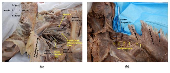
Two high-resolution cadaveric dissection photographs demonstrating the anatomical course and innervation of the dorsal scapular nerve (DSN). Image (a) presents an anterolateral view of the right neck in a supine position. Key structures identified include the phrenic nerve, anterior and middle scalene muscles, and the superior trunk of the brachial plexus. The DSN is labeled originating from the C4 root and is shown piercing the middle scalene muscle. Image (b) shows a posterior view in the prone position with the rhomboid major and minor muscles reflected laterally. This view clearly illustrates the posteroinferior trajectory of the DSN as it supplies the levator scapulae, rhomboid minor, and rhomboid major muscles. The dissections highlight the variability in nerve origin and its critical relationship to the scalene musculature and the posterior scapular muscle group. This material is designed for advanced anatomical study, specifically focusing on peripheral nerve pathways and surgical landmarks in the neck and upper back.
Dermatome and mayotomes of upper limb explain
dermatome upper limb cervical nerve sensory distribution diagram
Summary : This figure illustrates the dermatomal distribution patterns of the C4, C5, C6, and C7 spinal nerves on the human body, showing the specific skin areas innervated by each nerve root. illustration: # Overview : • Four schematic diagrams of the human upper body (front and back views) are shown, each labeled with a different cervical spinal nerve (C4, C5, C6, C7). • Each diagram highlights a distinct region of the skin corresponding to the sensory distribution (dermatome) of the respective nerve. # Dermatome Regions : • C4: – Shaded area covers the lower neck, upper shoulder, and extends over the clavicle region. • C5: – Shaded area includes the lateral (outer) aspect of the upper arm and shoulder, extending down to just above the elbow. • C6: – Shaded area runs from the lateral neck and shoulder, down the lateral arm and forearm, to include the thumb. • C7: – Shaded area extends from the posterior neck and shoulder, down the posterior arm and forearm, to include the middle finger. # Design Encodings : • Black shading is used to indicate the dermatome for each nerve root on a simplified human outline. • Each diagram is labeled in the upper left or right with the corresponding nerve root (C4, C5, C6, C7). # Spatial Relationships : • The diagrams are arranged in a 2x2 grid, with C4 and C5 on the top row, C7 and C6 on the bottom row. • Each dermatome is shown on one side of the body for clarity. # Analysis : • The figure visually distinguishes the unique, non-overlapping cutaneous territories supplied by each cervical nerve root. • The C4 dermatome is most superior and medial, while C7 is more posterior and extends to the middle finger. • The C6 dermatome uniquely includes the thumb, and C5 is limited to the upper lateral arm. • This layout aids in clinical localization of nerve root pathology based on sensory symptoms.
This medical illustration displays the sensory and motor innervation patterns of the human spinal nerves. Panel A is a dermatome map showing the horizontal and longitudinal sensory distribution of 31 pairs of spinal nerves (Cervical C2-C8, Thoracic Th1-Th12, Lumbar L1-L5, and Sacral S1-S5) across both anterior and posterior views of the human body. Panel B illustrates myotomal distributions through 14 distinct anatomical figures. The top row tracks cervical and first thoracic (C3-T1) root contributions to upper limb musculature, while the bottom row tracks lumbosacral (L1-S2) root contributions to the lower limbs. Specific muscle groups are highlighted with a color-coded heat map (0-100%) to represent response rates during intraoperative nerve root stimulation. This educational graphic serves as a clinical reference for mapping peripheral neurological deficits to their corresponding spinal levels, useful in neurology, physiatry, and neurosurgery for diagnosing nerve root entrapment or spinal cord injuries.
This clinical dermatome map, presented as an anatomical diagram overlaid on a human subject, illustrates the cutaneous sensory distribution of spinal nerves. The anterior trunk displays a sequential horizontal banding pattern representing thoracic dermatomes T2 through T12, with T4 roughly at the nipple line and T10 at the umbilicus. The upper extremities show cervical and upper thoracic distribution: C5 (lateral upper arm), C6 (lateral forearm and thumb), C7 (middle finger), C8 (medial hand and little finger), and T1 (medial forearm and arm). The lower extremity map details the lumbar and sacral levels: L1 (inguinal region), L2 (anterior mid-thigh), L3 (distal thigh and knee), L4 (medial lower leg), L5 (lateral lower leg and dorsal foot), and S1 (lateral foot and small toe). This resource is designed for primary care musculoskeletal medicine to assist in the neurological examination of sensory deficits, radiculopathy, or spinal cord injury level localization.
myotome upper limb nerve root muscle innervation table
Anatomical dissection of a right upper limb demonstrating a four-headed variant of the brachialis muscle and associated neural supply. Panel A shows the gross morphology of the brachialis muscle, subdivided into four distinct bellies: the main superficial head (MSH), main deep head (MDH), lateral superficial head (LSH), and lateral deep head (LDH). The bellies exhibit a light tan color with clear longitudinal striations and are retracted with surgical instruments to show their relative deep-to-superficial layering. Panel B illustrates the innervation pattern in the absence of the musculocutaneous nerve. The median nerve (MN), depicted as a white, linear trunk, is shown providing atypical muscular branches (marked with an asterisk) to the anterior compartment of the arm. This clinical image highlights rare anatomical variations in upper limb myology and neurovascular architecture, emphasizing the surgical significance of identifying accessory muscle heads and variant motor innervation from the median nerve during orthopedic or neurosurgical procedures.
This medical illustration displays the sensory and motor innervation patterns of the human spinal nerves. Panel A is a dermatome map showing the horizontal and longitudinal sensory distribution of 31 pairs of spinal nerves (Cervical C2-C8, Thoracic Th1-Th12, Lumbar L1-L5, and Sacral S1-S5) across both anterior and posterior views of the human body. Panel B illustrates myotomal distributions through 14 distinct anatomical figures. The top row tracks cervical and first thoracic (C3-T1) root contributions to upper limb musculature, while the bottom row tracks lumbosacral (L1-S2) root contributions to the lower limbs. Specific muscle groups are highlighted with a color-coded heat map (0-100%) to represent response rates during intraoperative nerve root stimulation. This educational graphic serves as a clinical reference for mapping peripheral neurological deficits to their corresponding spinal levels, useful in neurology, physiatry, and neurosurgery for diagnosing nerve root entrapment or spinal cord injuries.
This sequence of three clinical photographs documents the longitudinal outcomes of reconstructive nerve surgery (neurotization) for late-presentation brachial plexus injuries in pediatric patients. The images illustrate the progression of upper limb function and joint alignment over a four-year period. Initial frames show a patient with classic signs of C5-T1 nerve root involvement, including a severe supination deformity of the forearm, wrist extension deformity, and shoulder weakness characterized by biceps co-contraction during attempted abduction. Middle and final frames demonstrate varying degrees of recovery in the deltoid and infraspinatus muscles, leading to improved active shoulder abduction and some restoration of forearm pronation. The visual data highlights the clinical significance of nerve augmentation for improving muscle power in the biceps, deltoids, and wrist extensors, while also tracking the mitigation of pathological co-contractions. This case study serves as an educational reference for orthopedic and neurological specialists evaluating the efficacy of phrenic-to-suprascapular and contralateral C7 neurotization techniques in pediatric brachial plexus birth palsy (BPBP) or traumatic injury management.
Dermatomes and Myotomes of the Upper Limb
What is a Dermatome?
What is a Myotome?
DERMATOMES of the Upper Limb
| Nerve Root | Area of Skin Supplied |
|---|---|
| C4 | Lower neck, shoulder tip, over clavicle |
| C5 | Lateral (outer) aspect of upper arm and shoulder, lateral deltoid region |
| C6 | Lateral forearm, thumb, and index finger |
| C7 | Middle (long) finger; posterior arm and forearm |
| C8 | Ring finger, little finger, and medial forearm |
| T1 | Medial arm (inner upper arm) and axilla |
| T2 | Medial arm just below axilla (meets T1 territory) |
- C6 = Thumb (index + thumb)
- C7 = Middle finger (the "tallest" finger for C7 - the largest root)
- C8 = Little finger (ring + little)
- T1 = Medial arm
MYOTOMES of the Upper Limb
| Nerve Root | Key Movement | Key Muscle(s) | Peripheral Nerve |
|---|---|---|---|
| C5 | Shoulder abduction | Deltoid | Axillary nerve |
| C5-C6 | Elbow flexion | Biceps, Brachialis | Musculocutaneous nerve |
| C6 | Wrist extension, elbow supination | Extensor carpi radialis, Brachioradialis | Radial nerve |
| C6-C7 | Elbow extension, wrist extension | Triceps, Extensor carpi radialis | Radial nerve |
| C7 | Wrist flexion | Flexor carpi radialis | Median nerve |
| C7-C8 | Finger extension | Extensor digitorum | Radial nerve (posterior interosseous) |
| C7-C8 | Finger flexion (DIP joints) | Flexor digitorum profundus | Median + Ulnar nerve |
| C8 | Finger flexion | Flexor digitorum superficialis | Median nerve |
| C8-T1 | Thumb opposition | Opponens pollicis | Median nerve |
| C8-T1 | Finger abduction/adduction | Dorsal/palmar interossei | Ulnar nerve |
| T1 | Finger abduction (index) | Dorsal interosseous | Ulnar nerve |
- Neuroanatomy through Clinical Cases 3rd Edition, Table 3.4
REFLEXES (Quick Reference)
| Reflex | Root Level |
|---|---|
| Biceps jerk | C5-C6 |
| Brachioradialis jerk | C5-C6 |
| Triceps jerk | C6-C7 |
- Sabiston Textbook of Surgery
Combined Summary Table (Clinical Exam)
| Root | Motor (Myotome) | Sensory (Dermatome) | Reflex |
|---|---|---|---|
| C5 | Deltoid (shoulder abduction) | Lateral shoulder / lateral arm | Biceps |
| C6 | Biceps + wrist extension | Lateral forearm, thumb, index finger | Brachioradialis |
| C7 | Triceps + wrist flexion | Middle finger | Triceps |
| C8 | Hand intrinsics + finger flexors | Ring and little finger, medial forearm | None standard |
| T1 | Hand intrinsics + finger abduction | Medial arm / axilla | None standard |
- Rockwood and Green's Fractures in Adults 10th Ed 2025, Table 40-1
Clinical Importance
- Cervical disc prolapse (radiculopathy): A herniated C5-C6 disc compresses the C6 root - causing sensory loss in the thumb/index finger, weak elbow flexion, and reduced brachioradialis reflex.
- Spinal cord injury (ASIA grading): Myotomal and dermatomal testing determines the neurological level of injury.
- Brachial plexus injuries: Upper trunk injuries (C5-C6 = Erb's palsy) vs lower trunk injuries (C8-T1 = Klumpke's palsy) produce distinct deficits predictable from the tables above.
- Herpes zoster: Painful vesicular rash follows a dermatomal distribution - if it tracks down the lateral forearm to the thumb, the C6 ganglion is involved.
Tip to remember the dermatomes: Think of the arm reaching out with the thumb pointing up - sensory innervation runs from C5 (shoulder) → C6 (thumb side, lateral) → C7 (middle, posterior) → C8 (little finger side, medial) → T1 (inner arm) from lateral to medial.
Create a quick reference PDF of dermatomes and myotomes for clinical use
ls -la /tmp/workspace/ 2>/dev/null || mkdir -p /tmp/workspace && echo "workspace ready"