
Teach me leukemias and lymphomas in detail with pneumonics to learn it
leukemia blood smear lymphoblast
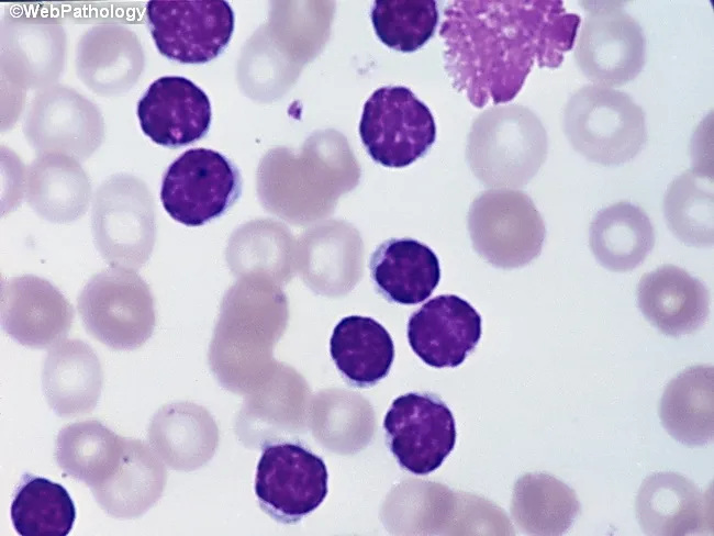
Peripheral blood smear from a patient with suspected chronic lymphocytic leukemia (CLL) shows a predominance of small, mature-appearing lymphocytes with scant cytoplasm and clumped nuclear chromatin, accompanied by fragile cells that readily disrupt during smear preparation, producing numerous smudge or basket cells visible on the slide. The image depicts a single smudge cell in the upper right, with numerous small darkly staining lymphocytes occupying the field. The smear was prepared with Wright-Giemsa stain and examined under light microscopy at high magnification (approximately 1000x total magnification with oil immersion). Clinically, CLL is defined by absolute mature lymphocytosis of ≥5.0 × 10^9/L sustained for ≥3 months, often asymptomatic and detected incidentally on complete blood count. Morphology alone is insufficient for diagnosis; immunophenotyping showing co-expression of CD5, CD19, CD23 with weak surface immunoglobulin and light chain restriction supports CLL diagnosis. Smudge cells result from fragile leukemic cells and reflect underlying cytoskeletal fragility, though they are not specific. The image illustrates typical features: monomorphic lymphocytes, scant cytoplasm, condensed chromatin, and absent prominent nucleoli. This finding has diagnostic significance, guiding further workup, including flow cytometry, cytogenetics, and molecular studies, to distinguish CLL from other chronic lymphoproliferative disorders such as small lymphocytic lymphoma (SLL), mantle cell lymphoma, prolymphocytic leukemia, and reactive lymphocytosis.
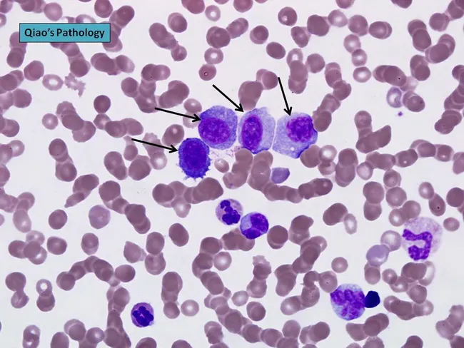
Peripheral blood smear prepared from a patient with suspected plasma cell leukemia (PCL) was examined by bright‑field light microscopy after Wright‑Giemsa staining. The smear shows several circulating plasma‑cell–like elements, including four conspicuous proplasmacytes with dispersed nuclear chromatin and prominent nucleoli, as indicated by arrows in the original figure. In addition, background erythrocytes exhibit rouleaux formation, a common feature in paraproteinemic states. The cells display eccentrically placed nuclei with pronounced basophilic cytoplasm and a coarse chromatin pattern; occasional cells show a perinuclear clearing (Golgi rim) suggestive of plasmacytic differentiation. Immunoglobulin light‑chain restriction is not determined on this smear alone but flow cytometry or immunohistochemistry would typically confirm clonal plasma cells. Relative proportions indicate that neoplastic plasma cells constitute a substantial fraction of leukocytes, consistent with plasma cell leukemia rather than reactive plasmacytosis. PCL is an aggressive myelomatous process often presenting with cytopenias, organomegaly, and sometimes lymphadenopathy; prognosis is poor, and treatment responsiveness is limited. This image illustrates characteristic morphologic features used for differential diagnosis against multiple myeloma, Waldenström macroglobulinemia, and other causes of plasmacytosis. Clinically relevant keywords include plasma cells, rouleaux, Wright‑Giemsa stain, 100X oil, peripheral blood, leukemia, myeloma, and hematologic malignancy.
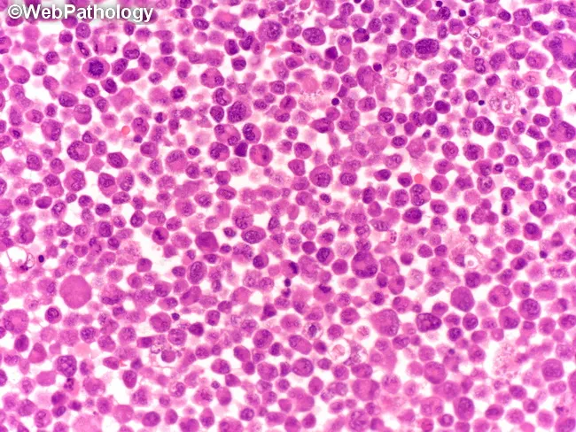
This brightfield light microscopy image depicts a dense bone marrow aspirate smear stained with hematoxylin and eosin. The cellularity is high with a uniform population of small to medium-sized lymphoid cells (lymphoblast-like cells) occupying much of the field. Nuclei are round to ovoid with finely dispersed, coarse chromatin, and scant basophilic cytoplasm; nucleoli are inconspicuous. The architecture is diffuse rather than follicular, with little evidence of erythroid or myeloid precursors; occasional mitotic figures may be present but are not prominent at this magnification. The histology shows a monotonous lymphoid infiltrate, a pattern that raises concern for a lymphoblastic process. Pathologic interpretation would emphasize lymphoid blasts consistent with acute lymphoblastic leukemia or lymphoblastic lymphoma; however, definitive classification requires ancillary studies such as flow cytometry immunophenotyping, immunohistochemistry, and genetic studies (cytogenetics/molecular). Clinically, such a pattern correlates with acute presentations: cytopenias, signs of marrow failure, fatigue, infections, or organomegaly. Potential uses include medical education for recognizing quantitative lymphoid blasts on bone marrow smears, training in differential diagnoses of monomorphic blue cell infiltrates, and optimizing search queries for pathology image datasets. Keywords include: lymphoblasts, pediatric ALL, bone marrow failure, marrow infiltration, hematopathology, immunophenotype, CD markers, prognosis guidance, diagnostic workflow, and educational value overall.
Hodgkin lymphoma Reed Sternberg cell histology
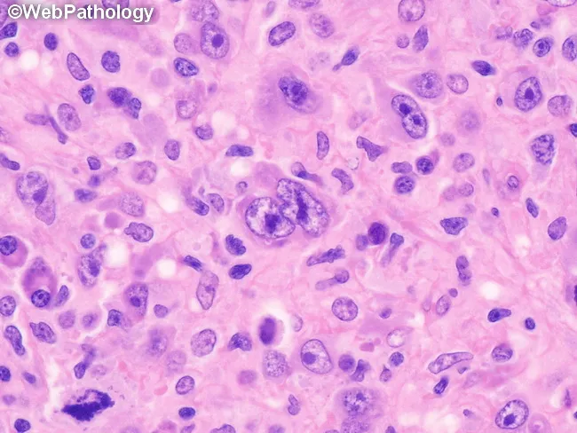
This histopathology image depicts a lymph node biopsy showing features diagnostic of lymphocyte-depleted classic Hodgkin lymphoma, reticular subtype. Using light microscopy on an H&E-stained paraffin section, the lymphoid architecture is markedly effaced by a cellular sheet of large, atypical mononuclear cells with prominent nucleoli. Occasional Reed-Sternberg cells are visible within the expansive background, including cells with multilobed or bilobed nuclei and prominent eosinophilic nucleoli, characteristic of Hodgkin lymphoma. The RS cells are scattered among numerous atypical mononuclear variants rather than forming a cohesive nodular structure. There is little-to-no fibrosis and only sparse non-neoplastic inflammatory cells, which is typical for the lymphocyte-depleted pattern. The background may show rimming by small lymphocytes and histiocytes in a reticular network, but overall cellularity is high. Immunophenotypic markers are not shown here, but in clinical practice RS cells typically express CD30 and CD15 with weaker PAX5 or B-cell markers. The diagnostic significance lies in recognizing the lymphocyte-depleted cHL morphology, which carries distinct clinical implications, often presenting with advanced stage disease and systemic symptoms. This image is useful for educational purposes, differential diagnosis conversation, and correlating histology with treatment planning (ABVD/BEACOPP regimens). Correlation with immunohistochemistry (CD30, CD15, PAX5) and EBV status further supports diagnosis in practice.
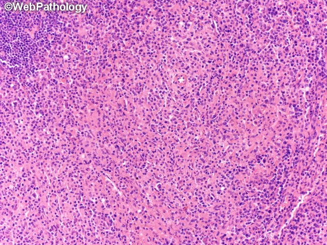
This brightfield histopathology image depicts a lymph node biopsy illustrating a histiocyte-rich variant of mixed cellularity classical Hodgkin lymphoma. The tissue is sectioned and stained with hematoxylin and eosin, viewed at low-to-intermediate magnification, revealing a densely cellular background with a prominent infiltrate of histiocytes and epithelioid macrophages forming aggregates, accompanied by scattered eosinophils, lymphocytes, plasma cells, and occasional neutrophils. The classic Reed-Sternberg cells are present but may be sparse within the abundant histiocytic milieu, with multilobed nuclei and prominent nucleoli. The architectural pattern is polymorphic rather than nodular, lacking prominent fibrous bands characteristic of nodular sclerosis. The histiocyte-rich variant features abundant macrophages mirroring granulomatous-like features, potentially mimicking inflammatory conditions; however, residual neoplastic Hodgkin cells in this setting bear the typical immunophenotype (CD30+, CD15+, PAX5 weak) in adjacent areas. Clinically, this histology correlates with mixed cellularity Hodgkin lymphoma, a B-cell origin neoplasm presenting with constitutional symptoms and lymphadenopathy. The image is relevant for educational demonstration of HL subtypes, differential diagnoses with non-Hodgkin lymphomas and granulomatous processes, and for training in histopathologic recognition, pattern recognition, and morphologic correlation with immunophenotype and clinical findings. Immunohistochemistry supports diagnosis by highlighting Reed-Sternberg cells (CD30+, CD15+, PAX5 weak) amid a histiocyte-rich background; EBV association may be variable; clinical staging and treatment follow Hodgkin lymphoma guidelines.
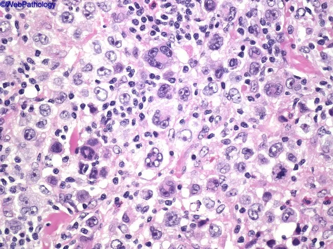
This high-power light micrograph of a lymph node biopsy, stained with Hematoxylin and Eosin, demonstrates classical Hodgkin lymphoma histology. The tissue is densely cellular with a polymorphic inflammatory background comprising small lymphocytes, eosinophils, plasma cells, and histiocytes. Scattered among the background are large binucleated Reed-Sternberg (RS) cells exhibiting prominent, mirror-image eosinophilic nucleoli and ample cytoplasm, giving an owl's eye appearance. Mononuclear RS variants with similar nuclear features are present, reflecting heterogeneity within the malignant cell population. The RS cells may appear with delicate nuclear membranes and occasional lobulation. The surrounding milieu often includes reactive T-lymphocytes and occasional eosinophils attracted by cytokines produced by RS cells. The overall architecture lacks well-formed nodules, and the background shows a mixed inflammatory infiltrate, characteristic of classical Hodgkin lymphoma subtypes such as mixed cellularity. Important differential considerations include infectious or benign reactive processes, but the presence of RS cells with classic morphology supports a diagnosis of classical HL. Clinically, these findings correlate with nodal enlargement and B symptoms in many patients, and histology guides staging and therapy decisions, including chemotherapeutic regimens such as ABVD and potential radiotherapy in select cases. Correlation with immunohistochemistry and clinical data improves diagnostic confidence and treatment planning for patient care.
CML Philadelphia chromosome BCR-ABL chronic myeloid leukemia
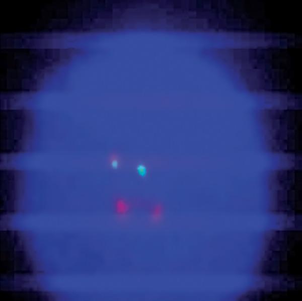
A diagnostic microphotograph of a peripheral blood interphase cell from a patient with Chronic Myeloid Leukemia (CML), analyzed via Fluorescence In Situ Hybridization (FISH). The image demonstrates an atypical BCR-ABL1 translocation pattern using a dual-color, dual-fusion probe. Against the dark blue DAPI-stained nuclear background, four distinct fluorescent signals are visible. The observed configuration is classified as 1F1G2R: one orange/yellow fusion signal (representing the BCR-ABL1 hybrid on the derivative chromosome 22), one green signal (representing the native BCR locus on chromosome 22), and two red signals (representing the native ABL1 locus on chromosome 9). This atypical pattern deviates from the standard 2F1G1R fusion signal usually seen in Philadelphia chromosome-positive cells, indicating clonal evolution or complex chromosomal rearrangements. This image serves as an educational example of cytogenetic variability in hematologic malignancies and the use of FISH for monitoring molecular response in patients undergoing tyrosine kinase inhibitor (TKI) therapy like imatinib.
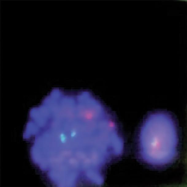
This diagnostic image displays a fluorescence in situ hybridization (FISH) analysis of human bone marrow cells, used primarily for detecting the BCR-ABL1 gene fusion associated with Chronic Myeloid Leukemia (CML). The image shows two blue-stained nuclei (DAPI) against a dark background. On the left, a larger metaphase cell exhibits a normal signal pattern with two distinct cyan/green signals and two distinct red signals (2G2R), representing the normal location of the ABL1 and BCR genes on their respective chromosomes. On the right, a smaller interphase cell demonstrates a fusion signal where a red and a cyan/green signal overlap or are closely apposed, indicative of a chromosomal translocation. This visual is a key educational tool for demonstrating molecular cytogenetics, specifically the dual-color, dual-fusion probe technique used in hematopathology to identify the Philadelphia chromosome.
🩸 Leukemias and Lymphomas: A Complete Teaching Guide
PART 1: THE BIG PICTURE - Classification
Mnemonic: "ALL AML CML CLL" = "A Marvelous Cancer Causes Long Lingering illness"
| Type | Cell Line | Acute/Chronic | Key Age |
|---|---|---|---|
| ALL | Lymphoid | Acute | Children (peak 2-3 yrs) |
| AML | Myeloid | Acute | Adults (also Down syndrome kids) |
| CML | Myeloid | Chronic | Adults 30-60 yrs |
| CLL | Lymphoid | Chronic | Elderly (median age 70) |
Quick rule: "A = young, C = old"
- Acute leukemias tend to be more aggressive, hit younger patients more (especially ALL in kids)
- Chronic leukemias tend to be indolent, hit older adults
PART 2: ACUTE LYMPHOBLASTIC LEUKEMIA (ALL)
Mnemonic for facts: "ALL CATS"
- A - Age peak: 2-3 years (most common childhood malignancy, 25% of all pediatric cancers)
- L - Lymphoblasts proliferate (bone marrow replaced)
- L - Low WBC, Low RBC, Low platelets on CBC (or HIGH WBC with blasts)
- C - CALLA positive (CD10+) = best prognosis in B-ALL
- A - Auer rods: ABSENT (Auer rods are in AML, not ALL)
- T - T-cell ALL = mediastinal mass (thymic) in teenage boys
- S - Survival 95% remission rate; 80% disease-free at 5 years
Immunophenotype of B-ALL: "19 good, 10 better"
- ALL B-cell subtypes co-express CD19 and/or CD22
- CALLA-positive = CD10+ = best prognosis
Chromosomal associations:
| Translocation | Significance |
|---|---|
| t(12;21) TEL-AML1 | Most common in children, GOOD prognosis |
| t(9;22) BCR-ABL (Ph+) | WORST prognosis in ALL; needs TKI + chemo |
| t(4;11) MLL-AF4 | Infant ALL, very poor prognosis |
| Hyperdiploidy (>50 chromosomes) | Good prognosis |
| Hypodiploidy (<44 chromosomes) | Poor prognosis |
Mnemonic for poor prognosis ALL: "Ph+ MLL = Poor"
- Philadelphia chromosome positive = bad
- MLL rearrangement (11q23) = bad
Clinical features: "BLAST"
- B - Bone pain
- L - Lymphadenopathy, hepatosplenomegaly
- A - Anemia (pallor, fatigue)
- S - Susceptibility to infection (neutropenia)
- T - Thrombocytopenic bleeding (petechiae, purpura)
PART 3: ACUTE MYELOID LEUKEMIA (AML)
Mnemonic: "AML = Auer Myeloblast Leukemia"
WHO/FAB Classification - Mnemonic: "My FAB 8 dishes"
- M0 = undifferentiated
- M1 = minimal differentiation
- M2 = t(8;21) - most common AML in young adults
- M3 = Acute Promyelocytic Leukemia (APL) - t(15;17) - treat with ATRA
- M4 = myelomonocytic
- M5 = monocytic
- M6 = erythroleukemia
- M7 = megakaryoblastic (Down syndrome!)
The Crown Jewel: APL = M3
Mnemonic: "15 to 17 = PML goes to RAR-α"
- t(15;17) fuses PML gene (chromosome 15) with RAR-α (chromosome 17)
- Treatment: ATRA (all-trans retinoic acid) - differentiates blasts into mature cells
- DIC (disseminated intravascular coagulation) is the deadly complication - watch for it!
- Auer rod bundles = "faggot cells" - pathognomonic for M3
Cytogenetic Risk Stratification (European LeukemiaNet):
- t(8;21) - RUNX1-RUNX1T1
- inv(16) or t(16;16) - CBFB-MYH11
- Mutated NPM1 without FLT3-ITD
- Biallelic mutated CEBPA
- inv(3), del(5q), monosomy 7 (-7), -5, complex karyotype
- FLT3-ITD high = bad
- Mutated RUNX1, ASXL1 = bad
Risk factors for AML: "BRAIN"
- B - Benzene exposure
- R - Radiation (ionizing)
- A - Alkylating chemotherapy (prior)
- I - Inherited syndromes (Down, Fanconi anemia, Noonan)
- N - Neurofibromatosis
PART 4: CHRONIC MYELOID LEUKEMIA (CML)
THE most testable leukemia fact: "CML = Philadelphia chromosome"
Mnemonic: "9 to 22 = Ph = CML"
- t(9;22) translocation = Philadelphia chromosome
- Fuses BCR (chromosome 22) with ABL (chromosome 9)
- BCR-ABL produces a constitutively active tyrosine kinase
Three Phases: "CAB"
- C - Chronic phase: Most patients here at diagnosis; indolent, splenomegaly, high WBC with all myeloid cell types
- A - Accelerated phase: Increasing blasts (10-19%), resistant disease
- B - Blast crisis: >20% blasts - acts like acute leukemia (can be myeloid or lymphoid)
Treatment: Tyrosine Kinase Inhibitors (TKIs)
Mnemonic: "Ima-Da-Nilo-Bo" = generations of TKIs
- 1st generation: Imatinib (Gleevec) - first approved, landmark drug
- 2nd generation: Dasatinib, Nilotinib - overcome most imatinib resistance
- 3rd generation: Bosutinib, Ponatinib - for T315I mutation ("gatekeeper" mutation)
Blood film in CML: "All stages on smear"
- WBC count massively elevated (50,000-200,000)
- Entire myeloid series visible: myeloblasts, promyelocytes, myelocytes, metamyelocytes, bands, segs
- Basophilia is characteristic (unique feature!)
- Low Leukocyte Alkaline Phosphatase (LAP) score - opposite of leukemoid reaction
PART 5: CHRONIC LYMPHOCYTIC LEUKEMIA (CLL)
Key facts: "CLL = Commoners' Leukemia (western world)"
- Most common leukemia in the Western world
- Median age 70 years; male > female (2:1)
- Rare in Asians (even after emigrating to the West - genetic predisposition)
Pathognomonic finding: Smudge cells (basket cells)
- Fragile CLL cells break during smear preparation
- Mnemonic: "CLL cells are fragile - they SMUDGE"
Immunophenotype: "CLL loves CD5"
Mnemonic: "5 in 19 AND 23"
- CD5+ (T-cell marker on B-cells - aberrant)
- CD19+, CD23+ (B-cell markers)
- Weak surface immunoglobulin (dim slg)
- Mnemonic: "CD5 is a traitor - a B-cell wearing a T-cell badge"
Staging - Rai (US) and Binet (Europe):
- Stage 0: Lymphocytosis only
- Stage I: + Lymphadenopathy
- Stage II: + Splenomegaly/hepatomegaly
- Stage III: + Anemia
- Stage IV: + Thrombocytopenia
Complications: "HART"
- H - Hypogammaglobulinemia (infections)
- A - Autoimmune hemolytic anemia (AIHA) - warm antibody
- R - Richter transformation (→ Diffuse Large B Cell Lymphoma - very poor prognosis)
- T - Thrombocytopenia (autoimmune)
Cytogenetics in CLL:
| Abnormality | Significance |
|---|---|
| del(13q14) | Most common; GOOD prognosis |
| del(11q) ATM | Intermediate |
| Trisomy 12 | Intermediate |
| del(17p) TP53 | WORST prognosis; resistant to chemo |
PART 6: HODGKIN LYMPHOMA (HL)
The Hallmark: Reed-Sternberg (RS) cells
Mnemonic: "RS = Really Scary Owl Eyes"
- Large binucleated cells with prominent eosinophilic nucleoli
- Classic "owl eye" appearance
- RS cells are CD15+, CD30+, PAX5 weak, CD20-
Epidemiology: Bimodal age distribution
Mnemonic: "Young and Old - biMODAL"
- Peak 1: Late teens/young adults (15-35 yrs)
- Peak 2: >55 years
- EBV associated in ~50% of cases in the USA
Subtypes: "NLMM" = "No Lymphs Make Me sad" (favorable to unfavorable)
| Subtype | Mnemonic | Key Feature | Prognosis |
|---|---|---|---|
| Nodular Lymphocyte Predominant (NLPHL) | "N = Nice" | LP cells (popcorn cells), CD20+, CD15/30- | Best |
| Nodular Sclerosis (NSHL) | "NS = Not Serious" | Most common (70%), lacunar cells, collagen bands | Good |
| Mixed Cellularity (MCHL) | "MC = Moderate Concern" | EBV+, diverse infiltrate | Intermediate |
| Lymphocyte Depletion (LDHL) | "LD = Lethal Danger" | Rarest, aggressive, HIV-associated | Worst |
Mnemonic for subtypes: "NNML" - "Nobody Notices Mixed Lesions" (N-S, N-LP, M-C, L-D)
Staging (Ann Arbor): "IALE"
- I = 1 lymph node region
- II = 2+ regions, same side of diaphragm
- III = Both sides of diaphragm
- IV = Extranodal/bone marrow involvement
Mnemonic: "B = Bad = Fever, Night sweats, Weight loss" = FNW
Treatment: "ABVD" (standard)
- A = Adriamycin (doxorubicin)
- B = Bleomycin
- V = Vinblastine
- D = Dacarbazine
PART 7: NON-HODGKIN LYMPHOMA (NHL)
Key principle: "NHL = No RS cells" (unlike Hodgkin)
Common NHL subtypes - Mnemonic: "DUMB-FL"
- D - Diffuse Large B Cell Lymphoma (DLBCL) - most common NHL in adults
- U - Unknown (many rare types)
- M - Mantle Cell Lymphoma (MCL)
- B - Burkitt Lymphoma
- F - Follicular Lymphoma (FL) - most common indolent NHL
- L - Lymphoblastic Lymphoma (T-cell)
1. DIFFUSE LARGE B CELL LYMPHOMA (DLBCL)
- Most common NHL (~30-40% of all NHL)
- Aggressive but potentially curable
- CD20+, CD19+
- Treatment: R-CHOP (Rituximab + Cyclophosphamide, Hydroxydaunorubicin, Oncovin, Prednisone)
Mnemonic: "R-CHOP = Really Cool Hospital Oncology Protocol"
2. FOLLICULAR LYMPHOMA
Mnemonic: "Follicular = t(14;18) = Bcl-2 turned ON = no apoptosis"
- t(14;18) = BCL-2 overexpression = cells don't die
- Indolent but incurable with standard chemotherapy
- "Waxing and waning" lymphadenopathy
- May transform to DLBCL (Richter-like transformation)
3. BURKITT LYMPHOMA
The fastest-growing cancer in humans
Mnemonic: "8 to 14 MYC = BURKing out"
- t(8;14) = c-MYC translocation
- "Starry sky" pattern on histology (macrophages engulfing apoptotic cells)
- Three types: Endemic (Africa, jaw, EBV-related), Sporadic (USA, abdomen), Immunodeficiency-related (HIV)
- Mnemonic: "EBV = Endemic/African jaw; HIV = Immunodeficiency"
- Tumor lysis syndrome risk is HIGH
4. MANTLE CELL LYMPHOMA
Mnemonic: "Mantle = t(11;14) = Cyclin D1 overexpressed = MANTLED in cyclin"
- t(11;14) = CCND1 gene overexpression (cyclin D1)
- CD5+, CD19+, CD20+, CD23- (differs from CLL which is CD23+)
- Mnemonic to distinguish MCL from CLL: "MCL = minus 23; CLL = plus 23"
- Poor prognosis
5. T-CELL LYMPHOBLASTIC LYMPHOMA
- Teenage boys, anterior mediastinal mass (SVC syndrome)
- Mnemonic: "T for Teenage, Thymus, Thorax"
PART 8: MASTER COMPARISON TABLE
| Feature | ALL | AML | CML | CLL |
|---|---|---|---|---|
| Peak age | Children 2-3 yrs | Adults | Adults 30-60 | Elderly >65 |
| Pathognomonic | - | Auer rods | Ph chromosome | Smudge cells |
| Key marker | CD10 (CALLA) | MPO+ | BCR-ABL | CD5+/CD19+/CD23+ |
| Translocation | t(9;22) worst; t(12;21) best | t(15;17) APL = ATRA | t(9;22) | del(13q) good, del(17p) bad |
| Cure | ~80% children | ~70% | TKIs - not curative | Incurable, but manageable |
| Blast crisis risk | - | - | YES (CML→blast crisis) | NO |
PART 9: MASTER MNEMONICS SUMMARY CARD
| Condition | Key Mnemonic |
|---|---|
| ALL | "ALL CATS" - Age, Lymphoblasts, Low counts, CALLA, Auer absent, T-cell mediastinal, Survival 95% |
| AML | "BRAIN" risk factors; Auer rods = AML |
| APL (M3) | "15 to 17 PML-RARA → ATRA" |
| CML | "9 to 22 = Ph = CML"; "CAB" phases; "Ima-Da-Nilo-Bo" TKIs |
| CLL | "CLL = CD5 traitor"; smudge cells; "13 lucky, 17 unlucky" |
| Hodgkin | "RS = Owl Eyes"; "ABVD = A Beautiful Vintage Dance" |
| DLBCL | "R-CHOP" |
| Follicular | "t(14;18) = BCL-2 on = can't die" |
| Burkitt | "8 to 14 MYC = fastest cancer"; starry sky |
| Mantle Cell | "t(11;14) = Cyclin D1; CD23 minus" |
PART 10: HIGH-YIELD EXAM TRAPS
- Auer rods = AML only (never ALL)
- Smudge cells = CLL
- Reed-Sternberg cells = Hodgkin lymphoma (but RS-like cells can appear in other conditions)
- Philadelphia chromosome = CML (also in Ph+ ALL = bad prognosis)
- ATRA treatment = APL (M3) only - do NOT use in other AML subtypes
- DIC in leukemia = think APL (M3)
- Starry sky pattern = Burkitt lymphoma
- Popcorn cells (LP cells) = Nodular Lymphocyte Predominant Hodgkin Lymphoma
- CD5+ B-cells = CLL or Mantle Cell Lymphoma (distinguish by CD23: CLL is CD23+, MCL is CD23-)
- Low LAP score = CML (high LAP = leukemoid reaction from infection)
- Jaw mass in African child = Endemic Burkitt lymphoma (EBV-related)
- Anterior mediastinal mass in teenage boy = T-cell ALL/lymphoblastic lymphoma
PART 11: BONUS - VISUAL MEMORY AID
       ALL (12 o'clock = young)
         |
CML -----+------ CLL
(3 o'clock         (9 o'clock
= middle-age)      = elderly)
         |
       AML (6 o'clock = any adult)
- Top = youngest patients (ALL)
- Going clockwise = getting older
- Ph chromosome connects CML and ALL (both can have t(9;22))
Teach me inflammation acute and chronic in detail and pneumonics to learn the chemical mediators
acute inflammation neutrophil emigration leukocyte rolling adhesion
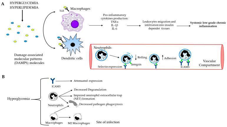
A two-part medical pathophysiology diagram illustrating the link between metabolic dysfunction and systemic inflammation. Part A depicts the cascade where hyperglycemia and hyperlipidemia generate Damage-Associated Molecular Patterns (DAMPs). These molecules bind to Toll-like receptors (TLRs) on macrophages and dendritic cells, triggering the release of pro-inflammatory cytokines (TNFͱ, IL-1̢, IL-6). This lead to leukocyte migration into insulin-dependent tissues and systemic low-grade chronic inflammation. An inset showing the vascular compartment details the neutrophil recruitment process, highlighting decreased rolling (mediated by selectin) and increased adhesion (mediated by integrins and ICAMs). Part B focuses on the cellular impact of hyperglycemia, showing attenuated ICAM expression, decreased neutrophil degranulation, impaired Neutrophil Extracellular Trap (NET) formation, and decreased phagocytosis by neutrophils and macrophages. It also notes the transition of macrophages to the M2 phenotype at sites of infection. This diagram is designed for advanced medical education regarding the immunology of metabolic diseases like diabetes and obesity.
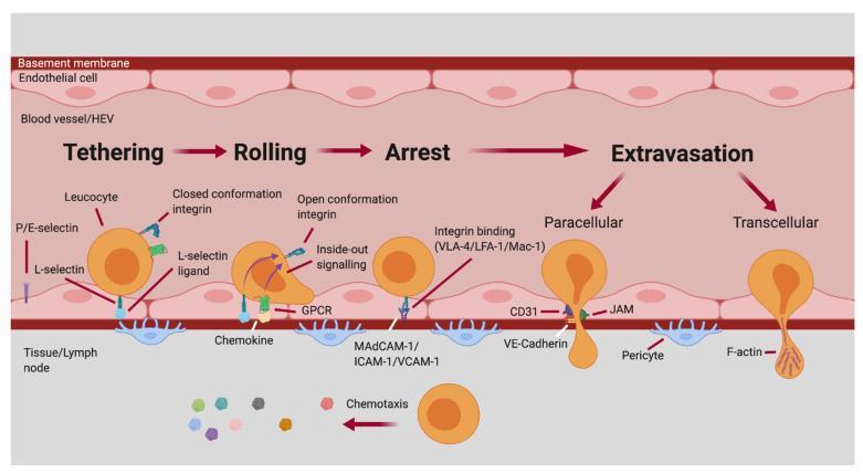
This pathophysiology diagram illustrates the sequential stages of the leukocyte adhesion cascade within a blood vessel or high endothelial venule (HEV). The process is divided into four main phases: Tethering, Rolling, Arrest, and Extravasation. 1. Tethering: Mediated by L-selectin and P/E-selectins interacting with their respective ligands. Leukocyte integrins are in a closed conformation. 2. Rolling: Leukocyte shape elongates as it interacts with chemokines via GPCRs. This triggers 'inside-out signaling,' shifting integrins to an open conformation. 3. Arrest: Firm adhesion is achieved through high-affinity integrin binding (VLA-4, LFA-1, Mac-1) to endothelial ligands (MAdCAM-1, ICAM-1, VCAM-1). 4. Extravasation: The leukocyte exits into the tissue/lymph node via two pathways: Paracellular migration (between endothelial cells, involving CD31, JAM, and VE-Cadherin) or Transcellular migration (through the endothelial cell, involving F-actin). The diagram concludes with the leukocyte passing the basement membrane and pericytes, directed by chemotaxis. This visual is designed for advanced immunology and medical students to understand cellular recruitment during inflammation or immune surveillance.
granuloma chronic inflammation macrophage epithelioid
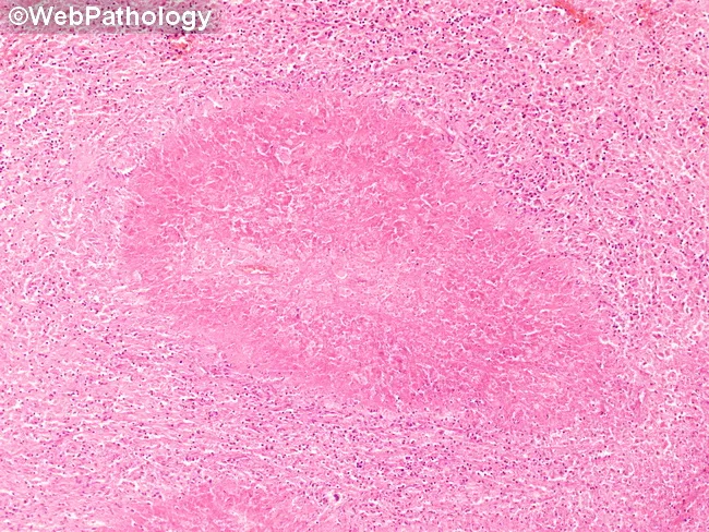
This is a low-magnification brightfield histology image of a tissue section stained with Hematoxylin and Eosin (H&E). The specimen shows granulomatous inflammation with aggregates of epithelioid macrophages forming a rounded granuloma, often with multinucleated giant cells, surrounded by a lymphocytic cuff. The central focus is well circumscribed within native parenchyma, suggesting a chronic, organized immune response to a persistent antigen. Necrosis is not clearly evident at this magnification, though subtle caseation cannot be excluded. Morphology is compatible with tuberculoid or non-caseating granulomas, and infectious versus noninfectious etiologies must be distinguished with ancillary studies. This pattern can occur in lymph nodes or solid organs and prompts a differential that includes tuberculous lymphadenitis, sarcoidosis, fungal granulomatous infections (Histoplasma, Coccidioides), and foreign-body reaction. Clinically, recognition of granulomas guides testing: acid-fast bacilli staining (Ziehl-Neelsen), fungal stains (GMS/PAS), cultures, PCR panels, and radiologic correlation. The image is educational for medical trainees, illustrating granuloma architecture, macrophage differentiation, and the spectrum of chronic inflammatory responses. Educational context.
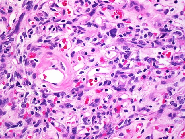
Imaging modality: Light microscopy; Technique: Hematoxylin and eosin staining; Specimen: Tissue biopsy; Magnification: 400x. The slide depicts a granular inflammatory tissue with chronic granulomatous features. Epithelioid histiocytes are arranged in nodular clusters, interspersed with multinucleated giant cells. The background shows a mixed inflammatory infiltrate dominated by small lymphocytes and occasional plasma cells. Blood vessels with mild perivascular inflammation are present, and the stromal framework appears fibrous in places. In several fields there are well-demarcated granuloma-like aggregates, while elsewhere the architecture is disrupted by focal necrotic debris and amorphous pink extracellular matrix. Notable features include coalescing macrophage-rich nodules and a few eosinophils; occasional foamy macrophages may be seen. The overall pattern is compatible with chronic granulomatous inflammation. Pathological significance: suggests a granulomatous process, prompting differential diagnoses such as infectious etiologies (mycobacterial, fungal) or noninfectious conditions like sarcoidosis or foreign-body reaction. Recommended ancillary studies: Ziehl-Neelsen or acid-fast stains, Gomori methenamine silver or PAS stains, fungal cultures, and targeted immunohistochemistry as indicated. Clinical correlation with patient history, imaging, and microbiology is essential for etiologic determination and guiding management. Clinical utility: this image serves as an educational reference for understanding granulomatous pathology in biopsy specimens and for teaching interpretation of epithelioid histiocytes, giant cells, and necrotic changes.
🔥 Inflammation: Acute & Chronic - Complete Teaching Guide with Mnemonics
PART 1: THE FOUNDATION - What is Inflammation?
The Cardinal Signs (Celsus + Virchow): "PRISH"
- R - Rubor (Redness) - from vasodilation
- P - (h)eat - Calor - from increased blood flow
- I - Increased blood flow = 2nd component
- S - Swelling - Tumor - from fluid exudation
- H - Heat - from vasodilation
- S - Swelling (Tumor)
- H - Heat (Calor)
- A - redness (ruboR)
- R - Pain (doloR) - added by Virchow
- P - Loss of function (added by Virchow) - Paralysis of function
PART 2: ACUTE INFLAMMATION
Definition
The Three Components: "FPL"
- F - Flow changes: vasodilation → increased blood flow (heat + redness)
- P - Permeability increase: protein-rich fluid exits vessels (swelling)
- L - Leukocyte emigration: neutrophils leave vessels, accumulate at injury site
2A: VASCULAR CHANGES
- Vasodilation - histamine acts on smooth muscle of post-capillary venules → increased blood flow (heat + redness)
- Stasis - increased permeability → fluid leaves → RBCs concentrate → flow slows
- Permeability increase - protein-rich exudate pours out → edema/swelling
Exudate vs Transudate:
| Feature | Exudate | Transudate |
|---|---|---|
| Protein | High | Low (mostly albumin) |
| Cause | Inflammation (increased permeability) | Hydrostatic/osmotic pressure |
| Example | Pus, fibrinous fluid | Heart failure edema |
Mnemonic: "Exudate = Exciting inflammation; Transudate = Tranquil, no inflammation"
2B: LEUKOCYTE RECRUITMENT - The 4-Step Cascade
| Step | Process | Key Molecules |
|---|---|---|
| 1. Rolling (Margination) | Neutrophils loosely tumble along endothelium | Selectins (P, E, L-selectin) bind sialyl-Lewis X |
| 2. Adhesion (Firm) | Tight binding to endothelium | Integrins (LFA-1, MAC-1) bind ICAM-1; VLA-4 binds VCAM-1 |
| 3. Migration (Transmigration/Diapedesis) | Squeezing through endothelial junctions | CD31 (PECAM-1) - homotypic interaction |
| 4. Taxis (Chemotaxis) | Directed movement toward injury | Complement C5a, LTB4, IL-8, bacterial products (fMLP) |
Selectin mnemonic: "PEL Selectins"
- P-selectin: expressed on Platelets and endothelium (stored in Weibel-Palade bodies, rapidly mobilized by histamine/thrombin)
- E-selectin: expressed on Endothelium only (activated by TNF, IL-1)
- L-selectin: expressed on Leukocytes (neutrophils, monocytes, T-cells)
Integrin mnemonic: "LFA MAC VLA"
- LFA-1 (CD11a/CD18): on neutrophils/T-cells → binds ICAM-1, ICAM-2
- MAC-1 (CD11b/CD18): on monocytes/DCs → binds ICAM-1
- VLA-4 (CD49d/CD29): on monocytes/T-cells → binds VCAM-1
2C: PHAGOCYTOSIS
- Recognition and attachment (opsonization by IgG, C3b)
- Opsonization - coating with antibodies/complement to enhance phagocytosis
- Kill (intracellular killing) - ROS, NO, lysosomal enzymes
Killing mechanisms: "RON"
- Reactive Oxygen Species (ROS) - "respiratory burst" via NADPH oxidase → H2O2 → HOCl (bleach!)
- Oxide - Nitric oxide (NO) - via iNOS
- Nitrogen + other mechanisms - MPO (myeloperoxidase), lysozyme, defensins
2D: OUTCOMES OF ACUTE INFLAMMATION: "RASH"
- R - Resolution (complete restoration - best outcome, minimal damage)
- A - Abscess formation (walled-off pus)
- S - Scarring/fibrosis (extensive necrosis → repair by fibrosis)
- H - leads to chronic inflammation (persistent stimulus)
PART 3: CHEMICAL MEDIATORS OF INFLAMMATION
Master Mnemonic: "PACK + BKP + CNOPF"
- P - Plasmin (fibrinolytic system)
- A - Alternative/Classical/Lectin pathways (Complement)
- C - Coagulation/Kinin system
- K - Kinins (Bradykinin)
- P - Prostaglandins
- B - Bradykinin
- K - Kinins
- P - PAF (Platelet Activating Factor)
MEDIATOR GROUP 1: VASOACTIVE AMINES - "His & Sero"
HISTAMINE - "His MAST"
- Source: MAST cells (mast cells, basophils, platelets)
- Stored preformed in granules - released FAST
- Triggers: Physical injury, IgE, C3a/C5a, IL-1
- Actions: Vasodilation + increased vascular permeability (postcapillary venules)
- Mnemonic: "Histamine Hates blood staying in vessels - it opens the gates"
SEROTONIN (5-HT)
- Source: Platelets (stored in dense granules)
- Similar actions to histamine
- Also a neurotransmitter
- Mnemonic: "Sero = Stored in platelet granules"
MEDIATOR GROUP 2: ARACHIDONIC ACID (AA) METABOLITES (EICOSANOIDS)
Membrane Phospholipids
        ↓ (Phospholipase A2 - blocked by CORTICOSTEROIDS via lipocortin)
  Arachidonic Acid
    /            \
COX pathway    LOX pathway
(Cyclooxygenase)  (Lipoxygenase)
    ↓                 ↓
Prostaglandins    Leukotrienes
Thromboxane A2    LTB4, LTC4, LTD4, LTE4
Prostacyclin (PGI2)
Master Mnemonic for AA metabolites: "COX Makes PTP; LOX Makes BDE"
- Prostaglandins (PGD2, PGE2, PGF2α)
- Thromboxane A2
- Prostacyclin (PGI2)
- B = LTB4 (chemotaxis of neutrophils)
- D = LTD4 (bronchoconstriction - main mediator in asthma)
- E = LTE4 (bronchoconstriction)
- (LTC4 also causes bronchoconstriction)
PROSTAGLANDINS - Mnemonic: "PGE2 = Pain, Fever, Vasodilate"
| Eicosanoid | Action | Mnemonic |
|---|---|---|
| PGI2 (Prostacyclin) | Vasodilation, ↑permeability, inhibits platelet aggregation | "PGI2 = Peace - no clots" |
| PGE2, PGD2 | Vasodilation, ↑permeability, PAIN, FEVER | "E2 = Everything bad (pain+fever)" |
| TXA2 (Thromboxane A2) | Vasoconstriction, platelet aggregation | "TX = TighteX blood vessels" |
| LTB4 | Chemotaxis of neutrophils | "B4 = Brings neutrophils Before others" |
| LTC4, LTD4, LTE4 (cysteinyl leukotrienes) | Bronchoconstriction, ↑vascular permeability | "CDE = Constrict During Exercise (asthma!)" |
Drug targets:
- NSAIDs (aspirin, ibuprofen): block COX → reduce prostaglandins → reduce pain, fever, inflammation
- Aspirin (irreversible COX inhibitor): also blocks TXA2 → antiplatelet effect
- Montelukast/Zafirlukast: block LTC4/LTD4 receptors → treat asthma
- Zileuton: blocks 5-lipoxygenase → blocks all leukotrienes
- Corticosteroids: block Phospholipase A2 → block ENTIRE AA pathway
Mnemonic: "Steroids Stop at the Source (Phospholipase A2)"
MEDIATOR GROUP 3: CYTOKINES - "TNF IL-1 IL-6 = The Terrible Inflammatory League"
TNF and IL-1: The "Dynamic Duo" of inflammation
- Endothelial activation → upregulate adhesion molecules (selectins, ICAM-1, VCAM-1)
- Increased permeability
- Stimulate prostaglandin production
- F - Fever (via PGE2 in hypothalamus)
- A - Acute phase proteins (CRP, fibrinogen, SAA - made by liver in response to IL-6!)
- S - Septic shock (massive TNF → vasodilation, DIC, multi-organ failure)
IL-6: "The Liver Lover"
- Made by macrophages
- Acts on liver → stimulates acute phase protein production (CRP, fibrinogen)
- Also involved in local and systemic inflammatory reactions
- Anti-IL-6 receptor (tocilizumab) is used in juvenile arthritis
IL-17: "The Neutrophil Caller"
- Produced by Th17 cells
- Promotes neutrophil recruitment (via chemokine production)
- Target in psoriasis treatment (secukinumab/ixekizumab block IL-17)
CHEMOKINES: "Directed Traffic Controllers"
- Chemokines are a subclass of cytokines that specifically drive chemotaxis
- IL-8 (CXCL8) = most important - made by macrophages/endothelium → recruits neutrophils
- CXCR4, CCR5: receptors that HIV uses to enter cells!
- Mnemonic: "Chemokines = Chemical GPS for leukocytes"
MEDIATOR GROUP 4: COMPLEMENT SYSTEM - "The C3 Pivot"
Three activation pathways: "CAL"
- Classical: antibody (IgG or IgM) binds antigen → activates C1 → C4/C2 → C3
- Alternative: microbial surfaces (LPS, endotoxin) spontaneously activate in absence of antibody
- Lectin: mannose-binding lectin (MBL) binds carbs on microbes → activates C3
Key complement fragments - Mnemonic: "C3b Coats, C5a Calls, MAC Kills"
| Fragment | Function | Mnemonic |
|---|---|---|
| C3b | Opsonization (coats microbes for phagocytosis) | "3b = 3 Bugs get Coated" |
| C3a, C5a | Anaphylatoxins → trigger mast cell degranulation → histamine release → ↑permeability, vasodilation | "3a/5a = Anaphylatoxins Alarm mast cells" |
| C5a | Chemotaxis for neutrophils (the most potent chemotactic complement fragment) | "5a = Five-alarm chemotaxis" |
| C5b-9 | MAC (Membrane Attack Complex) → lyses cell membranes | "5-9 = MAC = Murder All Cells" |
MEDIATOR GROUP 5: KININ SYSTEM - "Brady-kinin = Slow + Pain"
- Bradykinin is the main mediator
- Formed from kininogens by kallikrein (activated by Hageman factor/Factor XII)
- Actions: vasodilation, ↑permeability, pain (potentiated by PGE2), bronchospasm
- Very short half-life (degraded by kininase/ACE)
- Mnemonic: "Brady = Slow acting; 'kinin' = pain"
MEDIATOR GROUP 6: PLATELET ACTIVATING FACTOR (PAF)
- Source: many cells - mast cells, basophils, platelets, neutrophils
- Actions: platelet aggregation, bronchoconstriction, ↑permeability (10,000x more potent than histamine!), vasodilation
- Mnemonic: "PAF = Powerful Amplification Factor"
MEDIATOR GROUP 7: NITRIC OXIDE (NO)
- Produced by: endothelial cells (eNOS), macrophages (iNOS)
- Actions:
- Vasodilation (relaxes smooth muscle - same as nitroglycerin mechanism)
- Antimicrobial - kills microbes in macrophages
- Reduces platelet aggregation
- Mnemonic: "NO = Nasty to microbes, Nice to vessels (dilates them)"
MEDIATOR MASTER SUMMARY TABLE
| Mediator | Source | Vasodilation | ↑Permeability | Chemotaxis | Pain/Fever |
|---|---|---|---|---|---|
| Histamine | Mast cells, basophils | ✓ | ✓ | - | - |
| Serotonin | Platelets | ✓ | ✓ | - | - |
| PGE2/PGI2 | Many cells | ✓ | ✓ | - | ✓ (PGE2) |
| TXA2 | Platelets | ✗ (constricts) | - | - | - |
| LTB4 | Leukocytes | - | - | ✓✓✓ | - |
| LTC4/D4/E4 | Mast cells | - | ✓ | - | - |
| C3a/C5a | Plasma (complement) | ✓ | ✓ | ✓ (C5a) | - |
| IL-1, TNF | Macrophages | ✓ | ✓ | - | ✓ |
| IL-8 | Macrophages, endothelium | - | - | ✓✓ | - |
| Bradykinin | Plasma | ✓ | ✓ | - | ✓✓ |
| NO | Endothelium, macrophages | ✓ | - | - | - |
| PAF | Mast cells, platelets | ✓ | ✓✓✓ | - | - |
PART 4: MORPHOLOGICAL PATTERNS OF ACUTE INFLAMMATION
Mnemonic: "SUPF"
- Serous - watery exudate (e.g., skin blister, pleural fluid early viral infection); low protein
- Ulcer - epithelial defect from necrosis (e.g., peptic ulcer, pressure sore)
- Purulent/Suppurative - pus = neutrophils + necrotic cells + microbes (e.g., abscess, empyema)
- Fibrinous - fibrin-rich exudate (e.g., fibrinous pericarditis = "bread-and-butter" appearance)
PART 5: CHRONIC INFLAMMATION
Definition
Causes - Mnemonic: "PHA"
- P - Persistent infections (mycobacteria, fungi, parasites - organisms that resist eradication)
- H - Hypersensitivity/autoimmune diseases (RA, IBD, SLE, multiple sclerosis) - self-perpetuating T/B cell reaction
- A - Agents (toxic) - silica → silicosis; cholesterol → atherosclerosis
Morphological Features - Mnemonic: "MIT"
- Mononuclear infiltrate (macrophages, lymphocytes, plasma cells - NOT neutrophils!)
- Injury/tissue destruction (ongoing)
- Tissue repair by fibrosis/angiogenesis (simultaneous)
Key Cells: "MLEPO"
- Macrophages - dominant cell; the "directors" of chronic inflammation
- Lymphocytes (T and B cells)
- Eosinophils (especially in parasitic/allergic inflammation)
- Plasma cells (secrete antibodies)
- Other: mast cells, fibroblasts
THE MACROPHAGE: Director of Chronic Inflammation
| Type | Activation | Functions |
|---|---|---|
| M1 (classical) | IFN-γ (from Th1 cells), LPS | Kill microbes, produce ROS, NO, IL-1, TNF, IL-12 → pro-inflammatory |
| M2 (alternative) | IL-4, IL-13 (from Th2 cells) | Anti-inflammatory, tissue repair, fibrosis, produce IL-10, TGF-β |
Macrophage actions in chronic inflammation: "STACK"
- Secrete cytokines (TNF, IL-1, IL-12, IL-6)
- Tissue damage (proteases, elastase, collagenase)
- Antigen presentation (to T-cells via MHC)
- Chemokine secretion (recruit more leukocytes)
- Kill microbes (ROS, NO, lysosomal enzymes)
T LYMPHOCYTES IN CHRONIC INFLAMMATION
| T-helper | Cytokine secreted | Role |
|---|---|---|
| Th1 | IFN-γ | Activates M1 macrophages → kills intracellular bacteria; drives autoimmune disease |
| Th2 | IL-4, IL-5, IL-13 | Activates M2 macrophages; recruits eosinophils; drives allergy, asthma, helminth defense |
| Th17 | IL-17 | Recruits neutrophils + monocytes; drives psoriasis, some autoimmune diseases |
- Macrophage presents antigen → activates T-cells
- T-cells secrete IFN-γ → activates macrophages → more antigen presentation
- This SELF-PERPETUATING cycle maintains chronic inflammation
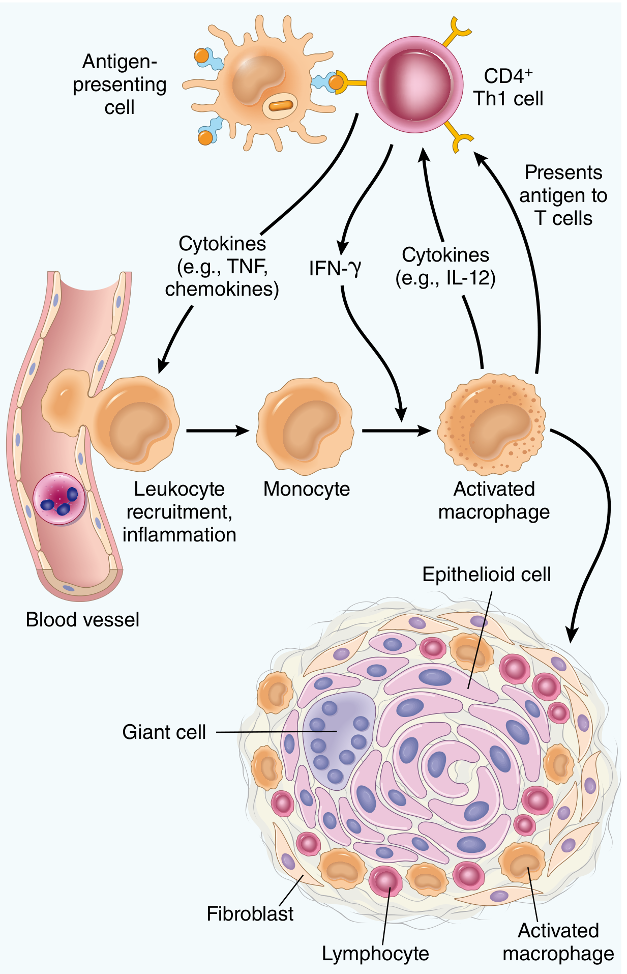
PART 6: GRANULOMATOUS INFLAMMATION
Definition
What triggers granuloma formation?
Components of a granuloma: "EAGLE"
- E - Epithelioid cells (activated macrophages - elongated, pink cytoplasm, look like squamous epithelium)
- A - Aggregate (central cluster of epithelioid cells)
- G - Giant cells (Langhans type: nuclei arranged in horseshoe; Foreign body type: nuclei scattered randomly)
- L - Lymphocytes (surrounding the granuloma)
- E - Extra fibrosis (surrounding cuff of fibroblasts)
Types of granulomas: "Case Closed vs Not Closed"
| Type | Features | Disease |
|---|---|---|
| Caseating (necrotizing) | Cheesy central necrosis (caseum) | Tuberculosis (most classic) |
| Non-caseating | No necrosis | Sarcoidosis, Crohn's disease, leprosy (tuberculoid), Berylliosis |
Diseases with granulomas - Mnemonic: "SCLBT-CGF"
- S - Sarcoidosis (non-caseating, unknown etiology)
- C - Crohn's disease (non-caseating, GI wall)
- L - Leprosy (non-caseating in tuberculoid type)
- B - Berylliosis (non-caseating)
- T - Tuberculosis (caseating, Langhans giant cells, acid-fast bacilli)
- C - Cat-scratch disease (stellate granuloma with central neutrophils)
- G - syphilis/Gumma (plasma cells prominent)
- F - Fungal infections (Histoplasma, Coccidioides)
PART 7: SYSTEMIC EFFECTS OF INFLAMMATION (Acute Phase Response)
Triggered by: TNF, IL-1, IL-6
Systemic manifestations - Mnemonic: "FLAP"
- F - Fever (IL-1/TNF → PGE2 in hypothalamus → raises set point)
- L - Leukocytosis (IL-1/TNF → bone marrow release of WBCs; left shift = band cells)
- A - Acute phase proteins (IL-6 → liver → CRP, fibrinogen, SAA, complement proteins)
- P - Protein catabolism (wasting, cachexia - TNF = "cachectin")
Acute Phase Proteins: "CFS" = CRP, Fibrinogen, SAA
- CRP (C-reactive protein): binds phosphocholine on microbes → opsonization + complement activation
- Fibrinogen: causes RBC rouleaux → elevated ESR
- SAA (serum amyloid A): can deposit as amyloid if overproduced chronically
PART 8: ACUTE vs CHRONIC INFLAMMATION - MASTER COMPARISON
| Feature | Acute | Chronic |
|---|---|---|
| Duration | Minutes to days | Weeks to months |
| Dominant cell | Neutrophil | Macrophage |
| Other cells | Mast cells, basophils, platelets | Lymphocytes, plasma cells, eosinophils |
| Vascular changes | Prominent (vasodilation, permeability) | Less prominent |
| Tissue injury | Usually less severe | Prominent, ongoing |
| Fibrosis | Absent | Common |
| Outcome | Resolution, abscess, scar, or → chronic | Fibrosis, functional loss |
Mnemonic: "Acute = Neutrophils; Chronic = Macrophages" - the single most important distinction.
PART 9: PHARMACOLOGY CONNECTIONS
| Drug | Target | What it blocks |
|---|---|---|
| Corticosteroids | Phospholipase A2 (via lipocortin) | Entire AA pathway (all prostaglandins + leukotrienes) |
| NSAIDs/COX inhibitors | Cyclooxygenase (COX-1/COX-2) | Prostaglandins + TXA2 |
| Aspirin (irreversible) | COX in platelets | TXA2 → antiplatelet effect |
| Montelukast, Zafirlukast | Leukotriene receptors (CysLT1) | LTC4/LTD4/LTE4 → for asthma |
| Zileuton | 5-Lipoxygenase | All leukotrienes |
| Antihistamines (H1 blockers) | H1 receptor | Histamine effects (allergy) |
| Anti-TNF (infliximab, adalimumab) | TNF-α | Chronic inflammation in RA, IBD, psoriasis |
| Tocilizumab | IL-6 receptor | Juvenile arthritis, rheumatoid arthritis |
| Secukinumab | IL-17A | Psoriasis |
PART 10: MASTER MNEMONICS FLASHCARD
| Topic | Mnemonic |
|---|---|
| Cardinal signs | SHARP (Swelling, Heat, redness, Pain, loss of function) |
| Acute inflammation components | FPL (Flow, Permeability, Leukocytes) |
| Leukocyte recruitment | RAMT (Rolling, Adhesion, Migration, Taxis) |
| Selectins | PEL (P on platelets, E on endothelium, L on leukocytes) |
| AA pathway | COX → "PTP" (Prostaglandins, TXA2, PGI2); LOX → LTBDE |
| Eicosanoid actions | TXA2 = "TighteX" (constricts/clots); PGI2 = "Peace" (dilates/anti-platelet); LTB4 = "Brings" neutrophils; CDE = "Constrict" (asthma) |
| Complement | C3b = Coats; C3a/C5a = Anaphylatoxins; C5a = Chemotaxis; C5b-9 = MAC |
| Kinin | Bradykinin = "Slow + Pain"; ACE inhibitor → bradykinin accumulates → cough |
| Cytokines | TNF/IL-1/IL-6 = "FAS" (Fever, Acute phase, Septic shock) |
| Chronic inflammation cells | MLEPO (Macrophages, Lymphocytes, Eosinophils, Plasma cells, Others) |
| Macrophage polarization | M1 = "Meanest Killer"; M2 = "Mellow Healer" |
| T-helper subsets | Th1 = IFN-γ + macrophage activation; Th2 = IL-4/5/13 + allergy; Th17 = IL-17 + neutrophils |
| Granuloma | EAGLE (Epithelioid, Aggregate, Giant cells, Lymphocytes, fEnce of fibroblasts) |
| Caseating vs non-caseating | TB = Caseating (necrosis); Sarcoid/Crohn = Non-caseating |
| Systemic effects | FLAP (Fever, Leukocytosis, Acute phase proteins, Protein catabolism) |
What is informed consent, autonomy, m- beneficence, justice, and other... Aspects of... Bioethics, define them.
bioethics four principles autonomy beneficence nonmaleficence justice Beauchamp Childress
⚖️ Bioethics: A Complete Guide
WHAT IS BIOETHICS?
- Ethics in biological research (on non-human organisms)
- Ethics in medical research (on humans)
- Animal ethics
- Environmental ethics
- Medical ethics
- Their patients
- Each other (colleagues)
- The State
The distinction: Medical etiquette = courtesy rules between professionals. Medical ethics = moral principles governing patient care.
THE FOUR PILLARS OF BIOMEDICAL ETHICS
Mnemonic: "ABCJ" = "A Beautiful Careful Journey"
- A - Autonomy
- B - Beneficence
- C - Non-maleficence (the "careful" part - do no harm)
- J - Justice
PILLAR 1: AUTONOMY
Definition
"Autonomy refers to the ability to choose without controlling interferences by others and without personal limitations that prevent meaningful choices, such as inadequate information or understanding." - Miller's Anesthesia, 10e
The landmark legal case:
- Schloendorff v. Society of New York Hospital (1914): Established that "it is the right of every adult person with capacity to determine what can be done to their own body."
- Salgo v. Trustees of Leland Stanford Hospital (1957): First use of the term "informed consent." Established that physicians have a duty to inform patients about risks, benefits, AND alternatives.
What autonomy requires of physicians: "RICE"
- R - Respect decisions made by capacitated patients (even "bad" decisions!)
- I - Inform patients fully and accurately
- C - Counter barriers to decision-making (language, pain, anxiety, literacy)
- E - Enable participation in decisions
Important nuances:
- A patient can refuse treatment even if that decision seems harmful to themselves
- Agreeing or disagreeing with the doctor is NOT by itself evidence of capacity or its absence
- Capacity is task-specific and time-variable - a person may have capacity for some decisions but not others
- Treatment refusal may simply reflect the patient prioritizing other values (dignity, privacy, independence) over medical outcomes
PILLAR 2: INFORMED CONSENT (Operationalizing Autonomy)
Definition
The three essential elements: "ICV"
| Element | What it means |
|---|---|
| I - Information | Patient receives adequate, accurate information about diagnosis, proposed treatment, risks, benefits, and alternatives |
| C - Capacity | Patient has the cognitive ability to understand and reason |
| V - Voluntariness | Decision is free from coercion, manipulation, or undue influence |
Components of information that must be disclosed: "DRABS"
- D - Diagnosis (what is wrong)
- R - Risks of the proposed treatment
- A - Alternatives (including doing nothing)
- B - Benefits of the proposed treatment
- S - Side effects and what to expect
Standards for information disclosure:
- Professional standard: What a reasonable physician would disclose (older standard, paternalistic)
- Reasonable patient standard: What a reasonable patient would want to know (more common today)
- Subjective patient standard: What THIS particular patient would want to know (most patient-centered)
Exceptions to informed consent: "ETIUP"
- E - Emergency (immediate threat to life)
- T - Therapeutic privilege (rare - disclosure itself would cause harm)
- I - Incompetence / lack of capacity
- U - Unconscious patient (implied consent)
- P - Patient waiver (patient explicitly waives the right to be informed)
Decision-Making Capacity vs. Competence:
| Term | Determined by | Meaning |
|---|---|---|
| Capacity | Clinician (bedside assessment) | Functional ability to make a specific medical decision |
| Competence | Court of law (legal determination) | Legal status to make binding decisions |
- C - Communicate a choice (can patient express a decision?)
- U - Understand information given
- R - Reason (appreciate how information applies to their situation)
- E - Express the logical reasoning behind their choice
Surrogate/Substitute Decision-Making (when capacity is absent):
- S - Spouse/partner
- S - Son/daughter (adult children)
- P - Parent
- C - Court-appointed guardian
- Substituted judgment: What would THIS patient have wanted? (preferred when prior wishes are known)
- Best interests: What would a reasonable person in this situation want? (when prior wishes unknown)
- Living will: Specifies what treatments to accept or refuse
- Durable Power of Attorney for Healthcare (DPAHC): Names a healthcare proxy
PILLAR 3: BENEFICENCE
Definition
This is the oldest medical principle - dating to the Hippocratic tradition. The physician's first duty is to serve the patient's wellbeing.
Types of beneficence:
- Positive beneficence: Taking positive steps to benefit the patient
- Utility: Balancing benefits against risks and harms
- Paternalism (strong): Overriding patient autonomy "for their own good" - generally considered unethical today
Conflict with autonomy - Paternalism:
PILLAR 4: NON-MALEFICENCE
Definition
- Avoid harm to patients (directly or through negligence)
- Weigh risks against potential benefits
- Not undertake procedures where harm outweighs benefit
Non-maleficence vs. Beneficence:
- Beneficence = actively do good
- Non-maleficence = do NOT do harm
The doctrine of double effect:
- The action itself is not intrinsically wrong
- The agent intends the good effect, not the bad
- The bad effect is not the means of achieving the good effect
- There is proportionate reason for accepting the bad effect
PILLAR 5: JUSTICE
Definition
"A concept that emphasizes fairness and equality among individuals." - Beauchamp & Childress
Types of justice in bioethics:
| Type | Meaning |
|---|---|
| Distributive justice | Fair allocation of resources, healthcare, and burdens in society |
| Procedural justice | Fair processes in decision-making |
| Restorative justice | Repairing harm done to patients |
| Rights-based justice | Respecting patients' legal and moral rights |
Criteria for distributive justice - "NFCME"
- N - Need (to each according to their need)
- F - Free-market exchange (to each according to what they can pay)
- C - Contribution (to each according to their contribution to society)
- M - Merit (to each according to their merit)
- E - Equal share (to each an equal share regardless of status)
Justice in research ethics:
- No group bears a disproportionate burden of research participation
- Benefits of research are fairly distributed
- Vulnerable populations (prisoners, children, the poor) are not exploited
OTHER KEY BIOETHICAL CONCEPTS
6. CONFIDENTIALITY
Definition
When can confidentiality be breached? "TRAD"
- T - Threat: Real and imminent threat of harm to patient or others (e.g., homicidal intent)
- R - Required by law: Notifiable diseases, gunshot wounds, child abuse reporting
- A - Authorized by patient: Patient consents to disclosure
- D - Defense: Physician needs to defend themselves in litigation
"It is ethical to disclose confidential information when the patient consents to it or when there is a real and imminent threat of harm to the patient or to others and this threat can be removed by a breach of confidentiality." - International Code of Medical Ethics, WMA 2006
7. VERACITY (TRUTHFULNESS)
Definition
- T - Truth-telling (disclosing accurate information)
- A - Absence of deception (not creating false impressions)
- C - Candor (volunteering information the patient would want to know)
- I - Integrity (consistency between beliefs, statements, and actions)
- T - Transparency (openness about reasoning)
8. FIDELITY (LOYALTY & PROMISE-KEEPING)
Definition
- Honoring commitments to patients
- Following through on referrals and follow-up plans
- Not abandoning a patient without adequate notice and transition of care
9. DIGNITY
- Age, disease, or disability
- Creed, ethnic origin, gender, nationality
- Political affiliation, race, sexual orientation
- Social standing or economic status
10. RESEARCH ETHICS - Additional Principles
- Beneficence (do good for research participants AND society)
- Respect for persons (autonomy in research = voluntary, informed consent)
- Assessment: Risk-benefit ratio must be favorable
- Truthjustice: Fair selection of research subjects
| Code | Year | Significance |
|---|---|---|
| Nuremberg Code (1947) | 1947 | First international code after Nazi experiments; voluntary consent is "absolutely essential" |
| Declaration of Helsinki | 1964 | WMA code for research; distinguishes therapeutic from non-therapeutic research |
| Belmont Report | 1979 | US framework for human subjects research; basis for federal regulations |
11. FUTILITY
- Cannot achieve its physiological objective, OR
- Will not benefit the patient in any meaningful way
- Physiological futility: Treatment cannot achieve its intended physiological effect
- Qualitative futility: Treatment achieves its goal but does not improve quality of life
- Quantitative futility: Chance of success is vanishingly small (often defined as <1%)
12. END-OF-LIFE ETHICS: "WPED"
- Withdrawal of treatment: Stopping treatment already started (ethically permissible with consent)
- Palliation: Prioritizing comfort and quality of life when cure is not possible
- Euthanasia: Intentionally ending a patient's life to relieve suffering (active = direct killing; passive = withholding treatment). Legal in some jurisdictions (Netherlands, Canada, Belgium)
- DNR orders (Do Not Resuscitate): Patient/surrogate directive to withhold CPR
| Type | Meaning | Legality |
|---|---|---|
| Active euthanasia | Administering a lethal agent (e.g., lethal injection) | Illegal in most countries |
| Passive euthanasia | Withholding/withdrawing life-sustaining treatment | Generally permissible |
| Physician-assisted suicide (PAS) | Physician provides means; patient self-administers | Legal in some jurisdictions (e.g., Oregon Death with Dignity Act) |
THE FOUR PRINCIPLES IN PRACTICE - How They Conflict
Common conflicts and how to resolve them:
| Scenario | Conflict | Resolution |
|---|---|---|
| Patient refuses life-saving blood transfusion (Jehovah's Witness) | Autonomy vs. Beneficence | Autonomy wins (if capacitated adult) |
| Telling a terminally ill patient their prognosis | Beneficence vs. Veracity | Veracity - patient has a right to know |
| Allocating one ICU bed to two equally sick patients | Justice vs. Beneficence | Triage by need, prognosis, or fairness criteria |
| Treating an unconscious patient in the ER without consent | Autonomy vs. Beneficence/Non-maleficence | Emergency exception: Beneficence prevails |
| Patient requests expensive treatment of marginal benefit | Justice (societal resources) vs. Beneficence | Shared decision-making; cost-effectiveness considered |
| Suicidal patient refuses psychiatric admission | Autonomy vs. Non-maleficence | Non-maleficence: involuntary hold may be warranted |
MASTER SUMMARY: BIOETHICS AT A GLANCE
| Principle | Core question | Latin/Greek root | Key duty |
|---|---|---|---|
| Autonomy | "Does the patient have the right to decide?" | autos (self) + nomos (law) | Respect choices of capacitated patients |
| Beneficence | "Are we doing good for the patient?" | bene (good) + facere (to do) | Promote patient welfare |
| Non-maleficence | "Are we causing unnecessary harm?" | primum non nocere | Avoid disproportionate harm |
| Justice | "Is this fair to everyone?" | justitia (fairness) | Equitable treatment and resource allocation |
| Confidentiality | "Are we protecting patient secrets?" | - | Disclose only with consent or legal requirement |
| Veracity | "Are we being truthful?" | veritas (truth) | Truth-telling to patients |
| Fidelity | "Are we keeping our promises?" | fides (faith/loyalty) | Honor commitments to patients |
MNEMONIC MASTER CARD
| Topic | Mnemonic |
|---|---|
| Four principles | ABCJ = "A Beautiful Careful Journey" (Autonomy, Beneficence, non-maleficence C, Justice) |
| Informed consent elements | ICV = Information, Capacity, Voluntariness |
| Content of disclosure | DRABS = Diagnosis, Risks, Alternatives, Benefits, Side effects |
| Exceptions to informed consent | ETIUP = Emergency, Therapeutic privilege, Incompetence, Unconscious, Patient waiver |
| Capacity test | CURE = Communicate, Understand, Reason, Express |
| Confidentiality breach | TRAD = Threat, Required by law, Authorized by patient, Defense |
| Distributive justice criteria | NFCME = Need, Free-market, Contribution, Merit, Equal share |
| Belmont principles | RAB = Respect, Assessment, Beneficence |
HISTORICAL TIMELINE
| Year | Event |
|---|---|
| 1750 BCE | Code of Hammurabi - earliest medical standards |
| 1500 BCE | Sushruta's Oath (India) |
| 200 BCE | Charaka's Oath (India) |
| 5th century BCE | Hippocratic Oath - primum non nocere |
| 1803 | Thomas Percival coins "medical ethics" |
| 1847 | AMA adopts first code of ethics |
| 1914 | Schloendorff case - right of self-determination |
| 1947 | Nuremberg Code - voluntary consent in research |
| 1948 | Declaration of Geneva (modern Hippocratic Oath) |
| 1957 | Term "informed consent" first used (Salgo case) |
| 1964 | Declaration of Helsinki (WMA) |
| 1976 | Tarasoff case - duty to warn |
| 1979 | Belmont Report |
| 2009 | Beauchamp & Childress formalize the four principles framework |