
Pcos to pmos
What does 'PCOS to PMOS' refer to in your question?
Newer updates in pcos and change in its name
polycystic ovary syndrome rename PMOS 2023 2024
PCOS renamed PMOS polycystic ovary syndrome new name 2023 2024 guidelines update
polycystic ovary syndrome guidelines 2023 update management
https://www.ajmc.com/view/pcos-renamed-pmos-in-landmark-shif…
PCOS Renamed PMOS: The Landmark 2026 Update
The Name Change
Why Was the Name Changed?
| Problem with "PCOS" | Explanation |
|---|---|
| "Cysts" is a misnomer | The ovarian morphology seen in PCOS is follicular (multiple small follicles), not true cysts. Not all patients have polycystic ovaries on ultrasound. |
| Ovary-centric framing | The condition is fundamentally a multi-system endocrine and metabolic disorder, not just an ovarian problem. |
| Missed diagnoses | Patients with irregular cycles, hyperandrogenism, and insulin resistance but no visible ovarian cysts were often not diagnosed. |
| Metabolic risks underplayed | The name failed to signal the significant risks of insulin resistance, type 2 diabetes, dyslipidemia, cardiovascular disease, and hypertension. |
| Stigma | Focus on "cysts" and "ovaries" caused reproductive stigma and undervalued the broader hormonal picture. |
Breaking Down the New Name: PMOS
- Poly-endocrine - Acknowledges involvement of multiple hormonal systems: insulin, androgens, LH/FSH dysregulation, neuroendocrine hormones. Infertility is driven by multiple interacting hormonal disturbances, not just an isolated ovarian issue.
- Metabolic - Highlights the central role of insulin resistance and metabolic dysfunction (present in the majority of patients, including lean women).
- Ovarian - Retained to reflect the reproductive and ovulatory effects that remain a key feature.
- Syndrome - Maintained as it still represents a constellation of features.
2023 International Evidence-Based Guideline Updates
Diagnostic Updates
- Rotterdam criteria retained (2 of 3: oligo/anovulation, clinical/biochemical hyperandrogenism, polycystic ovarian morphology - PCOM)
- Anti-Mullerian Hormone (AMH) can now replace ultrasound for PCOM assessment in adults (updated AMH thresholds provided based on assay type)
- Ultrasound follicle count threshold updated to ≥20 follicles per ovary (raised from ≥12) based on modern high-resolution probes
- In adolescents: diagnosis requires both irregular cycles AND hyperandrogenism (PCOM alone insufficient due to normal physiology)
Cardiovascular Risk (2024 Systematic Review - PMID: 39119982)
- PMOS patients have significantly elevated clinical cardiovascular disease risk - this was formally quantified
- Routine cardiometabolic screening (lipids, glucose, BP) recommended for all patients
Metabolic Management
- Metformin + combined oral contraceptive pills shown beneficial for metabolic and reproductive outcomes (PMID: 37554096)
- Inositol (myo-inositol) - now formally reviewed with evidence supporting use for metabolic and reproductive outcomes (PMID: 38163998)
- Lifestyle modification (diet + exercise) remains first-line for all phenotypes
Mental Health
- Anxiety, depression, and reduced quality of life formally recognized as core features requiring screening and management
Lifespan Approach
- 2023 guideline emphasizes care must address reproductive, metabolic, cardiovascular, dermatologic, sleep, and psychological features across the lifespan, not just fertility
Summary: Key Takeaway
- Improve diagnostic accuracy (especially in non-obese, non-cystic presentations)
- Direct treatment toward underlying metabolic drivers (insulin resistance)
- Reduce stigma
- Strengthen research consistency across the world
Post menopausal bleeding approach
"postmenopausal bleeding" endometrial cancer investigation guidelines
postmenopausal bleeding endometrial carcinoma approach algorithm

This diagnostic axial CT scan of the abdomen and pelvis illustrates the endometrial cavity in a postmenopausal patient presenting with vaginal bleeding. The primary focus is the uterus, which demonstrates a heterogeneous appearance within the endometrial cavity. A red arrow highlights a specific region of concern characterized by a soft-tissue density lesion. This area is slightly hyperdense compared to the adjacent myometrium but maintains irregular, poorly defined boundaries that blend into the surrounding uterine tissue. The heterogeneity suggests an intracavitary mass or thickening, which in this clinical context (elevated CA125 and postmenopausal status) is highly suspicious for malignancy, such as endometrial carcinoma or giant cell carcinoma. Other visible structures include the urinary bladder located anterior to the uterus and segments of the bowel within the pelvic cavity. No significant lymphadenopathy or distant visceral pathology is evident in this single slice. This image serves as a critical example of radiological findings in gynecologic oncology, emphasizing the role of CT in evaluating uterine masses and guiding diagnostic biopsy.

This composite figure displays pelvic Magnetic Resonance Imaging (MRI) findings in a 55-year-old patient with histopathologically confirmed endometrial endometrioid carcinoma (FIGO stage II). Panel A (Sagittal T2-weighted FR FSE) and Panel B (Axial T2-weighted FR FSE) reveal a prominent, heterogeneous endometrial mass (yellow arrows) distending the uterine cavity. Panel C shows an axial Diffusion-Weighted Image (DWI) at b=1200 s/mm2, where the lesion exhibits marked hyperintensity, indicating high cellularity. Panel D presents the corresponding Apparent Diffusion Coefficient (ADC) map, which shows localized signal loss (hypointensity) within the mass. A circular Region of Interest (ROI) measurement on the ADC map yields a value of 0.817 x 10^-3 mm2/s, confirming restricted diffusion characteristic of malignancy. These multi-parametric imaging findings are critical for the diagnostic workup, staging, and differentiation of malignant endometrial lesions from benign pathologies in patients presenting with postmenopausal bleeding.
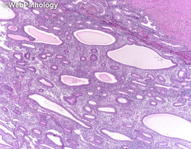
This hematoxylin and eosin stained section presents endometrial tissue from a perimenopausal patient with abnormal uterine bleeding. The architectural pattern shows cystically dilated glands with marked crowding and a back-to-back arrangement within fibromuscular stroma, yielding an overall increased gland-to-stroma ratio. Gland lumina are variably tortuous and irregular in contour, but the epithelial lining demonstrates only minimal cytologic atypia in this field. There is no evident stromal invasion or myometrial extension. The morphology is compatible with endometrial hyperplasia; features may reflect non-atypical hyperplasia or, if nuclear atypia is present in broader sections, atypical hyperplasia/EIN. In the clinical context provided, abnormal non-cyclical bleeding—postmenopausal bleeding, menorrhagia, dysmenorrhea, or intermenstrual bleeding—raises concern for progression to endometrial neoplasia. Differential considerations include complex hyperplasia with atypia, endometrioid intraepithelial neoplasia, and early endometrial carcinoma, particularly if atypia is evident on sampling. Pathologic significance lies in stratifying risk of progression and guiding management, which ranges from surveillance and hormonal therapy to surgical intervention depending on age, fertility wishes, and the presence of cytologic atypia. This image thus supports a radiologic-pathologic correlation for endometrial proliferative disorders in women presenting with abnormal uterine bleeding. Correlation with imaging and clinical data strengthens diagnostic confidence. These findings guide patient-centered treatment planning. decisions.
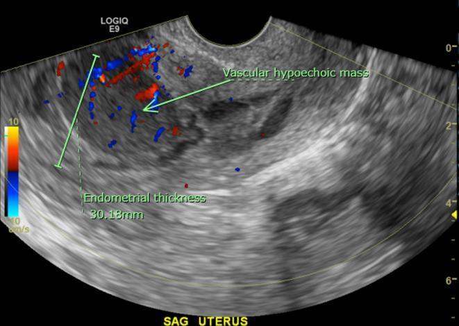
This diagnostic image is a transvaginal ultrasound of the uterus in the sagittal plane, featuring both gray-scale and color Doppler imaging. The uterine view reveals a markedly thickened endometrial stripe measuring approximately 30.18 mm, which is significantly above the normal postmenopausal range. The endometrial contour is irregular and is partially delineated by a trace amount of intrauterine fluid. Within the anterior endometrium, there is a distinct ovoid, homogeneous, hypoechoic mass measuring roughly 3.2 x 2.2 cm. Color Doppler overlay demonstrates internal vascularity within this mass, indicated by red and blue flow signals. Such findings are clinically significant in the context of postmenopausal bleeding, as they necessitate differentiation between endometrial carcinoma, polyps, or rarer entities like primary uterine lymphoma. The image illustrates key sonographic markers for endometrial pathology evaluation including thickness measurement, echogenicity of focal lesions, and assessment of vascular flow patterns.
Postmenopausal Bleeding (PMB) - Clinical Approach
Definition
Key principle: The severity of bleeding does NOT correlate with the risk of malignancy. Even a single spot must be investigated.
Causes (Differential Diagnosis)
| Cause | Frequency |
|---|---|
| Atrophic vaginitis / endometritis | ~60-80% (most common) |
| Endometrial polyp | ~10-12% |
| Endometrial hyperplasia / EIN | ~5-10% |
| Endometrial carcinoma | ~5-10% |
| Exogenous hormones (HRT, tamoxifen) | Variable |
| Cervical pathology (polyp, malignancy) | ~5% |
| Ovarian functional tumors (estrogen-secreting) | Rare |
| Other: coagulation disorders, trauma, urogenital atrophy | Uncommon |
Risk Factors for Endometrial Carcinoma in PMB
- Obesity (BMI >30)
- Diabetes mellitus (type 2)
- Nulliparity
- Chronic anovulation history (e.g., prior PCOS)
- Tamoxifen use
- Unopposed estrogen (HRT without progestogen)
- Hypertension (associated with malignant endometrial polyps)
- Late menopause / prolonged estrogen exposure
- Lynch syndrome / hereditary non-polyposis colorectal cancer (HNPCC)
Step-by-Step Clinical Approach
Step 1 - History
- Duration, frequency, amount of bleeding
- HRT or tamoxifen use
- Sexual history (new partner, trauma, infection)
- PMH: diabetes, hypertension, obesity
- Family history: Lynch syndrome, colorectal cancer
Step 2 - Physical Examination
- General: BMI, blood pressure
- Pelvic exam: vulva/vaginal atrophy, cervical lesions, uterine size
- Pap smear / cervical cytology - essential first step (note: Pap smear is insensitive for endometrial cancer; it can be negative even in invasive cervical cancer due to tumor necrosis)
- Cervical biopsy if gross lesion visible; colposcopy if abnormal Pap
Step 3 - Initial Investigations
- CBC (assess degree of blood loss)
- TSH (thyroid disease can mimic)
- Cervical swabs if indicated (STI screen)
- Transvaginal Ultrasound (TVUS) - first-line imaging investigation
The Endometrial Thickness Decision Cutoff
| Endometrial Stripe (TVUS) | Action |
|---|---|
| <4 mm | Low risk - endometrial biopsy may be deferred if no recurrent bleeding (Washington Manual cutoff) |
| <5 mm | High negative predictive value - can avoid biopsy in most cases (Berek & Novak / Swanson's cutoff) |
| ≥5 mm | Endometrial biopsy mandatory |
| Any thickness + recurrent bleeding | Biopsy regardless of thickness |
Meta-analysis evidence: 96% of women with endometrial cancer had an endometrial stripe >5 mm on TVUS. - Swanson's Family Medicine Review
Step 4 - Endometrial Sampling Methods
- Office endometrial biopsy (Pipelle) - first line, outpatient, ~90% sensitivity for endometrial cancer
- Sonohysterography (SIS) - saline infusion sonography - better for polyp delineation
- Hysteroscopy + directed biopsy - gold standard when office biopsy inconclusive or negative with persistent bleeding
- D&C (dilatation and curettage) - used when office biopsy not possible or non-diagnostic; more invasive/expensive, not first-line
Histopathology and Management
Endometrial Hyperplasia Nomenclature (WHO/EIN 2015 Update)
| Old WHO Classification | New EIN/ACOG 2015 Classification | Treatment |
|---|---|---|
| Simple / complex hyperplasia without atypia | Benign endometrial hyperplasia | Medical (progestogens) |
| Simple / complex atypical hyperplasia | EIN (Endometrial Intraepithelial Neoplasia) - premalignant | Surgical (hysterectomy preferred) or medical if poor candidate |
| Endometrial cancer (well differentiated) | Endometrial adenocarcinoma, endometrioid type | Surgical + staging |
Important: ~40-50% of women with EIN/atypical hyperplasia have concurrent carcinoma at hysterectomy. Risk of progression of hyperplasia WITHOUT atypia is low; with atypia it is ~30%.
Management by Cause
| Finding | Management |
|---|---|
| Atrophic vaginitis/endometritis | Topical or systemic estrogen (after excluding malignancy) |
| Cervical polyp | Office removal |
| Endometrial polyp | Hysteroscopic polypectomy (especially in postmenopausal - higher malignancy risk) |
| Benign hyperplasia | Cyclic/continuous progestogens; repeat biopsy in 3-6 months |
| EIN (postmenopausal) | Hysterectomy (rules out concurrent carcinoma) |
| Endometrial carcinoma | Surgical staging (TAH + BSO + pelvic/para-aortic LN dissection) +/- adjuvant therapy |
Endometrial Cancer Management Summary
- Stage IA (confined to endometrium or <50% myometrial invasion): Surgery alone; vaginal brachytherapy for select higher-risk cases
- Stage IB-IV: Surgery + radiation (vaginal brachytherapy for IB; external beam for stages II-IV)
- Stage III/IV: Surgery + systemic chemotherapy (carboplatin + paclitaxel is most common)
- HER2+ tumors: Add trastuzumab
- ER+ tumors with indolent course: Endocrine therapy (aromatase inhibitors, tamoxifen)
- MSI-H/dMMR tumors: Pembrolizumab ± lenvatinib
Special Situations
- Tamoxifen use: Increases endometrial polyps (cystic dilation + stromal condensation). These must be distinguished from malignancy - biopsy/hysteroscopy required.
- HRT-related bleeding: Endometrial biopsy recommended for any irregular bleeding after >6 months of HRT use, regardless of stripe thickness.
- Ovarian functional tumors: Estrogen-secreting tumors (granulosa cell tumors) cause endometrial hyperplasia/carcinoma via unopposed estrogen - must be excluded by pelvic TVUS.
Quick Algorithm Summary
PMB (any bleeding ≥12 months post-menopause)
        ↓
Pelvic exam + Pap smear + CBC + TSH
        ↓
Transvaginal Ultrasound (TVUS)
   ├── Endometrial stripe <4-5 mm → No biopsy if isolated episode; observe
   │                               Biopsy if recurrent bleeding
   └── Endometrial stripe ≥5 mm →
              ↓
         Endometrial Biopsy (Office Pipelle)
         ├── Benign / EH without atypia → Progestogens, follow-up
         ├── EIN → Hysterectomy (postmenopausal)
         ├── Endometrial carcinoma → Surgical staging + multidisciplinary team
         └── Inadequate/inconclusive → Hysteroscopy + directed biopsy