
Pathophysiological Sequence:Mechanical Delamination: Under the immense, multi-directional shear and compressive forces of mastication, the unreinforced Proplast-Teflon material undergoes structural breakdown and fragmenting.Macrophage Recruitment: Microscopic particulate polymer and metal debris flake off into the joint spaces, triggering a massive influx of foreign-body giant cells and macrophages attempting to phagocytose the non-degradable particles.Cytokine Cascade: Unable to digest the debris, the immune cells release a continuous wave of pro-inflammatory cytokines (such as TNF-$\alpha$, IL-1, and IL-6).Osteolysis: This localized inflammatory storm upregulates osteoclast activity, resulting in aggressive bone resorption, destruction of the glenoid fossa and condyle, and ultimate joint collapse. convert this into image
A detailed medical diagram showing the pathophysiological sequence of Proplast-Teflon TMJ implant failure in 4 sequential steps, clean scientific illustration style with labeled panels: Panel 1 - "Mechanical Delamination": Cross-section of a temporomandibular joint showing a Proplast-Teflon implant between the glenoid fossa and condyle, with arrows indicating multi-directional shear and compressive mastication forces causing the implant material to crack, fragment, and break apart into small particles. Panel 2 - "Macrophage Recruitment": Zoomed-in view of the joint space showing microscopic polymer and metal debris particles scattered in synovial tissue, surrounded by macrophages and multinucleated foreign-body giant cells attempting to engulf the non-degradable particles. Show cellular infiltration with purple-stained nuclei. Panel 3 - "Cytokine Cascade": Illustration of activated macrophages and giant cells releasing pro-inflammatory cytokines (TNF-alpha, IL-1, IL-6) shown as labeled arrows radiating outward from the frustrated immune cells, creating an inflammatory storm in the joint tissue. Panel 4 - "Osteolysis & Joint Collapse": Cross-section showing aggressive bone resorption at the glenoid fossa and mandibular condyle, with osteoclasts actively destroying bone, resulting in severe erosion, loss of joint architecture, and joint collapse. Show visible bone destruction and loss of normal anatomy. Layout: 2x2 grid of panels with a title at the top reading "Proplast-Teflon TMJ Implant Failure: Pathophysiological Sequence". Each panel has a bold numbered heading, descriptive label, and key cellular/molecular players annotated. Use a clean white background with color-coded panels, anatomical cross-sections, and cellular detail. Medical textbook illustration quality.
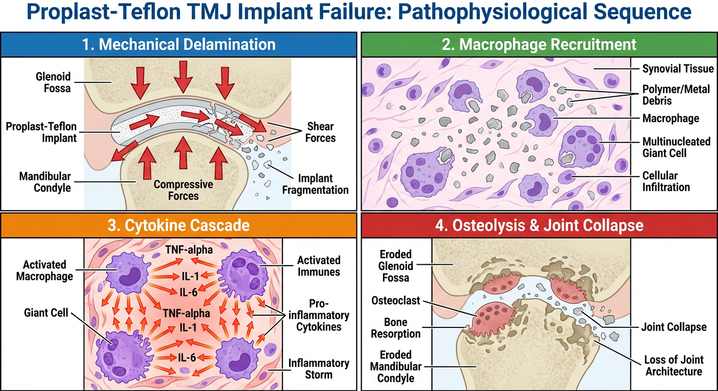
- Mechanical Delamination - Shows the TMJ implant fragmenting under mastication forces
- Macrophage Recruitment - Depicts foreign-body giant cells and macrophages swarming non-degradable debris
- Cytokine Cascade - Illustrates the release of TNF-alpha, IL-1, and IL-6 from frustrated immune cells
- Osteolysis & Joint Collapse - Shows osteoclast-driven bone resorption destroying the glenoid fossa and condyle