
Hyperventilation and hypoventilation during Anesthesia Write detailed answers for resident exam
Reading File
Now let me search the medical library in parallel for all relevant content.
Finding Sources
Finding Sources
Finding Sources
Excellent - found highly relevant content in Morgan & Mikhail and Miller's Anesthesia. Let me now read those sections in parallel.
Finding Sources
Reading File
Reading File
Reading File
Finding Sources
Finding Sources
Reading File
Finding Sources
Finding Sources
Reading File
Reading File
Finding Sources
Finding Sources
Reading File
Reading File
Reading File
Excellent - I now have comprehensive, authoritative content from Miller's Anesthesia, Barash's Clinical Anesthesia, and Katzung's Pharmacology. Let me compile this into a thorough resident exam answer.
Loading Image
Loading Image
Excellent! I now have all the content and key figures needed. Let me write the comprehensive resident exam answer.
Hyperventilation and Hypoventilation During Anesthesia
PART I: PHYSIOLOGY - THE FOUNDATION
Alveolar Ventilation and PaCO₂
The relationship between alveolar ventilation (VA) and arterial PaCO₂ is governed by:
PaCO₂ = k × VCO₂ / VA (where k = 0.86)
This inverse hyperbolic relationship means:
- Doubling VA halves PaCO₂ (hyperventilation)
- Halving VA doubles PaCO₂ (hypoventilation)
The figure below illustrates how alveolar ventilation determines both PAO₂ and PACO₂ simultaneously:
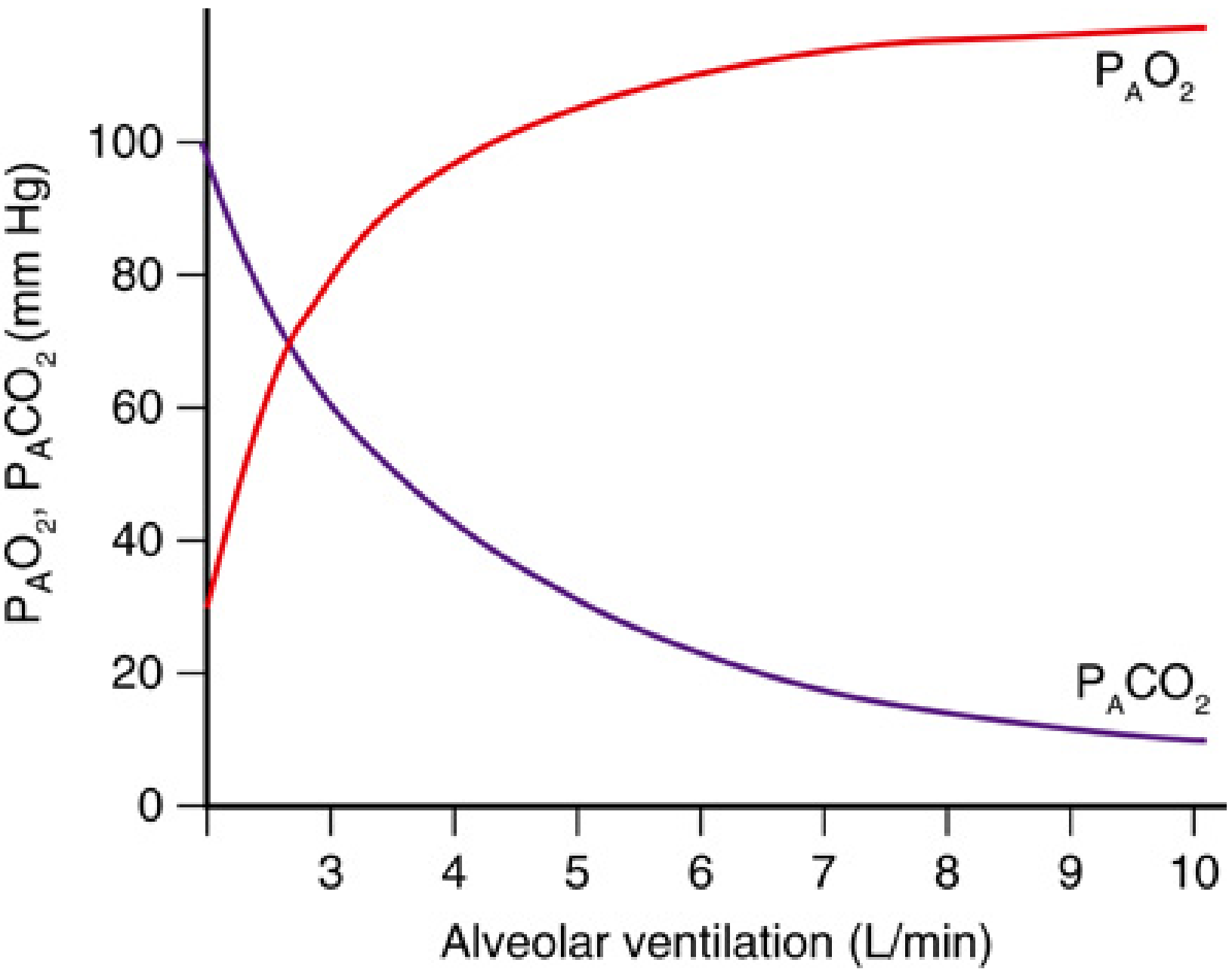
Fig: As alveolar ventilation falls (hypoventilation), PACO₂ rises steeply while PAO₂ drops. As ventilation increases (hyperventilation), PACO₂ falls and PAO₂ rises - but with diminishing returns. - Miller's Anesthesia, 10e
Normal Values (for reference)
| Parameter | Normal | Hypoventilation | Hyperventilation |
|---|---|---|---|
| PaCO₂ | 35-45 mmHg | >45 mmHg | <35 mmHg |
| pH | 7.35-7.45 | <7.35 (acidosis) | >7.45 (alkalosis) |
| PaO₂ | 80-100 mmHg | Decreased | Increased (on air) |
PART II: HYPOVENTILATION DURING ANESTHESIA
Definition
Alveolar ventilation less than that required to maintain PaCO₂ below 45 mmHg.
Mechanisms and Causes
1. Anesthetic Drug Effects on Respiratory Drive
Volatile Agents (Dose-Dependent Depression)
All inhaled volatile anesthetics cause a dose-dependent decrease in tidal volume (VT) with a compensatory but insufficient increase in respiratory rate, resulting in net hypoventilation and hypercapnia:
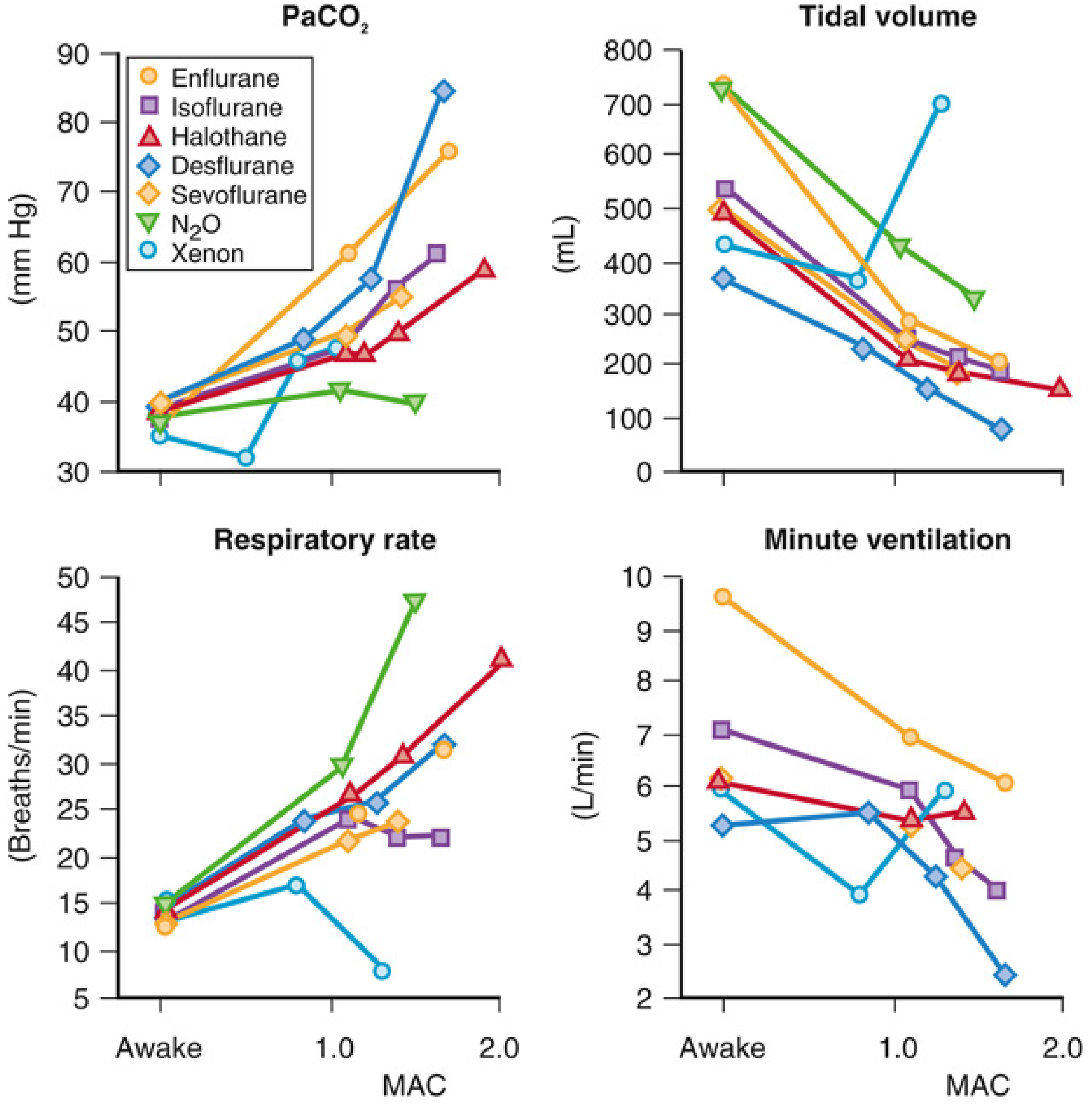
Fig: Dose-dependent effects of inhaled anesthetics. Note rising PaCO₂, falling tidal volume and minute ventilation despite increasing respiratory rate. Desflurane causes the greatest hypercapnia. - Miller's Anesthesia, 10e / Barash 9e
Key points:
- Volatile anesthetics depress both central and peripheral chemoreceptors
- At even 0.1 MAC, isoflurane and sevoflurane abolish the peripheral (carotid body) CO₂ chemoreflex
- Above 1 MAC, the peripheral chemoreflex is completely lost; only central chemoreflex remains
- In conscious humans, minute ventilation increases ~3 L/min per 1 mmHg rise in PaCO₂; this response is profoundly attenuated by volatile anesthetics
- Apneic threshold: The minimum PaCO₂ below which spontaneous ventilation ceases. It is normally 4-5 mmHg below the resting PaCO₂. Volatile anesthetics cause a rightward shift of this threshold - meaning spontaneous breathing efforts will not occur if controlled ventilation drives PaCO₂ below this shifted threshold (Barash, p. 1433)
Clinical Pearl: Assisting ventilation in a spontaneously breathing anesthetized patient may lower PaCO₂ to the apneic threshold, causing apnea and necessitating full controlled ventilation.
Opioids
- Cause respiratory depression through μ-receptor agonism in brainstem respiratory centers
- Decrease respiratory rate more than tidal volume (opposite of volatile agents)
- Diminish the CO₂ response curve (shift rightward AND decrease slope)
- Hypoxic ventilatory response abolished even at subanesthetic doses
- Synergistic respiratory depression when combined with volatile agents
Propofol and Other IV Agents
- Propofol produces dose-dependent respiratory depression and apnea on induction
- Benzodiazepines depress ventilatory response to CO₂; synergistic with opioids
2. Neuromuscular Blockade (NMB)
Residual NMB is a leading cause of postoperative hypoventilation:
- Incomplete reversal of NMB impairs the pressure-generating capacity of respiratory muscles
- According to the equation of motion: Pmus = V × R + V × E - reduced Pmus yields reduced tidal volume
- Decrements in Pmus initially trigger compensatory increases in rate, but hypercapnia develops when compensation is exhausted (Miller's Anesthesia, p. 1300)
- Upper airway muscle weakness is more sensitive to residual NMB than diaphragmatic weakness, predisposing to obstruction
3. Airway Obstruction
| Level | Cause |
|---|---|
| Upper airway | Loss of pharyngeal muscle tone, tongue falling back (supine position), laryngospasm |
| Lower airway | Bronchospasm, secretions, aspiration |
| Equipment | Kinked ETT, circuit obstruction, wrong-sized LMA, undetected esophageal intubation |
In obese patients and those with sleep apnea receiving monitored anesthesia care (MAC) with sedation: upper airway obstruction + hypoventilation + decreased FRC = rapid desaturation (Morgan & Mikhail, p. 69).
4. Increased Dead Space
VA = VE × (1 - VD/VT)
Conditions increasing dead space reduce alveolar ventilation even with unchanged total minute ventilation:
- Pulmonary embolism (most acute increase in dead space)
- Emphysema (chronic)
- Rapid shallow breathing pattern (increases VD/VT ratio)
- Mechanical ventilator circuit and HME filters add instrumental dead space
5. Increased CO₂ Production (Relative Hypoventilation)
If CO₂ production (VCO₂) rises without a corresponding increase in VA, PaCO₂ rises:
- Malignant hyperthermia - massive uncontrolled increase in metabolic rate; early sign is rising ETCO₂
- Thyroid storm, sepsis, fever, shivering
- Laparoscopic surgery - absorption of insufflated CO₂ from peritoneum (planned increase in ventilation required)
- High-carbohydrate TPN while on controlled ventilation
6. Altered Respiratory Mechanics
- Pneumothorax, hemothorax, pleural effusion
- Abdominal distension, prone positioning, obesity (reduced FRC, increased atelectasis)
- Pulmonary edema reducing compliance
- Bronchospasm increasing resistance
Consequences of Hypoventilation / Hypercapnia
| System | Effect |
|---|---|
| Respiratory | Hypoxemia (PAO₂ = [Pb - PH₂O] × FiO₂ - PACO₂/R - as PACO₂ rises, PAO₂ falls) |
| Cardiovascular | Tachycardia, hypertension, increased cardiac output (via catecholamine release); dysrhythmias; pulmonary vasoconstriction worsened |
| Cerebrovascular | Cerebral vasodilation → increased CBF → increased ICP (significant hypercapnia produces dramatic CBF increases) |
| Acid-base | Respiratory acidosis; rightward shift of oxyhemoglobin dissociation curve (Bohr effect) |
| Electrolytes | Hyperkalemia (H⁺/K⁺ exchange across cell membranes) |
| Renal | Bicarbonate retention (compensatory) with chronic hypercapnia |
Management
- Identify the cause - check ETT position, circuit integrity, capnograph waveform
- Controlled ventilation - if spontaneous breathing is inadequate
- Reverse residual NMB - neostigmine + glycopyrrolate or sugammadex
- Reverse opioids if appropriate - naloxone (titrate to avoid acute pain/hypertension)
- Treat bronchospasm - bronchodilators, deepen anesthesia
- Laparoscopy - increase minute ventilation to compensate for CO₂ absorption (typically increase RR or VT by 10-20%)
- Postoperatively - supplemental O₂, positioning (head-up), early extubation criteria assessment
PART III: HYPERVENTILATION DURING ANESTHESIA
Definition
Alveolar ventilation greater than metabolic needs, resulting in PaCO₂ <35 mmHg (hypocapnia) and respiratory alkalosis.
Causes
1. Iatrogenic (Most Common)
- Excessive mechanical ventilation settings during controlled ventilation
- Aggressive manual bag-mask ventilation during induction
- Over-zealous IPPV in response to perceived inadequacy
2. Neurogenic / Pain-Related
- Anxiety and pain before induction
- Light anesthesia / inadequate depth during surgical stimulation
- CNS stimulation (limbic system activation)
3. Metabolic Compensation
- Pre-existing metabolic acidosis - body attempts to compensate by blowing off CO₂
- Diabetic ketoacidosis, sepsis
4. Physiological Stimuli
- Hypoxia stimulates peripheral chemoreceptors → hyperventilation (though hypoxic ventilatory response is blunted by volatile agents)
- Pulmonary embolism - reflex hyperventilation before hypercapnia supervenes
5. Laparoscopy (Intraabdominal CO₂) - Patient
- Peritoneal stretch and diaphragmatic irritation can stimulate breathing in lightly anesthetized patients
Consequences of Hyperventilation / Hypocapnia
| System | Effect | Clinical Significance |
|---|---|---|
| Cerebrovascular | Vasoconstriction → reduced CBF | Used therapeutically to lower ICP; but <20 mmHg = cerebral ischemia |
| Cardiovascular | Decreased cardiac output; coronary vasoconstriction at extreme levels | Carotid surgery: eucapnia preferred to avoid both cerebral vasodilation AND over-vasoconstriction |
| Respiratory | Bronchoconstriction; shift of O₂-Hb curve leftward (Bohr effect - impairs O₂ off-loading to tissues) | |
| Neuromuscular | Decreased ionized calcium (alkalosis increases protein binding) → tetany, perioral tingling, Chvostek/Trousseau | Seen with severe hyperventilation in labor/anxiety |
| Cerebral | Apneic threshold approached - spontaneous breathing effort ceases | (See apneic threshold, above) |
| Uterine/Fetal | Extreme hypocapnia (PaCO₂ <20 mmHg) reduces uterine blood flow → fetal hypoxemia and acidosis | Especially relevant in obstetric anesthesia (Miller's Anesthesia, p. obstetric chapter) |
| Pulmonary vascular | Pulmonary vasoconstriction reduced → useful in right-to-left shunts | Hyperventilation + 100% O₂ is effective in lowering PVR (Morgan & Mikhail, p. Key Concepts) |
Therapeutic Uses of Controlled Hyperventilation
1. Raised Intracranial Pressure (ICP)
- Mechanism: CO₂ is a potent cerebral vasodilator. Hypocapnia causes cerebral arteriolar vasoconstriction → reduced CBV → reduced ICP
- CO₂ vasoreactivity is preserved under volatile anesthesia (isoflurane, sevoflurane, desflurane up to 1.5 MAC)
- Isoflurane-induced ICP rises can be blunted by hyperventilation
- Limitation: Not a definitive treatment; effect wanes over 6-8 hours (CSF pH equilibrates); extreme hypocapnia (<20 mmHg) can cause ischemia
- Desflurane: Vasoconstrictive response to hypocapnia is preserved - hyperventilation can prevent desflurane-induced ICP rises (Goodman & Gilman)
- Barash (p. 1415): "all three potent agents may be used at appropriate doses, especially with adjunctive and compensatory therapies, in nearly any neurosurgical procedure"
2. Pulmonary Hypertension / Right-to-Left Shunt
- Hyperventilation with hypocapnia reduces PVR
- Combined with 100% O₂, very effective in managing acute pulmonary hypertension intraoperatively
3. Malignant Hyperthermia (initial management)
- Hyperventilate with 100% O₂ to blow off accumulated CO₂ while dantrolene is drawn up
Risks of Therapeutic Hyperventilation
- Cerebral ischemia - PaCO₂ <20 mmHg can cause paradoxical cerebral vasoconstriction severe enough to produce focal ischemia
- Carotid endarterectomy - eucapnia preferred because hyperventilation causes vasoconstriction that may further reduce flow to an already ischemic hemisphere
- Obstetrics - fetal compromise from reduced uterine blood flow
- Cardiovascular - decreased venous return and cardiac output from high intrathoracic pressures with aggressive positive pressure ventilation
PART IV: APNEIC THRESHOLD - A CRITICAL CONCEPT
The apneic threshold is the minimum PaCO₂ required to stimulate spontaneous breathing. It sits 4-5 mmHg below the resting awake PaCO₂ (Barash, p. 1433).
Key facts:
- Volatile anesthetics cause a rightward shift of the apneic threshold - i.e., a higher PaCO₂ is needed to trigger breathing
- It is independent of the slope of the CO₂ response curve
- Clinical implication: If you assist/augment ventilation in a spontaneously breathing anesthetized patient and reduce PaCO₂ toward or below the apneic threshold, the patient becomes apneic, requiring full controlled ventilation
- N₂O added to a volatile anesthetic lessens this shift (Barash p. 1433)
PART V: MONITORING
| Monitor | What it tells you |
|---|---|
| Capnography (ETCO₂) | Gold standard for detecting hypoventilation/hyperventilation intraoperatively; ETCO₂ normally 2-5 mmHg below PaCO₂ |
| ABG | PaCO₂, pH, HCO₃⁻ - definitive assessment; essential when ETCO₂-PaCO₂ gap is unexpectedly wide |
| Pulse oximetry | Detects hypoxemia resulting from hypoventilation (late sign; especially unreliable on supplemental O₂) |
| Neuromuscular monitoring | TOF ratio to detect residual NMB (target TOF ratio >0.9 before extubation) |
| Respiratory rate and pattern | Tachypnea = shallow breathing; watch for paradoxical movement |
ETCO₂ - PaCO₂ Gradient: Normally 2-5 mmHg. An increased gradient means more dead space ventilation (PE, low cardiac output, emphysema). A negative gradient (ETCO₂ > PaCO₂) suggests artifact or sampling error.
PART VI: SUMMARY TABLE FOR EXAM
| Feature | Hypoventilation | Hyperventilation |
|---|---|---|
| PaCO₂ | >45 mmHg | <35 mmHg |
| pH | <7.35 (acidosis) | >7.45 (alkalosis) |
| PAO₂ | Decreased | Increased |
| CBF | Increased | Decreased |
| ICP | Increased | Decreased |
| PVR | Increased | Decreased |
| Ionized Ca²⁺ | Increased | Decreased |
| K⁺ | Increased | Decreased |
| Oxyhemoglobin curve | Rightward shift | Leftward shift |
| ETCO₂ | Elevated | Reduced |
| Common causes | Opioids, volatile agents, residual NMB, obstruction, laparoscopy CO₂ | Excessive controlled ventilation, pain/anxiety, light anesthesia, therapeutic (neurosurgery, pulm HTN) |
REFERENCES
- Miller's Anesthesia, 10e - Mechanisms of Hypoxemia and Hypercapnia; Effects of Inhaled Anesthetics on Hypercapnic Ventilatory Response (pp. 1287-1301, 2135-2137)
- Barash, Cullen, and Stoelting's Clinical Anesthesia, 9e - Response to Carbon Dioxide and Hypoxemia; Intracerebral Pressure (pp. 1433-1435, 1415)
- Katzung's Basic and Clinical Pharmacology, 16e - Respiratory Effects of Volatile Anesthetics (p. 702)
- Goodman & Gilman's The Pharmacological Basis of Therapeutics - Desflurane: vasoconstrictive response to hypocapnia
- Morgan & Mikhail's Clinical Anesthesiology, 7e - MAC/Oxygen supplementation; Key Concepts on PVR management
This is a shared conversation. Sign in to Orris to start your own chat.