
Types of thyroid nodules,diagnostic and management
Reading File
Now let me search the medical library and PubMed in parallel for comprehensive coverage.
Finding Sources
Searching PubMed
"thyroid nodule" AND management
Searching the Web
thyroid nodule types classification TIRADS diagnosis management 2024
Good - now let me read the key textbook sections in parallel.
Reading File
Reading File
Reading File
Reading File
Excellent content retrieved. Let me now get the management sections and fetch the diagnostic flowchart image.
Reading File
Reading File
Loading Image
Finding Sources
Now let me get the Bethesda classification and TIRADS details from Harrison's (2025) and Current Surgical Therapy.
Reading File
Reading File
Excellent content. Let me get the Bethesda table from Harrison's and the ATA sonographic pattern image.
Reading File
Loading Image
Reading File
I have comprehensive data. Let me also fetch the ACR TI-RADS image and the Harrison's approach flowchart.
Loading Image
Loading Image
I now have excellent, comprehensive data from multiple authoritative textbooks. Let me compose the full answer.
Thyroid Nodules: Types, Diagnosis, and Management
A thyroid nodule is a discrete lesion within the thyroid gland. Palpable nodules occur in up to 5% of women and 1% of men in iodine-replete areas, but high-resolution ultrasound detects nodules in 19-68% of the population ("incidentalomas"). While common, only ~5% are malignant. The three main reasons to intervene are: (1) concern for malignancy, (2) hyperfunction, and (3) compressive symptoms. - Sabiston Textbook of Surgery, p. 1502
1. Types of Thyroid Nodules
A. Benign Nodules (majority)
| Type | Key Features |
|---|---|
| Colloid/Adenomatous nodule | Most common; hyperplastic accumulation of colloid; spongiform on US |
| Follicular adenoma | Encapsulated benign neoplasm; cannot be distinguished from follicular carcinoma by FNA alone - requires surgical excision |
| Oncocytic (Hürthle cell) adenoma | Distinct WHO 2022 classification; "Hürthle cell" now discouraged as a misnomer |
| Simple/colloid cyst | Anechoic, completely cystic; malignancy risk <1% |
| Nodular hyperplasia | Part of multinodular goiter; multiple nodules |
| Focal thyroiditis | Inflammatory; may mimic malignancy clinically |
"The majority of thyroid nodules are benign neoplasms and include colloid nodules, degenerative cysts, nodular hyperplasia and follicular adenomas." - Sabiston Textbook of Surgery, p. 1503
B. Malignant Nodules (in decreasing frequency)
| Type | Key Features |
|---|---|
| Papillary thyroid carcinoma (PTC) | Most common (~80%); excellent prognosis; RET/PTC translocations; BRAF mutations |
| Follicular thyroid carcinoma (FTC) | Requires capsular/vascular invasion on histology to distinguish from adenoma |
| Oncocytic carcinoma (OCA) | Previously Hürthle cell carcinoma; distinct entity per WHO 2022 |
| Medullary thyroid carcinoma (MTC) | Arises from parafollicular C cells; secretes calcitonin; sporadic or familial (MEN 2A/2B); does NOT take up radioiodine |
| Poorly differentiated thyroid cancer (PDTC) | Aggressive; intermediate between DTC and ATC |
| Anaplastic thyroid carcinoma (ATC) | Most aggressive; rapidly fatal; does not respond to RAI |
| Primary thyroid lymphoma | Often arises in background of Hashimoto's thyroiditis; diffuse large B-cell commonest; avoid surgery as initial therapy |
| Metastasis to thyroid | Rare; from renal cell, breast, lung, melanoma |
"The most common malignant diagnosis is PTC, and in decreasing order of frequency also includes FTC, oncocytic carcinoma (OCA), MTC, PDTC, and ATC; other rare malignancies including primary thyroid lymphoma and metastasis to the thyroid gland can occur." - Sabiston Textbook of Surgery, p. 1503
C. Functional Status (Scintigraphic Classification)
| Type | Radioiodine Uptake | Malignancy Risk |
|---|---|---|
| Hot nodule (hyperfunctioning) | Increased | Almost never malignant; no FNA needed |
| Warm nodule | Same as surrounding | Low |
| Cold nodule (non-functioning) | Decreased | Higher (~5-15%); most warrant FNA |
2. Diagnostic Approach
Step 1: History & Physical Examination
High-risk clinical features suggesting malignancy:
- Age <20 or >70 years
- Male sex
- History of neck irradiation (risk increases linearly from 6.5-2000 cGy; peaks 20-30 years post-exposure)
- Rapid growth, hoarseness, dysphagia, dyspnea (compressive/infiltrative signs)
- Firm, fixed, immobile nodule >3-4 cm
- Cervical lymphadenopathy
- Family history of thyroid cancer or MEN syndrome
- Pheochromocytoma or hyperparathyroidism (suggests MEN 2)
"Patients younger than 20 years have an approximately 20-50% incidence of malignancy when presenting with a solitary thyroid nodule." - Cummings Otolaryngology, p. 3544
Step 2: Serum TSH
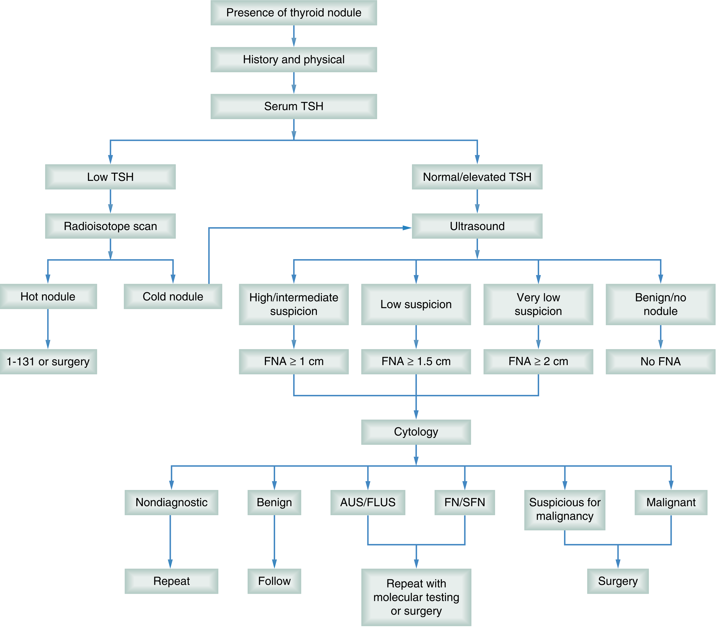
The first blood test. A suppressed TSH triggers radionuclide scanning to identify a hot nodule (virtually never malignant; treat hyperthyroidism - I-131 or surgery). A normal/elevated TSH proceeds to ultrasound.
Step 3: Neck Ultrasound
The most important radiographic study. Documents:
- Parenchymal pattern and gland size
- Nodule size, location, and characteristics
- Cervical lymph node status (central and lateral compartments)
Benign US features: Spongiform (>50% small cystic spaces), purely cystic, comet-tail artifacts
Malignant US features (in decreasing specificity):
- Microcalcifications
- Hypoechoic solid nodule
- Taller-than-wide shape
- Irregular/microlobulated margins
- Extrathyroidal extension
- Increased intranodular vascularity
- Associated abnormal lymph nodes
ATA Sonographic Risk Stratification (from Current Surgical Therapy 14e):
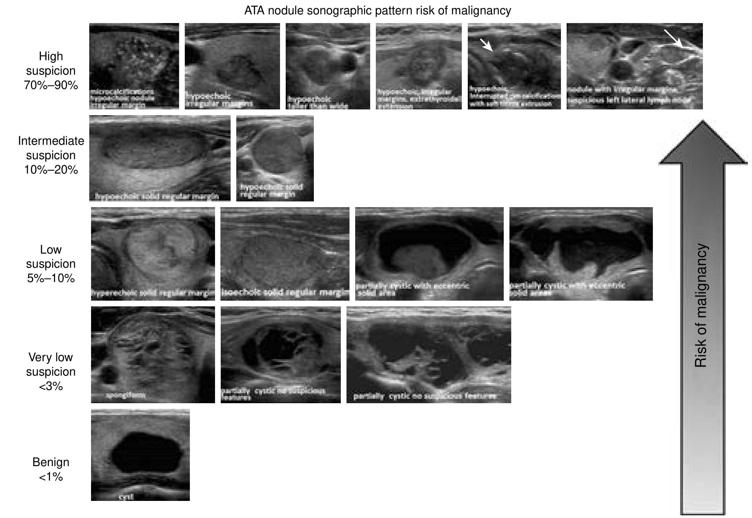
| ATA Pattern | Malignancy Risk | FNA Threshold |
|---|---|---|
| High suspicion | 70-90% | ≥ 1 cm |
| Intermediate suspicion | 10-20% | ≥ 1 cm |
| Low suspicion | 5-10% | ≥ 1.5 cm |
| Very low suspicion | <3% | ≥ 2 cm |
| Benign (cyst) | <1% | No FNA |
ACR TI-RADS System
A point-based system assessing five features: composition, echogenicity, shape, margin, and echogenic foci:
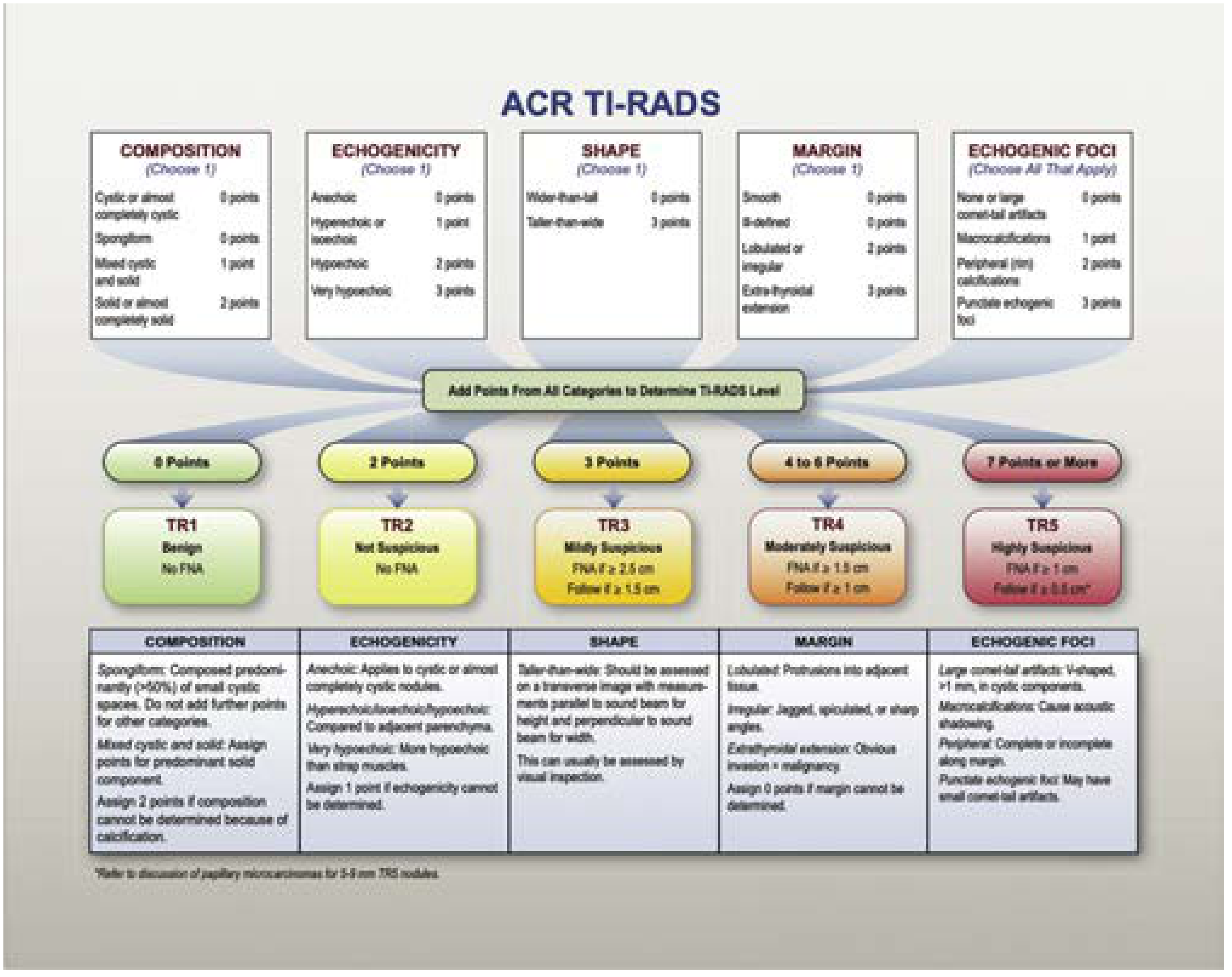
| Level | Points | Category | FNA Threshold |
|---|---|---|---|
| TR1 | 0 | Benign | No FNA |
| TR2 | 2 | Not suspicious | No FNA |
| TR3 | 3 | Mildly suspicious | FNA ≥ 2.5 cm |
| TR4 | 4-6 | Moderately suspicious | FNA ≥ 1.5 cm |
| TR5 | ≥7 | Highly suspicious | FNA ≥ 1 cm |
FDG-PET incidental thyroid nodules carry a higher cancer risk - FNA recommended at >1 cm threshold. - Current Surgical Therapy 14e, p. 1705
Step 4: Fine-Needle Aspiration (FNA) Biopsy
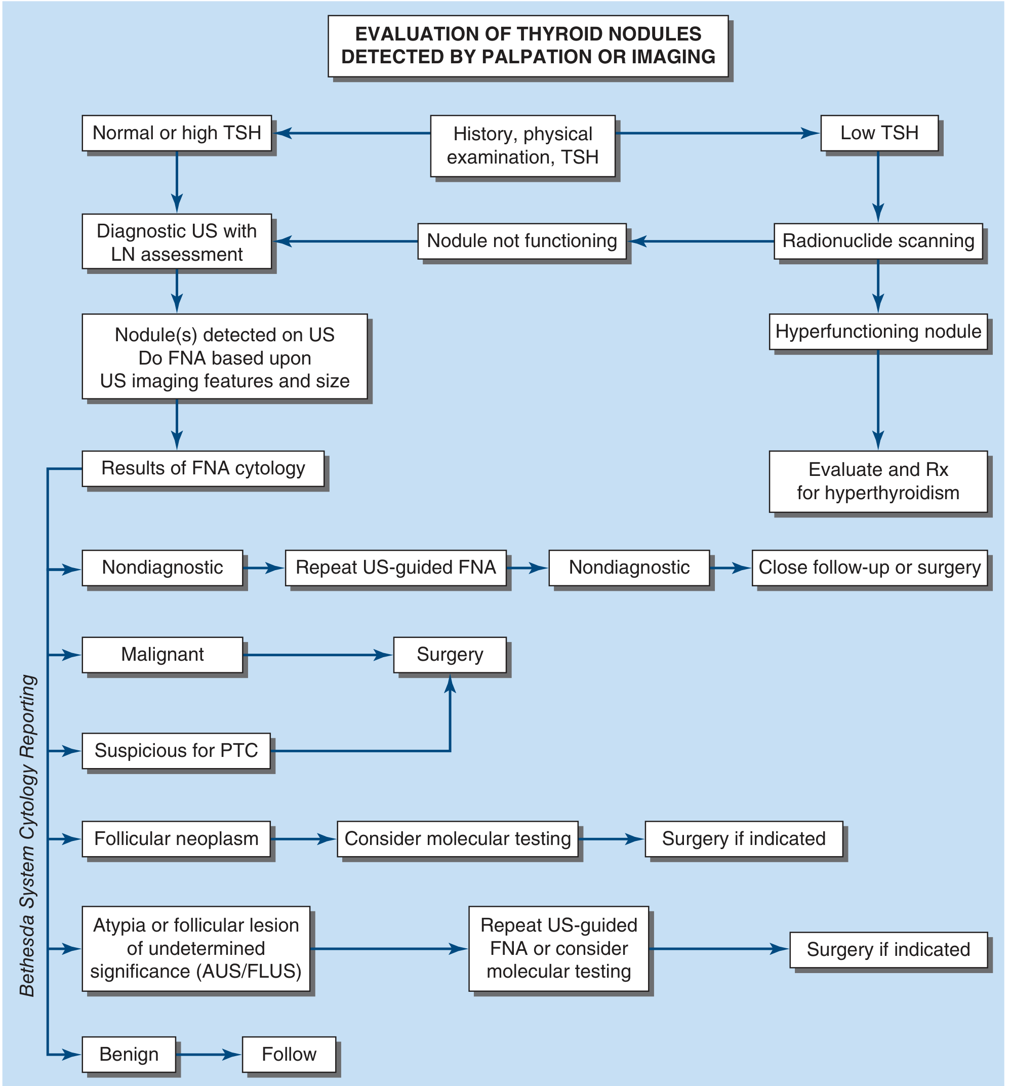
US-guided FNA is preferred (fewer non-diagnostic samples). Results are reported using the Bethesda System (Version 3, 2023) - Harrison's Principles of Internal Medicine 22E, p. 3092:
| Bethesda Category | Diagnosis | Risk of Malignancy | Usual Management |
|---|---|---|---|
| I | Nondiagnostic/Unsatisfactory | 13% (5-20%) | Repeat US-guided FNA |
| II | Benign | 4% (2-7%) | Follow with US |
| III | Atypia of Undetermined Significance (AUS/FLUS) | 22% (13-30%) | Repeat FNA or molecular testing |
| IV | Follicular Neoplasm (FN/SFN) | 30% (23-34%) | Molecular testing ± surgery |
| V | Suspicious for Malignancy | 74% (67-83%) | Surgery |
| VI | Malignant | 97% (97-100%) | Surgery |
NIFTP (noninvasive follicular thyroid neoplasm with papillary-like nuclear features) is no longer classified as malignant - it is a low-risk neoplasm that still requires surgical excision for definitive diagnosis.
Step 5: Molecular Testing (for Bethesda III-IV)
For indeterminate nodules (Bethesda III/IV), molecular tests help guide surgical vs. surveillance decisions:
- Afirma Gene Expression Classifier (GEC/GSC) - "rule-out" test (high NPV)
- ThyroSeq v3 - Next-generation sequencing of 112 genes; analyzes mutations, gene fusions, copy number alterations - both rule-out and rule-in
- ThyGenX/ThyraMIR and Rosetta GX Reveal - Less clinical data
Step 6: Additional Tests
- Serum calcitonin - Useful for diagnosis/surveillance of MTC; routine screening not standard in the US
- Serum thyroglobulin - Useful for post-treatment surveillance of differentiated thyroid cancer, NOT for initial diagnosis
- CT/MRI - Reserved for locally advanced disease, substernal extension, or suspected metastases
- Genetic testing (RET mutations) - Mandatory in all MTC patients; enables family screening
3. Diagnostic Algorithm
4. Management
Benign Nodules (Bethesda II)
- Observation with periodic US (typically 6-18 months, then every 2-3 years)
- No role for T4 suppression (ineffective in iodine-replete populations and carries cardiovascular/skeletal risks)
- Indications for repeat FNA: growth >20% in two dimensions, or change in ultrasound features
- Surgical indications for confirmed benign nodules: cosmetic concern, compressive symptoms (dysphagia, dyspnea), or patient preference
Hot/Toxic Nodule
- Radioiodine (I-131) or surgical resection (lobectomy)
- Antithyroid drugs as bridge to definitive treatment
Indeterminate Nodules (Bethesda III-IV)
- Molecular testing first if available - guides whether to observe or operate
- If molecular testing suggests low risk: surveillance with repeat US/FNA
- If molecular testing suggests high risk, or testing unavailable: diagnostic lobectomy
- A diagnosis of follicular neoplasm (Bethesda IV) always requires surgery (cannot distinguish adenoma from carcinoma by FNA alone)
Malignant/Suspicious Nodules (Bethesda V-VI)
- Surgical resection is the primary treatment
- Lobectomy vs. total thyroidectomy: Decision based on cancer size, risk features, patient factors; ATA guidelines support lobectomy for low-risk PTC <1 cm without extrathyroidal extension or lymph node involvement
- Preoperative US of cervical lymph nodes is mandatory before any thyroid cancer surgery
- Radioactive iodine (RAI) ablation - for differentiated thyroid cancer with high-risk features post-thyroidectomy
- TSH suppression therapy - Post-operative levothyroxine to maintain TSH below normal range in high-risk DTC
- External beam radiation - For anaplastic carcinoma, lymphoma (often highly radiosensitive), or cases with gross residual disease
- Targeted therapy - Kinase inhibitors (sorafenib, lenvatinib) for radioiodine-refractory metastatic DTC; vandetanib/cabozantinib for MTC; BRAF/MEK inhibitors for ATC with BRAF V600E mutations
Active Surveillance for Low-Risk PTC
Increasingly used for papillary microcarcinomas (<1 cm, intrathyroidal, node-negative) - an approach validated particularly in Japan and now endorsed by 2023 ETA guidelines and the 2024 Japan Association of Endocrine Surgery guidelines (PMID: 40058844).
5. Special Considerations
- Multinodular goiter: Risk of malignancy per nodule is the same as for a solitary nodule; dominant nodule or those with suspicious US features should be biopsied
- Substernal goiter: Most can be removed via cervical incision; posteriorly extending or infraaortic goiters may require sternotomy
- Pediatric nodules: 20-50% malignancy risk; treat more aggressively
- Pregnancy: US-guided FNA acceptable; surgery deferred to second trimester if possible; RAI is contraindicated
Sources:
- Sabiston Textbook of Surgery, 21st ed., pp. 1502-1503
- Harrison's Principles of Internal Medicine, 22nd ed. (2025), pp. 3091-3093
- Cummings Otolaryngology Head and Neck Surgery, pp. 3535-3674
- Current Surgical Therapy, 14th ed., pp. 1713-1755
- Schwartz's Principles of Surgery, 11th ed., p. 1669
- 2023 ETA Clinical Practice Guidelines (PMID: 37358008)
- 2024 Japan Endocrine Surgery Guidelines (PMID: 40058844)
This is a shared conversation. Sign in to Orris to start your own chat.