
General Pathology LQ 1. Define inflammation.Describe the Vascular and Cellular events of Inflammation 2. Define Shock . Mention types of Shock . Describe pathogenesis of septic shock
Reading File
Now I'll search the medical library for both topics in parallel.
Finding Sources
Finding Sources
Finding Sources
Finding Sources
Finding Sources
Reading File
Reading File
Reading File
Reading File
Reading File
I have excellent content. Now let me get the cellular events section and the definition/overview of inflammation and shock.
Reading File
Finding Sources
Finding Sources
Reading File
Reading File
Finding Sources
Finding Sources
Reading File
Loading Image
Loading Image
I now have all the content needed. Let me compile the comprehensive answers.
General Pathology Long Questions
Q1. Define Inflammation. Describe the Vascular and Cellular Events of Inflammation
Definition of Inflammation
Inflammation is a response of vascularized tissues that delivers leukocytes and molecules of host defense from the circulation to sites of infection and cell damage. It is a protective response essential for survival, serving to rid the host of the initial cause of cell injury (microbes, toxins) and the consequences of that injury (necrotic cells and tissues).
- Robbins, Cotran & Kumar Pathologic Basis of Disease: "Inflammation is a response of vascularized tissues that delivers leukocytes and molecules of host defense from the circulation to sites of infection and cell damage."
The cardinal signs of inflammation are:
- Calor - Heat
- Rubor - Redness
- Tumor - Swelling
- Dolor - Pain
- Functio laesa - Loss of function (added by Rudolf Virchow)
Types of Inflammation
| Feature | Acute Inflammation | Chronic Inflammation |
|---|---|---|
| Onset | Fast: minutes to hours | Slow: days |
| Cellular infiltrate | Mainly neutrophils | Monocytes/macrophages and lymphocytes |
| Tissue injury | Usually mild and self-limited | May be significant |
| Fibrosis | None | May be severe and progressive |
| Local/systemic signs | Prominent | Variable, usually modest |
The Five R's of Inflammation
- Recognition of the offending agent
- Recruitment of blood cells and proteins to the tissue site
- Removal of the offending agent
- Regulation of the reaction
- Repair of injured tissue
I. Vascular Events of Acute Inflammation
Acute inflammation has three major vascular components:
- Dilation of small vessels
- Increased permeability of the microvasculature
- Emigration of leukocytes from the microcirculation
Most of these changes occur in postcapillary venules at the site of infection or injury.
1. Vasodilation (Changes in Vascular Flow and Caliber)
- Vasodilation is one of the earliest reactions of acute inflammation and is responsible for the externally visible redness (erythema) and warmth.
- The most important chemical mediator of vasodilation is histamine.
- The sequence of events:
- Transient vasoconstriction (lasting only seconds)
- Followed by vasodilation - affecting arterioles first, then opening of new capillary beds
- Results in increased blood flow (hyperemia) - causes heat and redness
2. Increased Vascular Permeability (Exudation)
- Vasodilation is quickly followed by increased permeability of the microvasculature.
- Exudate = extravascular fluid with high protein concentration and cellular debris (implies increased vascular permeability during inflammation).
- Transudate = fluid with low protein content (essentially ultrafiltrate of plasma - NOT associated with inflammation).
- Edema = excess fluid in interstitial tissue or serous cavities.
- Pus = purulent exudate rich in leukocytes, dead cell debris, and microbes.
Mechanisms of increased vascular permeability:
| Mechanism | Details |
|---|---|
| Contraction of endothelial cells (most common) | Creates interendothelial gaps; elicited by histamine, bradykinin, leukotrienes; occurs within 15-30 min and is usually short-lived |
| Direct endothelial injury | Seen in burns; leakage starts immediately and is sustained for hours |
| Leukocyte-mediated injury | Activated leukocytes release toxic oxygen species and proteases, damaging endothelium |
| Increased transcytosis | Increased transport of vesicles across endothelial cells |
| Angiogenesis | New blood vessel growth with leaky and incompetent junctions |
3. Stasis
- Loss of fluid and increased vessel diameter lead to slower blood flow and higher concentration of red cells in small vessels, raising blood viscosity.
- Small vessels become engorged with slowly moving red cells - a condition termed stasis.
- Stasis is marked by vascular congestion and localized redness.
II. Cellular Events of Acute Inflammation
Leukocyte Recruitment - The Multi-Step Process
The journey of leukocytes from the vessel lumen to the tissue involves:
- Margination and rolling
- Adhesion to endothelium
- Transmigration through the vessel wall
- Migration in the tissues toward the stimulus (chemotaxis)
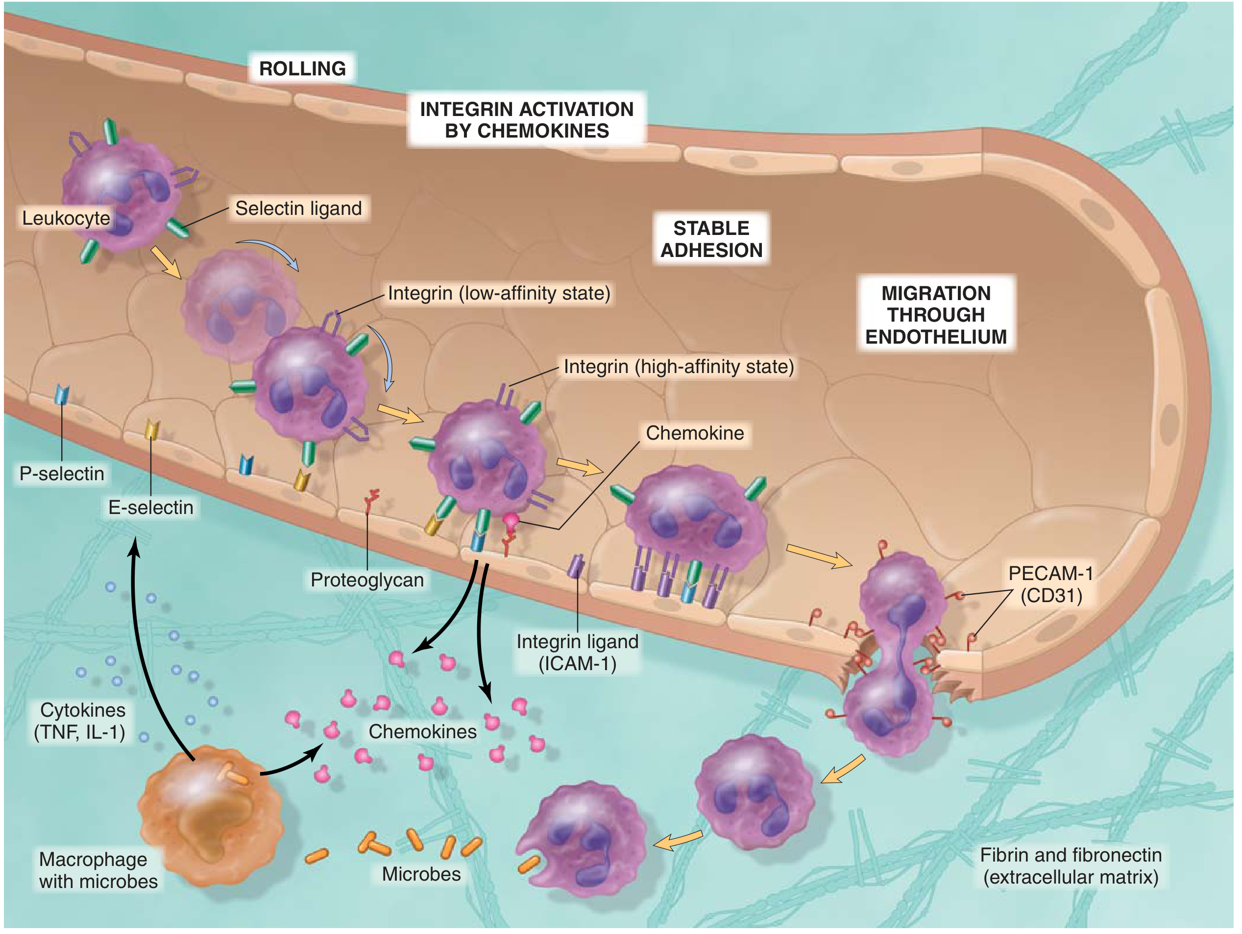
Step 1: Margination and Rolling
- Under normal laminar flow, red cells are central and leukocytes are peripheral.
- As flow slows (stasis), leukocytes assume a peripheral position - margination.
- Leukocytes then begin to roll along the endothelial surface, mediated by selectins:
- P-selectin - stored in Weibel-Palade bodies; rapidly traffics to surface within minutes on exposure to histamine or thrombin
- E-selectin - expressed on activated endothelium (upregulated by cytokines like TNF, IL-1)
- L-selectin - expressed on leukocytes
Step 2: Firm Adhesion (Stable Adhesion)
- Rolling cells fully stop and adhere firmly via integrins on leukocytes binding to ICAM-1 (Intercellular Adhesion Molecule-1) and VCAM-1 on endothelium.
- Chemokines displayed on the endothelial surface activate integrins from a low-affinity to a high-affinity state (conformational change).
- Key integrins: LFA-1 (CD11a/CD18) and Mac-1 (CD11b/CD18) → bind ICAM-1.
Step 3: Transmigration (Diapedesis)
- After firm adhesion, leukocytes move through the endothelium - a process called transmigration or diapedesis.
- Occurs primarily at interendothelial junctions in postcapillary venules.
- PECAM-1 (CD31), expressed on both leukocytes and endothelial cells, facilitates this process.
- After crossing the endothelium, leukocytes breach the basement membrane using collagenases.
Step 4: Chemotaxis
- After exiting the vessel, leukocytes migrate toward the site of injury following a chemical gradient (chemotaxis).
- Key exogenous chemoattractants: Bacterial products, N-formyl methionine peptides.
- Key endogenous chemoattractants:
- C5a (complement fragment)
- LTB4 (leukotriene B4)
- IL-8 (CXCL8)
- Platelet-activating factor (PAF)
Step 5: Phagocytosis and Destruction
Once at the site, leukocytes (predominantly neutrophils in acute inflammation) carry out:
A. Recognition and attachment - aided by opsonins (IgG, C3b), which enhance phagocytosis via Fc receptors and complement receptors.
B. Engulfment - leukocyte cytoplasm extends pseudopods around the particle forming a phagosome, which fuses with lysosomes to form a phagolysosome.
C. Killing and degradation - by two mechanisms:
- Oxygen-dependent mechanisms (most important):
- Reactive oxygen species (ROS) via NADPH oxidase (oxidative burst)
- Myeloperoxidase (MPO) - produces hypochlorous acid (HOCl), the most potent bactericidal agent
- Oxygen-independent mechanisms:
- Bactericidal permeability-increasing protein (BPI)
- Lysozyme, lactoferrin, defensins, elastase, collagenase
Key Inflammatory Mediators Summary
| Mediator | Source | Effect |
|---|---|---|
| Histamine | Mast cells, platelets | Vasodilation, increased permeability |
| Serotonin | Platelets | Vasodilation, increased permeability |
| Prostaglandins (PGE2, PGI2) | Mast cells, macrophages, endothelium | Vasodilation, pain, fever |
| Leukotrienes (LTC4, LTD4) | Mast cells | Vasoconstriction, bronchospasm, increased permeability |
| LTB4 | Neutrophils, macrophages | Chemotaxis |
| C3a, C5a | Complement activation | Vasodilation, chemotaxis |
| TNF, IL-1 | Macrophages | Fever, leukocytosis, endothelial activation |
| IL-8 (CXCL8) | Macrophages, endothelium | Chemotaxis of neutrophils |
| Bradykinin | Plasma kinin system | Vasodilation, pain, increased permeability |
| PAF | Leukocytes, mast cells | Platelet aggregation, chemotaxis |
| Nitric Oxide (NO) | Endothelium, macrophages | Vasodilation, microbicidal |
Q2. Define Shock. Mention Types of Shock. Describe Pathogenesis of Septic Shock
Definition of Shock
Shock is a state in which diminished cardiac output or reduced effective circulating blood volume impairs tissue perfusion and leads to cellular hypoxia. At the outset, cellular injury is reversible; however, prolonged shock eventually leads to irreversible tissue injury and is often fatal.
(Robbins & Kumar Basic Pathology)
Types of Shock
| Type of Shock | Clinical Examples | Principal Pathogenic Mechanisms |
|---|---|---|
| Cardiogenic | Myocardial infarction, Ventricular rupture, Arrhythmia, Cardiac tamponade, Pulmonary embolism | Failure of myocardial pump resulting from intrinsic myocardial damage, extrinsic pressure, or obstruction to outflow |
| Hypovolemic | Hemorrhage, Water loss (vomiting, diarrhea, burns) | Inadequate blood or plasma volume |
| Septic | Overwhelming microbial infections, Bacterial/fungal sepsis, Superantigens (toxic shock syndrome) | Peripheral vasodilation and pooling of blood; endothelial activation/injury; leukocyte-induced damage; DIC; activation of cytokine cascades |
| Neurogenic | Anesthesia, Spinal cord injury | Loss of vascular tone |
| Anaphylactic | IgE-mediated hypersensitivity (bee sting, drugs) | Systemic vasodilation and increased vascular permeability |
Pathogenesis of Septic Shock
Septic shock is triggered by microbial infections (most commonly gram-positive bacteria, followed by gram-negative bacteria and fungi) and is associated with severe systemic inflammatory response syndrome (SIRS). Mortality remains 20-30%.
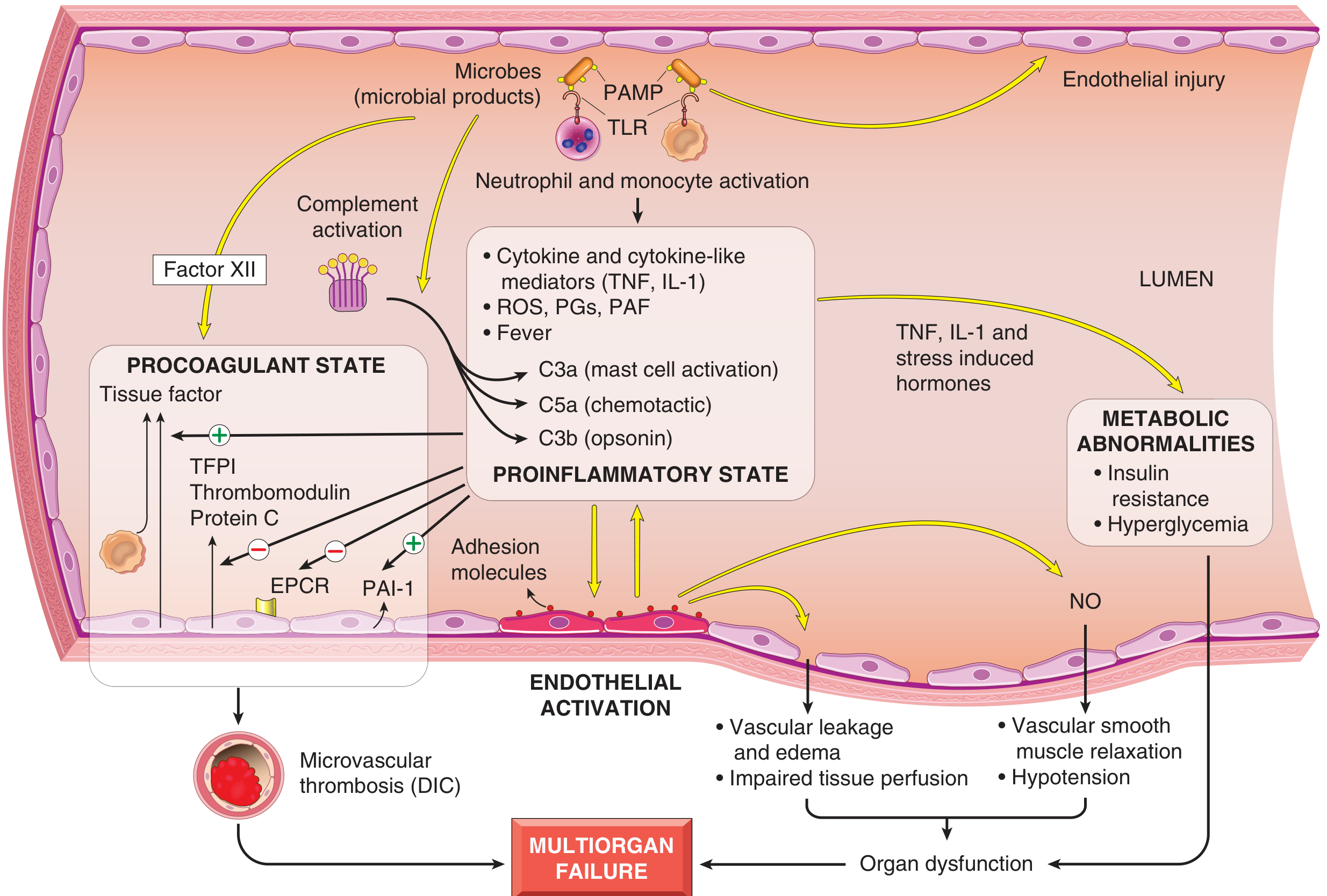
Key Pathogenic Mechanisms:
1. Inflammatory and Counterinflammatory Responses
- Microbial cell wall constituents engage receptors on innate immune cells:
- Toll-like receptors (TLRs) - recognize PAMPs (pathogen-associated molecular patterns)
- G-protein coupled receptors - detect bacterial peptides
- C-type lectin receptors (dectins) - recognize fungal cell wall components
- On activation, innate immune cells produce cytokines: TNF, IL-1, type I interferon, IL-12, IL-18
- Reactive oxygen species and lipid mediators (prostaglandins) are elaborated
- These upregulate adhesion molecules and further stimulate cytokine and chemokine production
- The complement cascade is activated, producing:
- C3a, C5a (anaphylatoxins) - proinflammatory
- C5a (chemotactic fragment)
- C3b (opsonin)
- With time, the hyperinflammatory state triggers counterregulatory immunosuppression (Th1 → Th2 shift, IL-10, IL-1 receptor antagonist, soluble TNF receptor, lymphocyte apoptosis)
- Patients oscillate between hyperinflammatory and immunosuppressed states
2. Endothelial Activation and Injury
- Proinflammatory cytokines cause widespread vascular leakage and tissue edema
- Cytokines loosen endothelial tight junctions → protein-rich edema throughout the body
- Impairs tissue perfusion and nutrient delivery
- Activated endothelium upregulates nitric oxide (NO) and other vasoactive mediators → vascular smooth muscle relaxation → systemic hypotension
3. Induction of a Procoagulant State
- Sepsis alters the expression of coagulation factors to favor coagulation:
- Proinflammatory cytokines increase tissue factor expression on endothelial cells and monocytes (triggers extrinsic coagulation pathway)
- Simultaneously decrease thrombomodulin, protein C, and TFPI (natural anticoagulants)
- PAI-1 (plasminogen activator inhibitor-1) is upregulated, impairing fibrinolysis
- Result: Disseminated Intravascular Coagulation (DIC) in up to 50% of septic patients
- Widespread microvascular thrombi → impaired blood flow → organ ischemia
4. Metabolic Abnormalities
- TNF, IL-1, and stress-induced hormones cause:
- Insulin resistance
- Hyperglycemia
- These metabolic derangements further impair organ function
5. Organ Dysfunction (Multiorgan Failure)
- Systemic hypotension + interstitial edema + small vessel thrombosis → decreased oxygen and nutrient delivery
- Mitochondrial damage from oxidative stress impairs oxygen utilization (even delivered O2 is not used)
- High cytokines diminish myocardial contractility and cardiac output
- Increased vascular permeability → Acute Respiratory Distress Syndrome (ARDS)
- Failure of multiple organs - kidneys, liver, lungs, heart → death
Superantigens and Toxic Shock Syndrome
- Certain secreted bacterial proteins (superantigens) can cause a syndrome similar to septic shock (e.g., toxic shock syndrome).
- Superantigens are polyclonal T-lymphocyte activators that induce high levels of cytokines, causing diffuse rash, vasodilation, hypotension, shock, and death.
Stages of Shock (Common to All Types)
-
Non-progressive (Compensated) Stage
- Reflex compensatory mechanisms are activated
- Baroreceptor reflexes, catecholamines, ADH release, RAAS activation, sympathetic stimulation
- Net effect: tachycardia, peripheral vasoconstriction, renal fluid conservation
- Blood shunted away from skin to vital organs (heart, brain)
-
Progressive Stage
- Widespread tissue hypoxia
- Intracellular aerobic respiration replaced by anaerobic glycolysis → lactic acidosis
- Worsening circulatory and metabolic derangement
- Cellular dysfunction becomes widespread
-
Irreversible Stage
- Cellular and tissue injury so severe that even if hemodynamic defects are corrected, survival is not possible
- Widespread cellular necrosis, failure of multiple organs
- Death
Sources: Robbins & Kumar Basic Pathology (9780323790185); Robbins, Cotran & Kumar Pathologic Basis of Disease (9780443264528)
This is a shared conversation. Sign in to Orris to start your own chat.