
Interstitial brachytherapy in detail according to standard books
Reading File
Now let me search the library for interstitial brachytherapy content in parallel:
Finding Sources
Finding Sources
Finding Sources
Reading File
Let me now read the dedicated brachytherapy sections from Grainger & Allison's (radiology textbook) and look for a dedicated radiation oncology textbook:
Reading File
Finding Sources
Now let me read the most comprehensive brachytherapy sections - Campbell Walsh Urology (prostate BT) and Grainger & Allison (general BT), plus Berek & Novak's Gynecology:
Reading File
Reading File
Let me now read the dedicated brachytherapy section in Campbell-Walsh (prostate) and the Grainger & Allison brachytherapy section more specifically:
Reading File
Reading File
Reading File
Finding Sources
I found very good content. Let me now read the Smith and Tanagho Urology section (which has a great classification) and the Campbell-Walsh HDR section:
Reading File
Reading File
Reading File
Excellent - I now have very rich content. Let me also get the LDR prostate brachytherapy technique section and fetch the key images:
Reading File
Loading Image
I now have comprehensive content from multiple authoritative textbooks. Let me compile the detailed response.
Interstitial Brachytherapy - Detailed Overview
(Sourced from Campbell-Walsh-Wein Urology, Smith & Tanagho's General Urology, Berek & Novak's Gynecology, and Grainger & Allison's Diagnostic Radiology)
1. Definition and Basic Principle
Brachytherapy refers to a treatment technique that places radioactive sources in close proximity to or directly into the tumor (from the Greek brachy = short distance). Interstitial brachytherapy specifically involves the placement of radioactive needles, afterloaded needles or catheters, or radioactive seeds directly into the tissue - as opposed to intracavitary brachytherapy, which places sources inside a body lumen or orifice.
A fundamental physical advantage is that radiation dose falls off rapidly according to the inverse square law - irradiation affects only a highly localized area around the source, meaning very high doses can be delivered to the target while healthy tissue further away receives minimal exposure.
- Smith & Tanagho's General Urology, p. 436
- Grainger & Allison's Diagnostic Radiology, p. 1726
2. Classification
Brachytherapy is classified along two axes:
| Axis | Categories |
|---|---|
| By placement | Interstitial vs. Intracavitary |
| By dose rate | Permanent/LDR vs. Temporary/HDR (vs. PDR) |
By Placement
- Interstitial: Radioactive needles, afterloaded catheters, or seeds placed directly into tissue (prostate, bladder, penis, periurethral soft tissues, gynecologic tumors)
- Intracavitary: Placement of sources into a lumen or orifice (uterine canal, vagina, urethra)
- Combined intracavitary/interstitial: Newer applicators that incorporate interstitial needles into standard intracavitary tandem-and-ring geometry
By Dose Rate
-
Low-dose rate (LDR): Continuous radiation over weeks-months via permanent seeds; dose rate <0.4 Gy/hour (technically, isotopes like Cs-137 fall here in traditional use)
-
High-dose rate (HDR): Short but high-dose radiation using temporary catheters; >12 Gy/hour with Ir-192 remote afterloading
-
Pulsed-dose rate (PDR): Uses an Ir-192 HDR source with a pulsed regimen designed to mimic the radiobiologic effects of LDR treatment; mostly seen at specialized academic centers
-
Smith & Tanagho's General Urology, p. 436
-
Berek & Novak's Gynecology, p. 2253
3. Isotopes Used
For Permanent (LDR) Interstitial Implants
| Isotope | Energy | Half-life | 90% dose delivered in | Monotherapy dose |
|---|---|---|---|---|
| ¹²⁵Iodine | 27 keV x-rays | 59.6 days | 204 days | 140-160 Gy |
| ¹⁰³Palladium | 21 keV x-rays | 17 days | 58 days | 110-125 Gy |
| ¹³¹Cesium (since 2004) | 30 keV x-rays | 9.7 days | 33 days | 100-115 Gy |
- ¹²⁵I is considered more "forgiving" dosimetrically due to slightly higher energy offering better prostatic coverage
- A randomized trial comparing ¹²⁵I vs. ¹⁰³Pd in low-risk disease found no difference in biochemical control; urinary symptoms were worse at 1 month with ¹⁰³Pd but resolved more rapidly
For Temporary (HDR) Interstitial Implants
-
Iridium-192 (¹⁹²Ir): 400 keV gamma radiation; used via remote afterloading systems; single source migrated along catheter length at variable positions and dwell times
-
Campbell-Walsh-Wein Urology, p. 4700-4701
4. Technique: LDR Permanent Seed Implant (Prostate Prototype)
Pre-procedure Planning
- Volume study using transrectal ultrasound (TRUS)
- Preoperative dosimetric planning determines number of seeds, seed activity, and needle/seed positions
- Seeds are peripherally loaded to minimize dose to the central urethra
Procedure
- Patient under spinal or general anesthesia
- TRUS probe placed transrectally for real-time imaging
- A perineal template grid is used to guide transperineal needle insertion
- Seeds are deposited at calculated positions as needles are withdrawn
- Typically 60-120 seeds placed using 15-25 needles
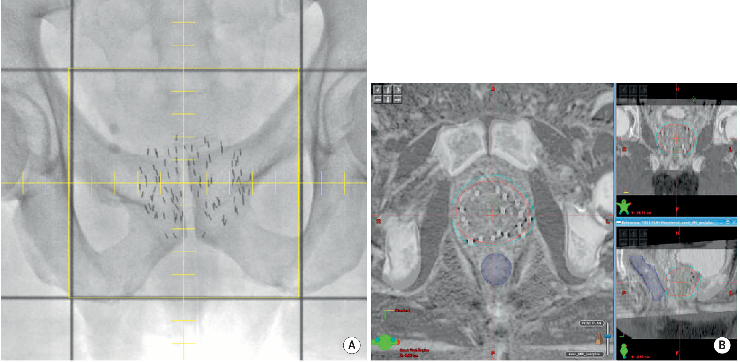
LDR brachytherapy for prostate cancer - plain radiograph and fused CT/MRI post-implant (Grainger & Allison)
Post-implant Dosimetry
-
CT with or without MRI fusion performed 4-6 weeks post-implant (not immediately) because seed migration occurs due to bleeding and swelling from the trauma of implantation
-
Key dosimetric parameters: D90 (dose covering 90% of prostate) and V100 (% of prostate volume receiving 100% prescribed dose)
-
MRI offers superior prostate delineation; CT better identifies metallic seed positions - fused images combine both advantages
-
Campbell-Walsh-Wein Urology, p. 4701-4702
-
Grainger & Allison's Diagnostic Radiology, p. 1729
5. Technique: HDR Interstitial Brachytherapy
Mechanism
- Hollow catheters are placed transperineally into the prostate (or through templates into other tissue targets) using the same grid-and-TRUS technique as LDR
- A single ¹⁹²Ir source is robotically driven ("afterloaded") along the length of each catheter
- The source dwells for seconds to minutes at 5 mm intervals - dwell times are modulated to optimize dose distribution
- All radioactive material is removed at the end of treatment - no sources remain in the patient
- Treatment is delivered in a shielded room via remote activation
Fractionation (Prostate HDR as Boost)
Common EBRT schedules combined with HDR boost include:
- 45 Gy in 25 fractions (5 weeks) + HDR
- 46 Gy in 23 fractions (4.5 weeks) + HDR
- 35.7 Gy in 13 fractions (2.5 weeks) + HDR
HDR monotherapy (without EBRT) is increasingly used for low-to-intermediate risk prostate cancer.
- Campbell-Walsh-Wein Urology, p. 4702-4703
6. Advantages and Disadvantages: HDR vs. LDR
Advantages of HDR over LDR
| Advantage | Explanation |
|---|---|
| Greater target flexibility | Catheter-based placement allows easier inclusion of extracapsular disease and seminal vesicles |
| Dose optimization | Modifying dwell times and positions allows real-time dose sculpting |
| Biological dose advantage | High dose per fraction exploits low alpha/beta ratio of prostate cancer |
| Radiation safety | No personnel handling of radioactive source; no sources left in patient |
| Cost-effectiveness | Single multipurpose ¹⁹²Ir source used for all patients |
| No post-implant dosimetry needed | Sources removed after treatment |
Disadvantages of HDR over LDR
| Disadvantage | Explanation |
|---|---|
| Treatment inaccuracies | Catheter/organ movement between imaging and source insertion; interobserver contouring variability |
| Patient convenience | Multiple fractions require repeated catheter placements or maintained catheter access |
| Shielded room required | High activity ¹⁹²Ir source requires dedicated infrastructure |
| Less pathologic response | Available post-treatment biopsy studies favor LDR |
| Limited phase III data | No comparable randomized data for HDR in high-risk prostate cancer |
- Campbell-Walsh-Wein Urology, p. 4703
7. Gynecologic Applications (Cervix Cancer)
Combined Intracavitary/Interstitial Brachytherapy
- Newer applicators incorporate interstitial needles into the standard tandem-and-ring or tandem-and-ovoids geometry (e.g., the Vienna applicator)
- Interstitial needles extend reach beyond what standard intracavitary geometry can dose
- Especially useful for bulky or irregularly shaped tumors that extend beyond the standard applicator coverage
- In the retroEMBRACE study: combined intracavitary/interstitial BT vs. intracavitary alone showed a 10% improvement in local control for larger tumors (cervix + tumor volume >30 cm³)
- Can often eliminate the need for more morbid techniques such as the Syed template or MUPIT template needle implants
2D vs. 3D Image-Guided Brachytherapy (IGA-BT)
- Classical 2D: Dose prescribed to "Point A" (2 cm superior to the cervical os, 2 cm lateral to the intrauterine canal); typical dose 7,000-8,000 cGy to Point A, limiting bladder/rectum to <6,000 cGy
- Modern 3D IGA-BT: MRI/CT-based volumetric planning; dose prescribed to the high-risk CTV D90 (8,500-9,500 cGy cumulative)
- The French STIC multicenter trial (705 patients) prospectively demonstrated 3D brachytherapy gave improved local control with half the toxicity of 2D brachytherapy
- Dose constraints (GEC-ESTRO): bladder D2cc <90 Gy EQD2; rectum D2cc <75 Gy EQD2; sigmoid D2cc <75 Gy EQD2
LDR vs. HDR for Cervix Cancer
-
LDR (Cs-137): Manual placement, requires inpatient admission 1-3 days, patient immobilization, DVT prophylaxis, radiation precautions for staff
-
HDR (Ir-192): Ambulatory, shorter total treatment time, remote afterloading eliminates staff exposure
-
A Cochrane review found no significant differences in OS, disease-specific survival, local control, or complications between LDR and HDR for locally advanced cervix cancer (based on 4 qualifying trials)
-
Berek & Novak's Gynecology, pp. 2253-2255
8. Other Interstitial Brachytherapy Applications
| Site | Details |
|---|---|
| Prostate cancer | LDR seed implant (low-risk) or HDR boost/monotherapy (intermediate/high-risk) |
| Penile/urethral cancer | Both EBRT and interstitial BT may be used; circumcision required prior to BT to expose the lesion and prevent inflammation-related swelling |
| Head and neck cancers | Interstitial implants for base of tongue, lip, floor of mouth; IMRT with or without BT boost |
| Breast cancer | Interstitial multi-catheter implant as partial breast irradiation (APBI) |
| Endobronchial tumors | Catheter-based bronchoscopic placement for airway tumors |
| Bladder cancer | Interstitial BT for selected Ta/T1 and T2 tumors; not yet standard of care |
| Gynecologic (vaginal/cervical) | Combined intracavitary/interstitial for bulky disease |
| Liver metastases | Interstitial BT as one of multiple local ablative options |
9. Dosimetry Principles
-
Dose from each source position is additive; superposition of contributions from all dwell positions defines the overall dose distribution
-
Inverse square law governs rapid dose fall-off away from each source
-
Imaging is mandatory: CT identifies applicator/seed positions precisely; MRI provides superior soft tissue definition (especially prostate and cervix)
-
For LDR prostate: post-implant CT/MRI fusion at 4-6 weeks - D90 and V100 are key quality indicators
-
For HDR: real-time or near-real-time planning at each fraction based on CT/MRI with applicators in situ
-
Grainger & Allison's Diagnostic Radiology, pp. 1726-1729
10. Comparison With External Beam Radiotherapy
| Feature | Interstitial BT | EBRT |
|---|---|---|
| Dose fall-off | Rapid (inverse square law) - spares adjacent normal tissue | Gradual; requires margins around target |
| Patient setup variation | Minimal - sources move with tumor | Significant daily repositioning needed |
| Tumor motion | Not a concern - sources within tumor | Major challenge (breathing, organ filling) |
| Dose per fraction | Can exploit high doses per fraction biologically | Typically 1.8-2 Gy fractions |
| Access | Invasive procedure required | Non-invasive |
| Suitable volumes | Small to medium tumors; precise geometry | Any volume; irregular shapes |
Sources:
- Campbell-Walsh-Wein Urology, 3-Volume Set - Chapters 157 & 158 (LDR/HDR Prostate Brachytherapy)
- Smith & Tanagho's General Urology, 19th Ed. - Chapter 26 (Brachytherapy classification and technique)
- Berek & Novak's Gynecology - Chapter on Primary Radiation Therapy (Cervix cancer brachytherapy)
- Grainger & Allison's Diagnostic Radiology - Chapter 67 (Principles of radiotherapy including brachytherapy)
Application in ent and head and neck surgery only
Searching sources...
Finding Sources
Finding Sources
Good - found key sources in Scott-Brown's, Cummings Otolaryngology, and Grainger & Allison. Let me read all of them in parallel:
Reading File
Reading File
Finding Sources
Excellent - found content in Scott-Brown's, Cummings, and KJ Lee's. Let me read the key sections in parallel:
Reading File
Reading File
Reading File
Now let me search for more specific ENT sites - floor of mouth, lip, tonsil, soft palate, and the Cummings brachytherapy section:
Reading File
Finding Sources
Finding Sources
Let me now read the floor of mouth / oral cavity sections and the tonsillar section from Scott-Brown's and Cummings:
Reading File
Reading File
Reading File
Now let me search for more specific content on floor of mouth BT, lip BT, and complications in H&N:
Finding Sources
Finding Sources
Reading File
Reading File
I now have comprehensive content from multiple ENT/H&N textbooks. Let me compile the full detailed answer.
Interstitial Brachytherapy in ENT and Head & Neck Surgery
(Sourced from Cummings Otolaryngology, Scott-Brown's Otorhinolaryngology Head & Neck Surgery, KJ Lee's Essential Otolaryngology, and Grainger & Allison's Diagnostic Radiology)
1. Historical Background and Basic Principles in H&N
Brachytherapy in head and neck cancer has deep roots - the first documented successful treatment was reported in 1899, when Thor Stenberg treated a nasal basal cell carcinoma with radium over 9 months, with the patient alive 30 years later. In 1901, Dr. Frand Williams in Boston reported successful treatment of a lip cancer. Radium was initially applied only to superficial sites because of severe toxicities; physicians later began inserting it directly into deep-seated tumors, founding the field of brachytherapy.
Core principle: Radiation dose from an implanted source obeys the inverse square law - it falls off rapidly with distance, allowing very high doses to be concentrated at the target (tumor) while sparing surrounding healthy tissues (including bone, nerve, major vessels).
Key advantage in H&N: Better dose localization → less radiation damage to surrounding healthy tissue, and prolonged time over which radiation is delivered.
Key disadvantage in H&N: Does not address subclinical nodal disease; requires general anesthesia in many cases; close proximity of mandible creates risk of osteoradionecrosis.
- Cummings Otolaryngology, p. 1343, 1361
2. Radioactive Sources Used in H&N Brachytherapy
| Source | Type | Notes |
|---|---|---|
| Iridium-192 (¹⁹²Ir) | LDR afterloading or HDR | Most widely used; dose rate 0.4-0.8 Gy/hour (LDR); or >12 Gy/hour (HDR remote afterloading) |
| Cesium-137 (¹³⁷Cs) | LDR needles | Traditional use; being replaced by ¹⁹²Ir |
| Radium-226 (²²⁶Ra) | Historical | Needles for implants; now largely obsolete |
| Gold-198 (¹⁹⁸Au) | Permanent grains | Used for nasopharyngeal recurrences (grain implantation) |
| Iodine-125 (¹²⁵I) | Permanent seeds | Low energy; used in selected cases |
A typical ¹⁹²Ir LDR implant delivers approximately 0.4-0.8 Gy/hour. HDR remote afterloading devices drive a single high-activity ¹⁹²Ir source through a set of interstitial catheters under computer control; typically 3.0-3.5 Gy is delivered to ~1 cm from the catheter periphery per treatment, with up to two daily treatments ~6 hours apart, each lasting 15-30 minutes depending on source strength and implant complexity.
- Cummings Otolaryngology, p. 1361
3. Site-by-Site Applications
A. Oral Tongue (Mobile Tongue)
Radiotherapy (brachytherapy preferred over EBRT) has been advocated as a primary treatment modality because it conserves tongue volume and morphology - an enormous functional advantage.
Indications:
- T1-T2 tumors of mobile tongue not suitable for surgery, or where organ preservation is prioritized
- Brachytherapy alone or as a boost after EBRT
Technique: ¹⁹²Ir interstitial implant through the tongue tissue, using afterloading catheters placed to encompass the tumor with adequate margins.
Outcomes: Brachytherapy is considered preferable to EBRT for tongue primaries in terms of dose localization and functional preservation.
Key limitation - Osteoradionecrosis (ORN): The mandible lies in close proximity to the tongue. Up to 9% of patients develop some form of osseous complication following tongue brachytherapy. This is a recognized and major concern that has led some centers to prefer surgery as primary treatment, keeping radiotherapy in reserve for:
- Poor pathologic prognostic indicators post-resection
- Recurrence
- Second primaries
An important additional consideration is that when surgery is not used as the primary modality, valuable histopathological prognostic information is lost - making decisions about elective neck dissection (END) more difficult.
Some authorities suggest surgery is superior to brachytherapy for Stage I/II tongue cancer for this reason (retaining radiotherapy as a reserve modality).
- Scott-Brown's Otorhinolaryngology, p. 6861
B. Floor of Mouth
Indications:
- T1/T2 floor-of-mouth carcinomas - brachytherapy (or EBRT) has been shown to give results comparable to surgery
Key limitation - ORN risk is particularly high here:
-
The floor of mouth lies in direct proximity to the mandible
-
Up to 8.5% of patients treated with brachytherapy for floor-of-mouth cancer require segmental mandibulectomy for osteoradionecrosis within 10 years (Gustave-Roussy Institute series; Pernot et al., Marsiglia et al.)
-
Several major units have changed practice from brachytherapy to surgery as the primary treatment modality for floor-of-mouth cancer, specifically because of this complication risk
-
T3/T4 lesions are best treated with surgery + post-operative radiotherapy (PORT)
-
Scott-Brown's Otorhinolaryngology, p. 6641-6652
C. Base of Tongue (Oropharynx)
Role of brachytherapy: RT for base of tongue is often performed as a combination of EBRT + interstitial ¹⁹²Ir implant.
Outcomes data (Cummings Otolaryngology):
| Treatment | T1 Local Control | T2 Local Control |
|---|---|---|
| EBRT alone (primary) | 78-96% | 47-88% |
| EBRT + brachytherapy implant | 71-100% | 71-100% |
| EBRT alone vs. combined | EBRT alone = unacceptably high failure rate (2× other groups) |
Houssef et al. compared surgery + adjuvant RT vs. EBRT + ¹⁹²Ir implant vs. EBRT alone for T1/T2 base-of-tongue cancers and found:
- Surgery + adjuvant RT ≈ EBRT + implant (comparable results)
- EBRT alone showed unacceptable failure rate - twice as high as the other two groups
For salvage/recurrent disease, afterloading techniques with ¹⁹²Ir have achieved local control of 59% and actuarial survival of 48%, though residual/recurrent tongue base disease remains a demanding problem.
- Cummings Otolaryngology, p. 1793
D. Tonsillar Region / Oropharynx
Salvage surgery after primary RT in the tonsillar region carries high mortality and low 5-year survival (Gehanno et al.: 5-year survival 24%, mortality as high as 8% in 120 patients).
Two investigations support brachytherapy salvage in the tonsillar region:
-
5-year survival rate: 64% (one series)
-
2-year survival rate: 42% (another series)
-
Cummings Otolaryngology, p. 1792
E. Nasopharynx (Recurrent/Salvage Setting)
Brachytherapy for nasopharyngeal carcinoma (NPC) is used specifically in the salvage setting for local failure following primary chemoradiation, as an alternative to re-irradiation with EBRT (which has poor outcomes due to proximity of brainstem, optic chiasm, and temporal lobe to the treatment field).
Rationale: Traditional 2D re-irradiation for NPC local failure has a 5-year survival of only 7.6% due to dose constraints protecting vital organs. Brachytherapy delivers a high dose with limited penetration - ideal for small recurrences without deep invasion.
Radioactive sources:
- Iridium-192 (¹⁹²Ir): The source is loaded into a tailor-made plastic mould fitted into the nasopharynx. The mould is placed transorally (via the oral cavity) under local anaesthesia.
- Gold-198 (¹⁹⁸Au) grains: Implanted directly into the nasopharynx after the soft palate is split open under general anaesthesia.
Patient selection: Both techniques are only suitable for small tumors <2 cm in maximal dimension (no deep invasion).
Outcomes:
- 5-year survival: 50-60% for salvage brachytherapy of NPC local recurrence
As an alternative to brachytherapy, stereotactic RT (3D/IMRT-based) for NPC local salvage has shown 5-year overall survival 40% and local control 57% (series of 30 patients) - brachytherapy outcomes are superior but patient selection is stricter (smaller tumors only).
Brachytherapy also has a potential role in nodal failure in NPC, used in conjunction with surgical resection of nodal metastasis (the limited penetration of brachytherapy makes it suitable as an adjunct after debulking surgery).
- Scott-Brown's Otorhinolaryngology, p. 8504-8520
F. Orbit and Paranasal Sinuses (Intracavitary, H&N)
The orbit and nasopharynx are the most common intracavitary head and neck brachytherapy sites. Custom PMMA (acrylic) stents are constructed around catheters into which radioisotopic seeds are inserted:
-
The radiation oncologist/physicist prescribes catheter positions in consultation with the maxillofacial prosthodontic team, after generating a master cast from an impression of the site
-
Stents are retained in position by anatomic soft tissue undercuts or alveolar structures
-
After maxillectomy, intracavitary void-filling with tissue-equivalent material improves dose distribution (preventing uneven dosing of peripheral tissues around the surgical defect)
-
Cummings Otolaryngology, p. 1702
G. Lip Cancer
One of the original sites treated with brachytherapy historically (1901 - Dr. Williams, Boston). Lip carcinoma remains amenable to interstitial brachytherapy, particularly for:
-
Small T1-T2 squamous cell carcinomas of the lip
-
As an organ-preserving alternative to surgery for cosmetically sensitive sites
-
Cummings Otolaryngology, p. 1343
4. Technique Summary for H&N Interstitial BT
LDR ¹⁹²Ir Interstitial Implant (Standard Technique)
- Planning phase: CT/MRI-based volumetric planning; catheter positions determined to achieve adequate dose to tumor with acceptable dose to adjacent mandible, vessels, nerves
- Implant procedure: Under general anaesthesia (most cases); hollow plastic catheters or metal needles inserted through the tumor-bearing tissue
- Afterloading: ¹⁹²Ir sources loaded into catheters; patient cared for in a shielded room during treatment
- Dose delivery: 0.4-0.8 Gy/hour continuously over 2-6 days
- Source removal: All sources and catheters removed at end of treatment
HDR Remote Afterloading (Modern Approach)
- Computer-controlled robotic ¹⁹²Ir source driven through catheters
- Dwells at 5 mm intervals; dwell times modulated to optimize dose distribution
- Each fraction: 3.0-3.5 Gy to ~1 cm from catheter periphery
- Up to 2 fractions per day (at least 6 hours apart)
- Each treatment: 15-30 minutes
- Advantages: Eliminates radiation exposure to nursing/medical staff; outpatient delivery possible
5. Combinations with EBRT
In head and neck cancer, interstitial brachytherapy is frequently used as a boost following a course of EBRT:
- EBRT addresses the primary tumor plus regional lymphatics (elective or therapeutic nodal irradiation)
- Brachytherapy delivers an additional dose increment to the primary tumor bed, achieving higher total doses than either modality alone
- This combination is the standard approach for base-of-tongue carcinoma and is used in other oral cavity sites
6. Complications Specific to H&N Brachytherapy
| Complication | Details |
|---|---|
| Osteoradionecrosis (ORN) of mandible | Most feared; up to 9% for tongue BT, up to 8.5% requiring segmental mandibulectomy for floor-of-mouth BT; high local doses from BT increase incidence; risk further raised by microvascular disease (atherosclerosis, diabetes) and post-RT dental extractions |
| Soft tissue necrosis | Histologically: epithelial hyperplasia → dermal fibrosis → necrosis; Marx characterized tissue as "hypoxic, hypocellular, hypovascular" |
| Mucositis | Acute; self-limiting; universal to varying degree with all H&N RT |
| Xerostomia | From inclusion of salivary glands in the radiation field |
| Trismus | Radiation fibrosis of masticator muscles |
| Radiation-related fistulae | In high-dose zones near mucosa |
| Radioprotection requirements | LDR implants require patient isolation in shielded room; HDR remote afterloading eliminates this (no radiation exposure to staff during treatment) |
ORN pathophysiology (Scott-Brown's Vol. 2):
- Impaired tissue repair capacity
- Vasculitis → obliteration of blood vessels → avascular necrosis
- Marx triad: hypoxic + hypocellular + hypovascular tissue
- Macroscopically: loss of skin/soft tissue exposing bone, bony sequestration, secondary infection
Note on temporal bone: The compact (non-cancellous) nature of the petrous temporal bone and poor blood supply of the tympanic ring make this region particularly susceptible to ORN from brachytherapy of adjacent tumors.
- Scott-Brown's Vol. 2, p. 1011
- Cummings Otolaryngology (Key Points, p. 1702)
7. Contraindications and Patient Selection
| Factor | Implication |
|---|---|
| Tumor > 2 cm in nasopharynx | Contraindication to NPC salvage BT |
| Deep invasion of tumor | Contraindication to BT (limited penetration cannot cover deep extension) |
| Prior high-dose RT to site | Increased ORN risk; careful re-treatment planning needed |
| Poor dental hygiene | Must optimize dental care pre-BT to reduce ORN risk |
| Diabetes / atherosclerosis | Increased ORN risk; relative contraindication to BT near mandible |
| Need for nodal treatment | BT alone insufficient; must add EBRT or neck dissection |
8. Summary Table: BT Applications by H&N Site
| Site | Role | Source/Technique | Key Outcome Data | Main Risk |
|---|---|---|---|---|
| Oral tongue (T1-T2) | Primary or EBRT boost; organ preservation | ¹⁹²Ir LDR/HDR interstitial | Comparable to surgery for T1/T2 | ORN 9% |
| Floor of mouth (T1-T2) | Primary (results = surgery); T3/T4 → surgery + PORT | ¹⁹²Ir LDR interstitial | ~= surgery; many units switching to surgery | ORN up to 8.5% → mandibulectomy |
| Base of tongue | EBRT + BT boost (standard); salvage afterloading | ¹⁹²Ir LDR/HDR interstitial | T1: 71-100%; T2: 71-100% (EBRT + BT) | Functional swallowing |
| Tonsillar region | Salvage after RT failure | ¹⁹²Ir afterloading | 5-yr survival 64%; 2-yr survival 42% | High operative mortality if surgical salvage used |
| Nasopharynx (recurrent) | Salvage for small (<2 cm) recurrences | ¹⁹²Ir mould or ¹⁹⁸Au grains | 5-yr survival 50-60% | Limited to tumors without deep invasion |
| Orbit/sinus (intracavitary) | Post-maxillectomy/targeted | PMMA stent with ¹²⁵I seeds | Site-specific | Dosimetric consistency |
| Lip (T1-T2) | Primary; cosmetic preservation | Interstitial needles | Historically effective (first H&N site treated, 1901) | ORN (low risk, away from mandible) |
Sources:
- Cummings Otolaryngology Head and Neck Surgery - Chapter 75 (Radiation Therapy Principles) & Chapter 96 (Oropharyngeal Cancers)
- Scott-Brown's Otorhinolaryngology Head & Neck Surgery - Oral tongue, floor of mouth, and nasopharynx chapters
- Scott-Brown's Vol. 2 - Osteoradionecrosis chapter
- KJ Lee's Essential Otolaryngology - Oral tongue cancer chapter
- Grainger & Allison's Diagnostic Radiology - Brachytherapy principles chapter
This is a shared conversation. Sign in to Orris to start your own chat.