I now have all the information needed to write a comprehensive ENT Assessment for Allergic Rhinitis. Let me compile the complete assessment.
ENT Assessment: Allergic Rhinitis
I. BIOGRAPHIC DATA
| Field | Details |
|---|
| Patient Name | Mr. Rajan Mehta |
| Age | 28 years |
| Sex | Male |
| Date of Birth | July 15, 1997 |
| Marital Status | Single |
| Occupation | Software Engineer (works indoors, air-conditioned office) |
| Religion | Hindu |
| Nationality | Indian |
| Address | 14/B, Green Park Colony, Hyderabad, Telangana |
| Date of Admission | July 25, 2026 |
| Ward / Room | ENT OPD - Room No. 3 |
| Attending Physician | Dr. Suresh Anand, ENT Specialist |
| Informant | Patient himself (reliable) |
| Source of Referral | General Practitioner |
| Mode of Admission | Outpatient (walk-in) |
II. CHIEF COMPLAINTS
The patient presents with the following complaints for the past 6 weeks, worsening over the last 2 weeks:
- Persistent sneezing (paroxysmal, especially in the morning)
- Watery, clear nasal discharge (rhinorrhea)
- Nasal congestion / stuffiness (bilateral)
- Nasal itching and itching of the palate
- Eye itching, redness, and tearing (bilateral)
- Headache (mild, frontal region)
- Fatigue and disturbed sleep due to nasal congestion
III. HISTORY OF PRESENT ILLNESS
Mr. Rajan Mehta, a 28-year-old male, presents to the ENT OPD with a 6-week history of recurrent sneezing, bilateral nasal obstruction, and profuse clear watery rhinorrhea. Symptoms began insidiously in early June and have progressively worsened. He reports that symptoms are worst in the morning upon waking and during periods of high dust exposure. He works in a building undergoing renovation near his office, which he identifies as a possible trigger.
He also complains of bilateral eye itching, redness, and lacrimation consistent with allergic conjunctivitis. He reports frequent headaches in the frontal region, which he attributes to nasal congestion. He has disturbed sleep due to difficulty breathing through the nose and snores at night.
He denies fever, facial pain, purulent nasal discharge, loss of smell, or blood in the nasal discharge. He does not report any wheezing, chest tightness, or difficulty breathing.
Aggravating Factors: Dust, strong odors, cold air, early morning exposure, proximity to flowers/plants
Relieving Factors: Over-the-counter antihistamine tablets (taken occasionally, partial relief)
Duration: 6 weeks (persistent/perennial pattern with seasonal exacerbation)
As noted in Goldman-Cecil Medicine, "The natural history of allergic rhinitis is for symptoms to worsen inexorably during several weeks in the presence of ongoing allergen exposure. Symptoms often do not peak until well after the peak in pollen counts." - Goldman-Cecil Medicine, p. 2388
IV. PAST MEDICAL HISTORY
| Category | Details |
|---|
| Previous similar episodes | Yes - patient had similar sneezing and runny nose episodes every year since age 15 |
| Childhood illnesses | Recurrent episodes of "cold" in childhood, eczema at age 7 (resolved) |
| Hospitalizations | None |
| Surgeries | Nil |
| Trauma / injury | Nil |
| Known allergies | Dust mites (suspected), pollen |
| Asthma | No diagnosed asthma; no wheezing reported |
| Medications (past/current) | OTC Cetirizine 10 mg occasionally (partial relief); no topical nasal sprays used previously |
| Immunizations | Up to date; no influenza vaccine this year |
| Family History | Father has bronchial asthma; mother has seasonal allergic rhinitis; elder sister has atopic dermatitis |
Note: The strong family history of atopy (asthma, eczema, allergic rhinitis in first-degree relatives) is consistent with the hereditary predisposition of atopic disease, as highlighted in K.J. Lee's Essential Otolaryngology: "Family history of allergic rhinitis, conjunctivitis, asthma" is a key component of allergy history-taking.
V. PHYSICAL EXAMINATION
General Survey
- Patient is a well-nourished young adult male, alert and oriented
- Appears mildly distressed due to nasal congestion and frequent sneezing during examination
- Mouth breathing noted
- No pallor, icterus, cyanosis, clubbing, or lymphadenopathy
- Vital signs:
- BP: 118/76 mmHg
- HR: 78 bpm, regular
- RR: 17 breaths/min
- Temperature: 37.1°C (afebrile - fever is NOT a feature of allergic rhinitis)
- SpO2: 99% on room air
As noted in Goldman-Cecil Medicine: "Fever is not a feature of allergic rhinitis, even though the lay term for this condition is hay fever." - Goldman-Cecil Medicine, p. 2390
Head and Face
- Allergic salute sign: Transverse crease across the bridge of the nose from habitual upward rubbing
- Allergic shiners: Dark, bluish-gray discoloration under both eyes (suborbital venous congestion)
- Dennie-Morgan lines: Infraorbital skin folds present bilaterally
- No sinus tenderness on palpation over frontal or maxillary sinuses
Eyes (Ocular Examination)
- Bilateral conjunctival injection (redness)
- Mild conjunctival edema and excessive lacrimation
- No purulent discharge
- Pupils equal, round, reactive to light (PERRL)
- No photophobia
Nose (Anterior Rhinoscopy)
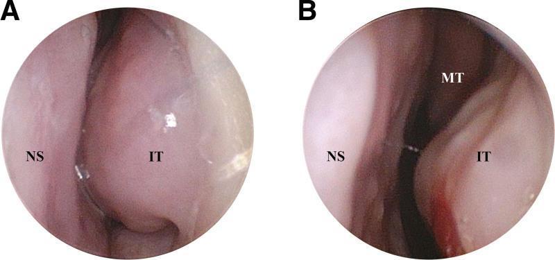
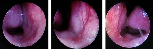
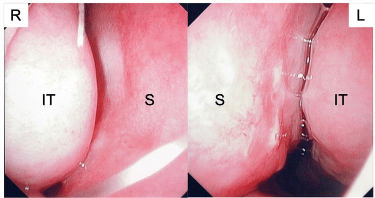
- Inferior turbinates: Swollen and hypertrophied bilaterally
- Nasal mucosa: Pale, bluish-grey/edematous appearance (classic allergic appearance)
- Nasal discharge: Clear, watery, profuse
- Nasal septum: Midline, no deviation noted
- Nasal polyps: Absent
- No purulent or blood-stained discharge
Tintinalli's Emergency Medicine describes: "Patients with allergic rhinitis report symptoms of paroxysmal sneezing, nasal pruritus, rhinorrhea, oropharyngeal pruritus, hyperemia, and ocular pruritus. On physical examination, there may be hypertrophy and edema of the nasal turbinates with associated pale, bluish hue or pallor." - Tintinalli's, p. 894
Throat / Oropharynx
- Mildly injected posterior pharyngeal wall
- Mucoid postnasal drip visible along posterior pharyngeal wall
- Tonsils: Grade I, non-inflamed
- No exudate
Ears (Otoscopic Examination)
- Bilateral tympanic membranes intact, pearly gray, light reflex present
- No middle ear effusion noted
- Hearing: Normal by finger rubbing test
Neck
- No cervical lymphadenopathy
- Thyroid: Not enlarged
Chest / Respiratory
- Chest: Clear to auscultation bilaterally
- No wheeze, crackles, or added sounds
- Normal vesicular breath sounds
VI. LABORATORY INVESTIGATIONS
| Investigation | Ordered Value / Finding | Clinical Relevance |
|---|
| Complete Blood Count (CBC) | WBC: 8,200/µL; Eosinophils: 6% (0.49 × 10⁹/L) - mildly elevated; RBC, Hb, Platelets: Normal | Peripheral eosinophilia is a supportive finding in allergic rhinitis |
| Absolute Eosinophil Count (AEC) | 490 cells/µL (slightly elevated; normal <450) | Supports allergic/atopic state |
| Total Serum IgE | 320 IU/mL (elevated; normal <100 IU/mL) | Elevated total IgE indicates atopic state; not diagnostic alone |
| Nasal Smear for Eosinophils (NSE) | Abundant eosinophils on nasal cytology | Highly specific for allergic rhinitis when positive |
| Blood Glucose (RBS) | 96 mg/dL | Normal; baseline before steroid prescription |
| Liver Function Test (LFT) | Within normal limits | Baseline prior to medication |
| Kidney Function Test (KFT) | Within normal limits | Baseline |
Harrison's Principles of Internal Medicine states: "The nasal secretions of allergic patients can be rich in eosinophils, and a modest peripheral eosinophilia can be observed... Total serum IgE may be moderately elevated." - Harrison's, p. 2853
VII. SPECIAL INVESTIGATIONS
1. Skin Prick Test (SPT) - GOLD STANDARD
- Purpose: Identifies specific allergens responsible for symptoms
- Method: Extracts of common aeroallergens (house dust mite, cockroach, grass pollen, tree pollen, cat dander, mold spores, fungal spores) applied on the forearm with a lancet prick
- Result (expected/typical): Positive wheal-and-flare reaction (>3mm) to house dust mite (Dermatophagoides pteronyssinus) and grass pollen
- Interpretation: A positive intracutaneous skin test with 1:10-1:20 weight/volume extract has high predictive value
Goldman-Cecil Medicine: "Prick skin testing is safe, specific, and rapid, and it is the diagnostic test of choice for identifying relevant allergens." - Goldman-Cecil Medicine, p. 2495
2. Serum Specific IgE (RAST / ImmunoCAP Assay)
- ELISA-based measurement of serum allergen-specific IgE
- Ordered for: House dust mite, grass pollen, cockroach, pet dander
- Result: Elevated specific IgE to dust mite (Class III: 3.5-17.4 kUA/L)
- Indicated when skin testing cannot be performed (e.g., dermographism, patient on antihistamines)
3. Nasal Endoscopy
- Flexible fiberoptic nasal endoscopy performed to rule out nasal polyps, sinusitis, or anatomical abnormality
- Findings: Pale, edematous inferior turbinates bilaterally; clear mucus; septum midline; no polyps; no purulent discharge
4. CT Scan of Paranasal Sinuses (if indicated)
- Not immediately required in uncomplicated allergic rhinitis
- Indicated if: Patient shows signs of chronic rhinosinusitis, nasal polyps, or failure to respond to initial therapy
- Would reveal mucosal thickening or opacification of sinuses if sinusitis present
5. Peak Flow / Spirometry
- Ordered given strong family history of asthma
- Rationale: "Patients with persistent allergic rhinitis should be screened for asthma, and patients with asthma should be evaluated for allergic rhinitis." - Murray & Nadel's Textbook of Respiratory Medicine
VIII. MEDICATIONS PRESCRIBED
| Drug | Class | Dose & Route | Frequency | Duration | Purpose |
|---|
| Cetirizine 10 mg | 2nd gen. oral H1-antihistamine | 10 mg PO | Once daily (evening) | 4 weeks | Reduces sneezing, itching, rhinorrhea, and ocular symptoms |
| Fluticasone Furoate Nasal Spray | Intranasal glucocorticoid | 2 sprays per nostril | Once daily (morning) | 4-6 weeks | Most effective drug for nasal congestion; up to 70% overall symptom relief |
| Normal Saline Nasal Irrigation (Jal Neti / NaSal wash) | Saline (non-pharmacologic) | 240 mL per nostril | Twice daily | Ongoing | Mechanical clearance of allergens and secretions |
| Olopatadine 0.1% Eye Drops | Topical antihistamine (ocular) | 1 drop each eye | Twice daily | 2-4 weeks | Relief of allergic conjunctivitis (itching, redness) |
| Montelukast 10 mg | Leukotriene receptor antagonist | 10 mg PO | Once daily (night) | 4 weeks (if incomplete response) | Additional relief of nasal congestion; approved for both seasonal and perennial AR |
| Pseudoephedrine 60 mg (PRN) | Oral alpha-adrenergic decongestant | 60 mg PO | As needed (max 3x/day) | Short course only (3-5 days) | Rapid relief of severe nasal congestion (avoid in hypertension) |
Harrison's Principles of Internal Medicine: "Intranasal high-potency glucocorticoids are the most effective drugs available for the relief of persistent rhinitis, seasonal or perennial, and are effective in relieving nasal congestion as well as ocular symptoms." - Harrison's, p. 304
"Oral long-acting H1 antihistamines, such as fexofenadine, loratadine, desloratadine, cetirizine, and levocetirizine, are effective for nasopharyngeal itching, sneezing, and watery rhinorrhea." - Harrison's, p. 304
Allergen Immunotherapy (future consideration): If symptoms are inadequately controlled with pharmacotherapy or if the patient wishes to reduce long-term medication dependence, subcutaneous immunotherapy (SCIT) or sublingual immunotherapy (SLIT) for dust mite and grass pollen allergens may be considered.
IX. NURSING CARE PLAN (NCP)
NCP 1: Ineffective Airway Clearance
| Component | Details |
|---|
| Nursing Diagnosis | Ineffective Airway Clearance related to nasal mucosal edema, hypersecretion, and turbinate hypertrophy secondary to allergic rhinitis, as evidenced by nasal congestion, mouth breathing, and disturbed sleep |
| Goal | Patient will demonstrate improved nasal airway patency and report decreased nasal congestion within 72 hours of treatment initiation |
| Nursing Interventions | 1. Elevate head of bed to 30-45° to facilitate nasal drainage and reduce mucosal congestion 2. Teach and demonstrate proper nasal saline irrigation (NaSal wash) technique twice daily 3. Administer intranasal fluticasone furoate as prescribed; instruct patient on correct spray technique (avoid spraying directly on septum) 4. Encourage adequate oral hydration (2-3 L/day) to thin secretions 5. Monitor respiratory rate and oxygen saturation 6. Advise patient to avoid known triggers (dust, strong odors, cold air) |
| Rationale | Elevated positioning reduces venous pooling and mucosal edema. Saline irrigation mechanically removes allergens and thick mucus. Topical steroids reduce mucosal inflammation, the primary cause of congestion. Hydration thins secretions and aids mucociliary clearance. |
| Evaluation | Patient reports subjective improvement in nasal breathing; bilateral nasal passages are more patent on rhinoscopy; snoring and mouth breathing reduced |
NCP 2: Disturbed Sleep Pattern
| Component | Details |
|---|
| Nursing Diagnosis | Disturbed Sleep Pattern related to nasal congestion, sneezing, and post-nasal drip secondary to allergic rhinitis, as evidenced by patient's report of difficulty breathing at night and fatigue |
| Goal | Patient will report improved sleep quality and reduced nocturnal awakenings within 1 week |
| Nursing Interventions | 1. Administer cetirizine in the evening as prescribed (minimizes drowsiness interference with daytime activities; reduces nocturnal symptoms) 2. Educate patient on allergen reduction measures in the bedroom: use dust-mite-proof pillow and mattress covers, wash bed linen in hot water (>60°C) weekly, keep pets out of the bedroom 3. Instruct patient to perform nasal saline irrigation before bedtime 4. Keep bedroom temperature moderate (not too warm or cold) and well ventilated 5. Assess and document sleep quality daily using a simple sleep diary 6. Avoid sedating antihistamines (first-gen) that can disrupt REM sleep architecture |
| Rationale | Nighttime antihistamine use reduces nocturnal symptom burden. Allergen reduction in the sleeping environment decreases overnight allergen exposure, which directly reduces symptom intensity. Saline wash before bed clears accumulated secretions. |
| Evaluation | Patient reports sleeping without frequent awakening; fatigue levels decreased; no mouth breathing at rest |
NCP 3: Acute Pain (Headache)
| Component | Details |
|---|
| Nursing Diagnosis | Acute Pain related to frontal sinus pressure secondary to nasal mucosal congestion, as evidenced by patient's complaint of frontal headache (rated 4/10 on NRS) |
| Goal | Patient will report pain reduction to ≤2/10 within 48 hours |
| Nursing Interventions | 1. Assess pain: location, intensity (NRS 0-10), quality, duration, and aggravating/relieving factors 2. Apply warm compresses over the frontal and maxillary sinus areas for 5-10 minutes, 3x/day 3. Encourage use of prescribed nasal saline rinse to decompress nasal passages and relieve sinus pressure 4. Administer analgesics as ordered (e.g., Paracetamol 500-1000 mg PO if needed) 5. Instruct patient to avoid bending forward or straining, which increases sinus pressure 6. Monitor for worsening headache, fever, or purulent discharge which could indicate sinusitis requiring escalation |
| Rationale | Nasal congestion obstructs the ostiomeatal complex, causing sinus pressure buildup and headache. Warm compresses promote local vasodilation and sinus drainage. Saline rinse reduces mucosal edema, relieving blockage. Positioning reduces gravity-dependent pressure. |
| Evaluation | Patient reports headache intensity decreased; no new signs of acute sinusitis; facial tenderness on palpation resolved |
NCP 4: Deficient Knowledge
| Component | Details |
|---|
| Nursing Diagnosis | Deficient Knowledge regarding allergic rhinitis, allergen avoidance, and medication use, as evidenced by patient's irregular use of OTC antihistamines and inability to identify specific triggers |
| Goal | Patient will verbalize understanding of allergic rhinitis, demonstrate correct use of nasal spray and saline rinse, and identify at least 3 allergen-avoidance strategies before discharge |
| Nursing Interventions | 1. Explain the pathophysiology of allergic rhinitis in simple terms (immune overreaction to harmless substances like dust mites or pollen) 2. Teach proper technique for intranasal steroid spray (tilt head slightly forward, aim spray toward outer eye, avoid septum) 3. Demonstrate nasal saline irrigation technique; have patient return-demonstrate 4. Provide written allergy action plan: what to do when symptoms worsen 5. Educate on allergen reduction: dust-mite covers, HEPA air filters, limiting outdoor exposure during high pollen seasons, avoiding tobacco smoke and strong perfumes 6. Emphasize adherence to intranasal steroid (takes 1-2 weeks for full effect; do not stop when symptoms improve) 7. Explain the side effect profile of each medication and when to seek emergency care |
| Rationale | Allergen avoidance is the most cost-effective strategy for managing allergic rhinitis. Correct spray technique maximizes drug deposition and minimizes septal side effects. Written action plans improve self-management. Medication adherence education reduces treatment failure. |
| Evaluation | Patient correctly demonstrates nasal spray technique and saline rinse; lists specific triggers and 3 avoidance strategies; verbalizes when to return to clinic |
NCP 5: Risk for Ineffective Self-Health Management
| Component | Details |
|---|
| Nursing Diagnosis | Risk for Ineffective Self-Health Management related to chronic nature of allergic rhinitis, complexity of multi-drug regimen, and risk of developing complications (sinusitis, asthma) |
| Goal | Patient will adhere to treatment regimen and attend follow-up appointment in 4 weeks |
| Nursing Interventions | 1. Simplify medication regimen: provide a written schedule for each drug 2. Counsel regarding the chronic, recurring nature of allergic rhinitis and the importance of maintenance therapy 3. Screen for asthma: provide peak flow meter if available; instruct on its use (given strong family history of asthma) 4. Refer to allergist/immunologist for formal allergy testing and discussion of immunotherapy options 5. Schedule follow-up in 4 weeks to assess treatment response, medication tolerance, and need for step-up therapy 6. Provide contact details and instruct patient to report: development of fever, purulent nasal discharge, new-onset wheezing, loss of smell, or severe facial pain |
| Rationale | Persistent allergic rhinitis is a significant risk factor for development of asthma and chronic rhinosinusitis. "Patients with persistent allergic rhinitis should be screened for asthma, and patients with asthma should be evaluated for allergic rhinitis." (Murray & Nadel's). Immunotherapy offers a disease-modifying option that reduces medication burden over time. |
| Evaluation | Patient attends follow-up; no new complications; adherent to medication schedule; understands asthma warning signs |
Summary of Diagnosis
ARIA Classification (Allergic Rhinitis and its Impact on Asthma):
- Persistent (symptoms >4 days/week AND >4 weeks duration)
- Moderate-Severe (sleep disturbance, impairment of daily activities, fatigue present)
- Comorbidity: Allergic conjunctivitis
Likely Sensitizing Allergens: House dust mite (primary), grass pollen (secondary), possibly workplace construction dust
Management Plan:
- Intranasal glucocorticoids (first-line, most effective for nasal congestion and overall symptoms)
- Second-generation oral antihistamine (for itching, sneezing, rhinorrhea, ocular symptoms)
- Allergen avoidance measures
- Allergen-specific IgE testing / Skin prick test to confirm sensitizers
- Asthma screening (spirometry/peak flow) given family history
- Consider allergen immunotherapy if pharmacotherapy inadequate
Sources: Cummings Otolaryngology Head and Neck Surgery; Harrison's Principles of Internal Medicine 22E; Goldman-Cecil Medicine; K.J. Lee's Essential Otolaryngology; Murray & Nadel's Textbook of Respiratory Medicine; Tintinalli's Emergency Medicine; Scott-Brown's Otorhinolaryngology; Robbins Pathologic Basis of Disease