
Cervical carcinoma complications and treatments. Make the latest guidelines and make a ppt
latest cervical cancer treatment guidelines 2024 2025 FIGO NCCN staging management
"cervical cancer" AND "guideline"[pt]
https://www.mdpi.com/2072-6694/16/14/2541
cervical cancer FIGO staging diagram
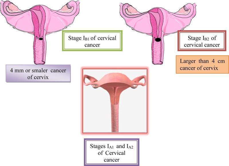
A multi-panel medical illustration and anatomical diagram depicting the FIGO staging of cervical cancer (Stages IA and IB). The central bottom panel displays a 3D-style anatomical diagram representing Stages IA1 and IA2, where the cervix appears macroscopically normal, indicating microinvasive disease that is not visible to the naked eye. The top left panel illustrates Stage IB1, showing a cross-section of the uterus and cervix with a small, localized dark lesion on the cervical canal; it is annotated as a cancer of 4 mm or smaller. The top right panel illustrates Stage IB2, showing a significantly larger, darker, and more prominent exophytic/ulcerative lesion on the cervix, annotated as being larger than 4 cm. All panels show relevant reproductive anatomy including the uterus, fallopian tubes, and ovaries. This educational graphic serves to distinguish clinical stages of cervical carcinoma based on tumor size and macroscopic visibility, relevant for gynecologic oncology and diagnostic pathology training.
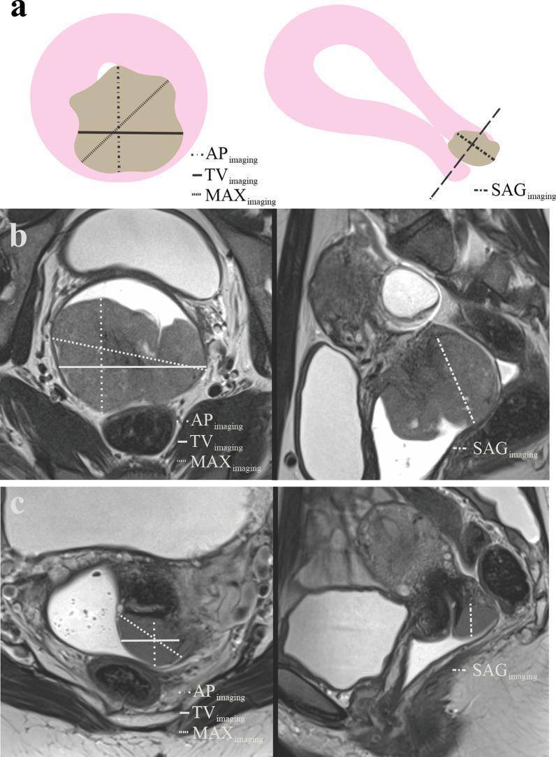
This educational image details the radiological assessment and measurement protocols for cervical cancer using MRI.
"cervical cancer" AND pembrolizumab AND chemoradiotherapy
mkdir -p /tmp/workspace/cervical-cancer-ppt/assets && ls -la /tmp/workspace/
python /tmp/skills/shared/scripts/fetch_images.py "https://cdn.orris.care/cdss_images/pmc_clinical_VQA_0f6bdae723d69486aec56dcf57534eadd94b434749c8714e7cb38ba36ac392d1.jpg" > /tmp/workspace/cervical-cancer-ppt/assets/staging_img.json 2>/tmp/workspace/cervical-cancer-ppt/assets/err.log; echo DONE; wc -c /tmp/workspace/cervical-cancer-ppt/assets/staging_img.json
~/cervical-cancer-ppt/build_ppt.py
import json, base64
from io import BytesIO
from pptx import Presentation
from pptx.util import Inches, Pt, Emu
from pptx.dml.color import RGBColor
from pptx.enum.text import PP_ALIGN, MSO_ANCHOR
from pptx.enum.shapes import MSO_SHAPE
from pptx.oxml.ns import qn

# ---------- Colors ----------
NAVY = RGBColor(0x1B, 0x2A, 0x4A)
TEAL = RGBColor(0x0E, 0x76, 0x7A)
ACCENT = RGBColor(0xC2, 0x38, 0x5A)   # magenta/pink accent (gyn-onc theme)
LIGHT = RGBColor(0xF4, 0xF6, 0xF8)
GRAY = RGBColor(0x5A, 0x63, 0x6E)
WHITE = RGBColor(0xFF, 0xFF, 0xFF)
DARK = RGBColor(0x22, 0x22, 0x22)

prs = Presentation()
prs.slide_width = Inches(13.333)
prs.slide_height = Inches(7.5)
BLANK = prs.slide_layouts[6]

SW, SH = prs.slide_width, prs.slide_height

def add_slide():
    return prs.slides.add_slide(BLANK)

def bg(slide, color=WHITE):
    slide.background.fill.solid()
    slide.background.fill.fore_color.rgb = color

def rect(slide, x, y, w, h, color, line=False):
    shp = slide.shapes.add_shape(MSO_SHAPE.RECTANGLE, x, y, w, h)
    shp.fill.solid()
    shp.fill.fore_color.rgb = color
    if line:
        shp.line.color.rgb = color
    else:
        shp.line.fill.background()
    shp.shadow.inherit = False
    return shp

def textbox(slide, x, y, w, h, text, size=18, color=DARK, bold=False, italic=False,
            align=PP_ALIGN.LEFT, font="Calibri", anchor=None, line_spacing=None):
    tb = slide.shapes.add_textbox(x, y, w, h)
    tf = tb.text_frame
    tf.word_wrap = True
    if anchor:
        tf.vertical_anchor = anchor
    tf.margin_left = 0; tf.margin_right = 0; tf.margin_top = 0; tf.margin_bottom = 0
    lines = text.split("\n")
    for i, line in enumerate(lines):
        p = tf.paragraphs[0] if i == 0 else tf.add_paragraph()
        p.text = line
        p.alignment = align
        if line_spacing:
            p.line_spacing = line_spacing
        r = p.runs[0] if p.runs else p.add_run()
        r.font.size = Pt(size)
        r.font.bold = bold
        r.font.italic = italic
        r.font.name = font
        r.font.color.rgb = color
    return tb

def bullet_block(slide, x, y, w, h, items, size=15, color=DARK, bullet_color=None,
                  space_after=8, bold_first=False, font="Calibri"):
    """items: list of (text, level, bold) or plain strings"""
    tb = slide.shapes.add_textbox(x, y, w, h)
    tf = tb.text_frame
    tf.word_wrap = True
    tf.margin_left = 0; tf.margin_right = 0; tf.margin_top = 0; tf.margin_bottom = 0
    bullet_color = bullet_color or ACCENT
    first = True
    for item in items:
        if isinstance(item, tuple):
            text, level, bold = (item + (False,))[:3] if len(item) == 2 else item
        else:
            text, level, bold = item, 0, False
        p = tf.paragraphs[0] if first else tf.add_paragraph()
        first = False
        p.level = level
        p.space_after = Pt(space_after)
        marker = "‣ " if level == 0 else "–  "
        r = p.add_run()
        r.text = (marker + text) if level <= 1 else text
        r.font.size = Pt(size - (2 if level > 0 else 0))
        r.font.bold = bold
        r.font.name = font
        r.font.color.rgb = color if level > 0 else DARK
    return tb

def header(slide, kicker, title, num, total=15):
    rect(slide, 0, 0, SW, Inches(1.15), NAVY)
    rect(slide, 0, Inches(1.15), SW, Pt(3), ACCENT)
    textbox(slide, Inches(0.55), Inches(0.14), Inches(9), Inches(0.35), kicker.upper(),
            size=13, color=RGBColor(0x9F, 0xC9, 0xCB), bold=True)
    textbox(slide, Inches(0.55), Inches(0.42), Inches(11.5), Inches(0.65), title,
            size=27, color=WHITE, bold=True)
    textbox(slide, Inches(12.55), Inches(7.12), Inches(0.6), Inches(0.3), f"{num}",
            size=11, color=GRAY, align=PP_ALIGN.RIGHT)
    textbox(slide, Inches(0.55), Inches(7.12), Inches(6), Inches(0.3),
            "Cervical Carcinoma: Complications & Treatment Guidelines", size=9, color=GRAY)

def footer_note(slide, text):
    textbox(slide, Inches(0.55), Inches(6.85), Inches(12.2), Inches(0.35), text,
            size=10.5, color=GRAY, italic=True)

def card(slide, x, y, w, h, title, body_items, title_color=TEAL, size=13.5):
    rect(slide, x, y, w, h, LIGHT)
    rect(slide, x, y, Inches(0.08), h, title_color)
    textbox(slide, x + Inches(0.25), y + Inches(0.12), w - Inches(0.4), Inches(0.4),
            title, size=15.5, color=title_color, bold=True)
    bullet_block(slide, x + Inches(0.25), y + Inches(0.58), w - Inches(0.45), h - Inches(0.7),
                 body_items, size=size, bullet_color=DARK, space_after=4)

# =========================================================
# SLIDE 1 - TITLE
# =========================================================
s = add_slide()
bg(s, NAVY)
rect(s, 0, Inches(5.7), SW, Inches(1.8), TEAL)
rect(s, 0, Inches(5.62), SW, Pt(3), ACCENT)
textbox(s, Inches(0.9), Inches(2.1), Inches(11.5), Inches(0.5), "GYNECOLOGIC ONCOLOGY",
        size=16, color=RGBColor(0x9F, 0xC9, 0xCB), bold=True)
textbox(s, Inches(0.9), Inches(2.6), Inches(11.5), Inches(1.6), "Cervical Carcinoma",
        size=54, color=WHITE, bold=True)
textbox(s, Inches(0.9), Inches(3.75), Inches(11.5), Inches(1.0),
        "Complications and Current Treatment Guidelines", size=26, color=RGBColor(0xE8,0xE8,0xE8))
textbox(s, Inches(0.9), Inches(5.95), Inches(11), Inches(0.5),
        "NCCN v2.2026  |  FIGO 2018 Staging  |  ESGO / ESMO / SEOM 2023-2025  |  KEYNOTE-A18 / KEYNOTE-826",
        size=15, color=WHITE, bold=True)
textbox(s, Inches(0.9), Inches(6.45), Inches(11), Inches(0.4),
        "A clinical overview for education and practice reference", size=13, color=RGBColor(0xD8,0xE8,0xE8), italic=True)

# =========================================================
# SLIDE 2 - AGENDA
# =========================================================
s = add_slide(); bg(s)
header(s, "Overview", "Agenda", 2)
agenda = [
    ("01", "Disease Overview & Risk Factors", "Epidemiology, HPV etiology, screening context"),
    ("02", "FIGO 2018 Staging", "How stage drives treatment selection"),
    ("03", "Complications of Disease", "Local invasion, obstruction, hemorrhage, metastasis"),
    ("04", "Complications of Treatment", "Surgical and radiotherapy/chemotherapy morbidity"),
    ("05", "Treatment by Stage", "Latest NCCN / ESGO / ESMO / FIGO guideline recommendations"),
    ("06", "Advanced / Recurrent Disease", "Chemo-immunotherapy: pembrolizumab, bevacizumab, ADCs"),
    ("07", "Follow-up & Key Takeaways", "Surveillance schedules and summary"),
]
y = Inches(1.55)
for num, title, sub in agenda:
    rect(s, Inches(0.55), y, Inches(0.75), Inches(0.72), TEAL)
    textbox(s, Inches(0.55), y, Inches(0.75), Inches(0.72), num, size=20, color=WHITE, bold=True,
            align=PP_ALIGN.CENTER, anchor=MSO_ANCHOR.MIDDLE)
    textbox(s, Inches(1.5), y + Inches(0.02), Inches(5.3), Inches(0.35), title, size=16.5, color=NAVY, bold=True)
    textbox(s, Inches(1.5), y + Inches(0.38), Inches(10.8), Inches(0.32), sub, size=12.5, color=GRAY)
    y += Inches(0.79)

# =========================================================
# SLIDE 3 - OVERVIEW & RISK FACTORS
# =========================================================
s = add_slide(); bg(s)
header(s, "Background", "Disease Overview & Risk Factors", 3)
card(s, Inches(0.55), Inches(1.55), Inches(6.0), Inches(5.1), "Epidemiology",
     [("4th most common cancer in women worldwide (GLOBOCAN)", 0),
      ("~660,000 new cases and ~350,000 deaths annually worldwide", 0),
      ("Marked disparity: incidence & mortality highest in low- and middle-income countries with limited HPV vaccination/screening access", 0),
      ("Median age at diagnosis 45-55 years; increasing incidence of adenocarcinoma in younger women", 0),
      ("Nearly all cases attributable to persistent high-risk HPV infection", 0)])
card(s, Inches(6.78), Inches(1.55), Inches(6.0), Inches(5.1), "Risk Factors & Etiology",
     [("Persistent high-risk HPV (16, 18 account for ~70%)", 0),
      ("Early coitarche, multiple sexual partners, high parity", 0),
      ("Cigarette smoking - cofactor for HPV persistence", 0),
      ("Immunosuppression (HIV infection, transplant recipients)", 0),
      ("Long-term combined oral contraceptive use (>5 yrs)", 0),
      ("Lack of HPV vaccination and inadequate cytology/HPV screening", 0),
      ("Histology: squamous cell carcinoma (~70-75%), adenocarcinoma (~20-25%), rarer neuroendocrine/small-cell types", 0)])
footer_note(s, "Sources: Robbins & Cotran Pathologic Basis of Disease; Berek & Novak's Gynecology; GLOBOCAN 2024 (CA Cancer J Clin 2026)")

# =========================================================
# SLIDE 4 - FIGO STAGING (with image)
# =========================================================
s = add_slide(); bg(s)
header(s, "Staging", "FIGO 2018 Staging System", 4)
stages = [
    ("I", "Confined to cervix", "IA: microscopic, ≤5mm depth  |  IB1 ≤2cm, IB2 >2-4cm, IB3 >4cm"),
    ("II", "Beyond uterus, not to pelvic wall/lower vagina", "IIA: upper 2/3 vagina  |  IIB: parametrial invasion"),
    ("III", "Extends to pelvic wall, lower 1/3 vagina, or nodes", "IIIA: lower vagina  |  IIIB: pelvic wall/hydronephrosis  |  IIIC: pelvic (C1)/para-aortic (C2) nodes*"),
    ("IVA", "Spread to bladder or rectal mucosa", "Biopsy-confirmed adjacent organ invasion"),
    ("IVB", "Distant metastasis", "Includes distant nodal spread, lung, liver, bone"),
]
x0 = Inches(0.55); y0 = Inches(1.55); w0 = Inches(7.4)
for stg, desc, detail in stages:
    rect(s, x0, y0, Inches(0.9), Inches(0.92), ACCENT)
    textbox(s, x0, y0, Inches(0.9), Inches(0.92), stg, size=19, color=WHITE, bold=True,
            align=PP_ALIGN.CENTER, anchor=MSO_ANCHOR.MIDDLE)
    textbox(s, x0 + Inches(1.05), y0 + Inches(0.02), w0 - Inches(1.1), Inches(0.35), desc, size=14.5, color=NAVY, bold=True)
    textbox(s, x0 + Inches(1.05), y0 + Inches(0.38), w0 - Inches(1.1), Inches(0.5), detail, size=11.5, color=GRAY)
    y0 += Inches(1.02)
textbox(s, x0, y0+Inches(0.02), w0, Inches(0.4), "*Stage IIIC designated r (imaging) or p (pathology) per 2018 revision",
        size=10.5, color=GRAY, italic=True)

# staging image
try:
    data = json.loads(open("/tmp/workspace/cervical-cancer-ppt/assets/staging_img.json").read())
    b64 = data[0]["base64"]
    raw = base64.b64decode(b64.split(",")[1] if "," in b64 else b64)
    img_stream = BytesIO(raw)
    rect(s, Inches(8.15), Inches(1.55), Inches(4.6), Inches(4.95), WHITE)
    s.shapes.add_picture(img_stream, Inches(8.25), Inches(1.65), width=Inches(4.4))
    textbox(s, Inches(8.15), Inches(6.35), Inches(4.6), Inches(0.35),
            "FIGO staging IA/IB illustration", size=10.5, color=GRAY, italic=True, align=PP_ALIGN.CENTER)
except Exception as e:
    textbox(s, Inches(8.15), Inches(1.55), Inches(4.6), Inches(1), f"[image unavailable]", size=12, color=GRAY)
footer_note(s, "Source: Berek & Novak's Gynecology, FIGO Staging 2018 revision; NCCN Cervical Cancer Guidelines v2.2026")

# =========================================================
# SLIDE 5 - COMPLICATIONS OF DISEASE
# =========================================================
s = add_slide(); bg(s)
header(s, "Complications I", "Complications of Disease Progression", 5)
card(s, Inches(0.55), Inches(1.55), Inches(4.0), Inches(5.1), "Local / Regional",
     [("Ureteral obstruction → hydronephrosis → post-renal (obstructive) renal failure - a leading cause of death in untreated/advanced disease", 0),
      ("Vesicovaginal and rectovaginal fistulae from direct tumor invasion", 0),
      ("Parametrial and pelvic sidewall invasion causing pelvic/sciatic-type pain", 0),
      ("Lymphatic obstruction → lower-limb lymphedema", 0)])
card(s, Inches(4.78), Inches(1.55), Inches(4.0), Inches(5.1), "Hemorrhage & Infection",
     [("Acute vaginal hemorrhage from friable exophytic tumor - can be life-threatening, may need emergent vaginal packing/embolization", 0),
      ("Pyometra and hematometra from cervical canal obstruction", 0),
      ("Secondary pelvic infection/abscess", 0),
      ("Anemia from chronic blood loss", 0)])
card(s, Inches(9.0), Inches(1.55), Inches(3.78), Inches(5.1), "Metastatic Spread",
     [("Pelvic and para-aortic lymph node metastases (nodal status = major prognostic factor)", 0),
      ("Hematogenous spread to lung, liver, bone", 0),
      ("Peritoneal/omental spread (less common)", 0),
      ("Bladder/rectal mucosal invasion (Stage IVA)", 0),
      ("Paraneoplastic thromboembolism (elevated VTE risk)", 0)])
footer_note(s, "Sources: Robbins & Cotran Pathologic Basis of Disease; Brenner & Rector's The Kidney; Berek & Novak's Gynecology")

# =========================================================
# SLIDE 6 - COMPLICATIONS OF TREATMENT (SURGERY)
# =========================================================
s = add_slide(); bg(s)
header(s, "Complications II", "Complications of Treatment - Surgery", 6)
card(s, Inches(0.55), Inches(1.55), Inches(6.0), Inches(5.1), "Radical Hysterectomy / Lymphadenectomy",
     [("Vesicovaginal fistula: ~0.3% in large series", 0),
      ("Ureterovaginal fistula: ~1% (higher with prior radiation)", 0),
      ("Bladder atony / voiding dysfunction from autonomic nerve injury (~4-10 weeks recovery, sometimes permanent)", 0),
      ("Ureteral injury or stricture", 0),
      ("Lower-limb and genital lymphedema after pelvic lymphadenectomy (up to 20-25%)", 0),
      ("Intraoperative hemorrhage, venous thromboembolism", 0),
      ("Sexual dysfunction, vaginal shortening", 0)])
card(s, Inches(6.78), Inches(1.55), Inches(6.0), Inches(5.1), "Fertility-Sparing Surgery (Trachelectomy/Conization)",
     [("Cervical stenosis, dysmenorrhea", 0),
      ("Second-trimester pregnancy loss, cervical incompetence requiring cerclage", 0),
      ("Preterm birth in subsequent pregnancies", 0),
      ("Infertility from cervical mucus factor", 0),
      ("Risk of occult nodal disease if inadequate staging performed first", 0)])
footer_note(s, "Sources: Berek & Novak's Gynecology; Campbell-Walsh-Wein Urology (Fistula chapter); ESGO 2023 Fertility-Sparing Guidelines")

# =========================================================
# SLIDE 7 - COMPLICATIONS OF TREATMENT (RT/CHEMO)
# =========================================================
s = add_slide(); bg(s)
header(s, "Complications III", "Complications of Treatment - Radiotherapy & Chemotherapy", 7)
card(s, Inches(0.55), Inches(1.55), Inches(4.0), Inches(5.1), "Acute RT Effects",
     [("Radiation cystitis, diarrhea, proctitis", 0),
      ("Skin desquamation in treatment field", 0),
      ("Myelosuppression (with concurrent chemo)", 0),
      ("Fatigue, nausea", 0)])
card(s, Inches(4.78), Inches(1.55), Inches(4.0), Inches(5.1), "Chronic RT Morbidity",
     [("Recto-vaginal fistula: ~1.4-5.3% after pelvic RT", 0),
      ("Bowel stricture/obstruction - risk roughly doubled vs. surgery alone", 0),
      ("Chronic radiation cystitis, hemorrhagic cystitis", 0),
      ("Vaginal stenosis and dyspareunia", 0),
      ("Ovarian failure / premature menopause if ovaries in field", 0),
      ("Secondary pelvic malignancy (rare, long-term)", 0)])
card(s, Inches(9.0), Inches(1.55), Inches(3.78), Inches(5.1), "Cisplatin-Based Chemo & Novel Agents",
     [("Nephrotoxicity, ototoxicity, peripheral neuropathy (cisplatin)", 0),
      ("Myelosuppression, nausea/vomiting", 0),
      ("Bevacizumab: hypertension, GI perforation, fistula, thromboembolism, impaired wound healing", 0),
      ("Immune-related adverse events with pembrolizumab (colitis, pneumonitis, endocrinopathy)", 0)])
footer_note(s, "Sources: Campbell-Walsh-Wein Urology; Yamada's Textbook of Gastroenterology; KEYNOTE-A18/826 safety data")

# =========================================================
# SLIDE 8 - TREATMENT OVERVIEW / ALGORITHM
# =========================================================
s = add_slide(); bg(s)
header(s, "Treatment", "Treatment Overview by Stage (NCCN v2.2026 / FIGO)", 8)
rows = [
    ("IA1", "Conization (fertility-sparing) or simple/extrafascial hysterectomy; sentinel node mapping if LVSI+", TEAL),
    ("IA2 - IB2", "Radical hysterectomy (Type C) + pelvic lymphadenectomy, OR radical trachelectomy if fertility desired & criteria met", TEAL),
    ("IB3 - IIA2", "Definitive concurrent chemoradiation (cisplatin) + brachytherapy preferred in US; radical surgery an alternative for select IB3", ACCENT),
    ("IIB - IVA", "Concurrent chemoradiation + brachytherapy; ADD pembrolizumab (high-risk LACC per KEYNOTE-A18)", ACCENT),
    ("IVB / Metastatic / Recurrent", "Systemic therapy: platinum-doublet + bevacizumab ± pembrolizumab (PD-L1+); tisotumab vedotin after progression", NAVY),
]
y = Inches(1.6)
for stage, plan, color in rows:
    rect(s, Inches(0.55), y, Inches(2.15), Inches(0.86), color)
    textbox(s, Inches(0.55), y, Inches(2.15), Inches(0.86), stage, size=14.5, color=WHITE, bold=True,
            align=PP_ALIGN.CENTER, anchor=MSO_ANCHOR.MIDDLE)
    rect(s, Inches(2.8), y, Inches(9.95), Inches(0.86), LIGHT)
    textbox(s, Inches(3.0), y+Inches(0.05), Inches(9.6), Inches(0.76), plan, size=13, color=DARK,
            anchor=MSO_ANCHOR.MIDDLE)
    y += Inches(0.98)
footer_note(s, "Radical hysterectomy Type C preferred over simple hysterectomy - wider parametrial/paracervical margin. Treatment decided by multidisciplinary tumor board.")

# =========================================================
# SLIDE 9 - EARLY STAGE TREATMENT DETAIL
# =========================================================
s = add_slide(); bg(s)
header(s, "Treatment Detail", "Early-Stage Disease: Surgery & Fertility Preservation", 9)
card(s, Inches(0.55), Inches(1.55), Inches(6.0), Inches(5.1), "Radical Hysterectomy (Type C)",
     [("Preferred for FIGO IB1-IB3 and IIA1-IIA2", 0),
      ("Wider excision of paracervix, includes cardinal/uterosacral ligaments, upper vagina, pelvic ± para-aortic nodes", 0),
      ("Sentinel lymph node (SLN) mapping increasingly used (NCCN, ESGO) in tumors <2cm", 0),
      ("Minimally invasive (laparoscopic/robotic) approach now discouraged for tumors ≥2cm after LACC trial showed inferior survival vs. open surgery - open radical hysterectomy is now standard of care", 0),
      ("Adjuvant chemoradiation added if high-risk features post-op: positive margins, parametrial invasion, positive nodes (Sedlis criteria)", 0)])
card(s, Inches(6.78), Inches(1.55), Inches(6.0), Inches(5.1), "Fertility-Sparing Options",
     [("Cone biopsy / simple trachelectomy: Stage IA1 without LVSI", 0),
      ("Radical trachelectomy + pelvic lymphadenectomy: select IA2-IB1 (<2cm), negative margins, no aggressive histology", 0),
      ("Negative pelvic nodal status is a precondition (ESGO 2023) - nodal assessment performed first", 0),
      ("Not recommended for small-cell neuroendocrine tumors, gastric-type adenocarcinoma, or minimal deviation adenocarcinoma", 0),
      ("Cerclage placement addressed by ESGO/BGCS guidance to reduce preterm birth risk", 0)])
footer_note(s, "Sources: NCCN Cervical Cancer v2.2026; ESGO 2023 Fertility-Sparing Guidelines; Cancers 2024;16:2541 (guideline comparison)")

# =========================================================
# SLIDE 10 - LOCALLY ADVANCED TREATMENT
# =========================================================
s = add_slide(); bg(s)
header(s, "Treatment Detail", "Locally Advanced Disease: Chemoradiation + Brachytherapy", 10)
card(s, Inches(0.55), Inches(1.55), Inches(6.0), Inches(5.1), "Standard of Care",
     [("Definitive concurrent chemoradiotherapy (CCRT): external beam RT + weekly cisplatin (40 mg/m2)", 0),
      ("Followed by intracavitary/interstitial brachytherapy boost to cervix/parametria", 0),
      ("Total treatment time goal: <=8 weeks (delays worsen local control)", 0),
      ("Image-guided adaptive brachytherapy (IGABT/MRI-based) now recommended over 2D planning where available (ESGO/ESTRO/ABS/GEC)", 0),
      ("Extended-field RT to para-aortic nodes if PET/CT-positive nodes", 0)])
card(s, Inches(6.78), Inches(1.55), Inches(6.0), Inches(5.1), "Practice-Changing Update: Immunotherapy Addition",
     [("KEYNOTE-A18/ENGOT-cx11/GOG-3047 (Lancet 2024): pembrolizumab + CCRT then maintenance pembrolizumab vs. CCRT alone in high-risk locally advanced cervical cancer", 0),
      ("Significant improvement in progression-free AND overall survival", 0),
      ("FDA-approved (Jan 2024); incorporated into NCCN v2.2026 as a category 1 option for high-risk LACC (node-positive or Stage III-IVA)", 0),
      ("Cost-effectiveness debated (JAMA Netw Open 2025) but panel consensus favors offering it", 0)])
footer_note(s, "Sources: Lorusso et al. Lancet 2024;403:1341 & Lancet 2024;404:1321 (KEYNOTE-A18); NCCN Cervical Cancer v2.2026")

# =========================================================
# SLIDE 11 - RECURRENT / METASTATIC
# =========================================================
s = add_slide(); bg(s)
header(s, "Treatment Detail", "Recurrent, Persistent & Metastatic Disease", 11)
card(s, Inches(0.55), Inches(1.55), Inches(4.0), Inches(5.1), "Central Recurrence",
     [("Pelvic exenteration if isolated central recurrence after RT, no distant disease", 0),
      ("5-year survival 30-60% in well-selected candidates", 0),
      ("Reirradiation ± surgery for select cases", 0)])
card(s, Inches(4.78), Inches(1.55), Inches(4.0), Inches(5.1), "Systemic First-Line",
     [("Platinum-based doublet (cisplatin/carboplatin + paclitaxel) + bevacizumab (GOG-240)", 0),
      ("Add pembrolizumab if PD-L1 CPS ≥1 (KEYNOTE-826, NEJM 2021) - now standard first-line regimen", 0),
      ("Improves OS and PFS vs. chemo ± bevacizumab alone", 0)])
card(s, Inches(9.0), Inches(1.55), Inches(3.78), Inches(5.1), "Beyond First-Line",
     [("Tisotumab vedotin (antibody-drug conjugate, tissue factor-directed) after progression on chemo", 0),
      ("Pembrolizumab monotherapy for MSI-H/dMMR or TMB-high tumors regardless of line", 0),
      ("Clinical trial enrollment strongly encouraged", 0)])
footer_note(s, "Sources: NCCN v2.2026; KEYNOTE-826 (NEJM 2021); GOG-240; Colombo et al.; SEOM-GEICO 2023 Guidelines")

# =========================================================
# SLIDE 12 - GUIDELINE COMPARISON
# =========================================================
s = add_slide(); bg(s)
header(s, "Guidelines", "How Major Societies Compare (2023-2026)", 12)
data = [
    ("NCCN (USA)", "v2.2026", "Open radical hysterectomy for early stage; CCRT+brachytherapy + pembrolizumab (cat.1) for high-risk LACC; SLN mapping accepted"),
    ("FIGO Cancer Report", "2021 (update pending)", "Staging reference (2018 revision); NACT favored where RT resources limited"),
    ("ESGO (Europe)", "2023", "Mandates negative pelvic nodes before fertility-sparing surgery; recommends SLN biopsy; individualized follow-up"),
    ("ESMO", "2023", "Surgery an option after neoadjuvant chemo in IB2-IIIB in select cases; pelvic exenteration option in IVA"),
    ("SEOM-GEICO (Spain)", "2023", "Aligns with NCCN on immunotherapy incorporation; detailed screening/HPV vaccination guidance"),
    ("Korean Society Gyn Onc", "2025", "Updated Delphi-based consensus incorporating pembrolizumab-CCRT data"),
]
y = Inches(1.55)
colw = [Inches(2.5), Inches(1.5), Inches(8.35)]
headers_ = ["Society", "Version", "Key Distinguishing Points"]
x = Inches(0.55)
for i, htext in enumerate(headers_):
    rect(s, x, y, colw[i], Inches(0.45), NAVY)
    textbox(s, x+Inches(0.1), y, colw[i]-Inches(0.2), Inches(0.45), htext, size=13, color=WHITE, bold=True, anchor=MSO_ANCHOR.MIDDLE)
    x += colw[i]
y += Inches(0.45)
for idx, (soc, ver, pts) in enumerate(data):
    rowcolor = LIGHT if idx % 2 == 0 else WHITE
    x = Inches(0.55)
    rh = Inches(0.83)
    for i, val in enumerate([soc, ver, pts]):
        rect(s, x, y, colw[i], rh, rowcolor)
        textbox(s, x+Inches(0.1), y+Inches(0.05), colw[i]-Inches(0.2), rh-Inches(0.1), val,
                size=11.5 if i==2 else 12.5, color=DARK, bold=(i==0), anchor=MSO_ANCHOR.MIDDLE)
        x += colw[i]
    y += rh
footer_note(s, "Source: Restaino et al. Management for Cervical Cancer Patients: Comparison of Guidelines. Cancers 2024;16(14):2541")

# =========================================================
# SLIDE 13 - FOLLOW-UP / SURVEILLANCE
# =========================================================
s = add_slide(); bg(s)
header(s, "Surveillance", "Follow-up After Primary Treatment", 13)
card(s, Inches(0.55), Inches(1.55), Inches(6.0), Inches(5.1), "Standard Follow-up Schedule",
     [("Physical & pelvic exam every 3-4 months for first 2 years", 0),
      ("Every 6-12 months from years 3-5", 0),
      ("Annual exam after year 5", 0),
      ("Cervical/vaginal cytology per society-specific interval - not all guidelines mandate routine cytology after treatment", 0),
      ("Patient education: symptoms of recurrence (bleeding, pain, leg swelling, weight loss)", 0)])
card(s, Inches(6.78), Inches(1.55), Inches(6.0), Inches(5.1), "Imaging & Special Considerations",
     [("Imaging only if clinically indicated (ESMO/ESGO) - not routine surveillance imaging in most", 0),
      ("NCCN: PET-CT at 3-6 months post-adjuvant therapy for Stage IB3+ or those requiring adjuvant treatment", 0),
      ("Pelvic organ toxicity screening emphasized in RT-treated patients (bladder, bowel, vaginal health)", 0),
      ("HPV vaccination counseling for partners/family; survivorship care planning", 0),
      ("Sexual health, lymphedema, and fertility counseling integrated into follow-up", 0)])
footer_note(s, "Sources: NCCN v2.2026; ESGO 2023; ESMO Clinical Practice Guidelines; Cancers 2024;16:2541")

# =========================================================
# SLIDE 14 - KEY TAKEAWAYS
# =========================================================
s = add_slide(); bg(s)
header(s, "Summary", "Key Takeaways", 14)
pts = [
    "Cervical carcinoma is an HPV-driven disease; prevention via vaccination and screening remains the highest-impact intervention.",
    "Complications range from local (fistula, ureteral obstruction, hemorrhage) to treatment-related morbidity (surgical fistula, radiation proctitis/cystitis, chemo/immunotherapy toxicity).",
    "FIGO 2018 staging (clinical + imaging/pathology) drives treatment selection.",
    "Early-stage disease: open radical hysterectomy (Type C) or fertility-sparing surgery in selected patients; minimally invasive surgery no longer preferred for tumors >=2cm.",
    "Locally advanced disease: concurrent cisplatin-based chemoradiation + brachytherapy remains standard; addition of pembrolizumab (KEYNOTE-A18) is the major 2024-2026 practice change for high-risk disease.",
    "Metastatic/recurrent disease: platinum + bevacizumab ± pembrolizumab (PD-L1+) first-line; tisotumab vedotin and clinical trials thereafter.",
    "Multidisciplinary care, individualized follow-up, and survivorship support are guideline-endorsed across NCCN, ESGO, ESMO, FIGO, and SEOM.",
]
y = Inches(1.6)
for i, pt in enumerate(pts, 1):
    rect(s, Inches(0.55), y, Inches(0.5), Inches(0.5), ACCENT)
    textbox(s, Inches(0.55), y, Inches(0.5), Inches(0.5), str(i), size=16, color=WHITE, bold=True,
            align=PP_ALIGN.CENTER, anchor=MSO_ANCHOR.MIDDLE)
    textbox(s, Inches(1.25), y+Inches(0.02), Inches(11.5), Inches(0.6), pt, size=13.5, color=DARK,
            anchor=MSO_ANCHOR.MIDDLE)
    y += Inches(0.72)

# =========================================================
# SLIDE 15 - REFERENCES
# =========================================================
s = add_slide(); bg(s)
header(s, "References", "Key Sources & Further Reading", 15)
refs = [
    "NCCN Clinical Practice Guidelines in Oncology: Cervical Cancer, Version 2.2026. J Natl Compr Canc Netw.",
    "Lorusso D, et al. Pembrolizumab + chemoradiotherapy for high-risk locally advanced cervical cancer (ENGOT-cx11/GOG-3047/KEYNOTE-A18). Lancet 2024;403:1341-1350; overall survival results Lancet 2024;404:1321-1332.",
    "Colombo N, et al. Pembrolizumab for persistent, recurrent, or metastatic cervical cancer (KEYNOTE-826). N Engl J Med 2021.",
    "Restaino S, et al. Management for Cervical Cancer Patients: Comparison of Guidelines (ESGO-NCCN-ASCO-AIOM-FIGO-BGCS-SEOM-ESMO-JSGO). Cancers 2024;16(14):2541.",
    "Manso L, et al. SEOM-GEICO Clinical Guidelines on Cervical Cancer (2023). Clin Transl Oncol 2024.",
    "Yoo JG, et al. Clinical Practice Guidelines for Cervical Cancer: Korean Society of Gynecologic Oncology update. J Gynecol Oncol 2025.",
    "Berek & Novak's Gynecology, 17th ed. - Cervical Cancer chapters (Staging, Treatment, Chronic Morbidity).",
    "Robbins, Cotran & Kumar - Pathologic Basis of Disease - Cervical Carcinoma.",
    "Goldman-Cecil Medicine, International Edition - Cervical Cancer Treatment and Prognosis.",
    "Campbell-Walsh-Wein Urology - Oncologic and Radiation Fistulae; Combination Therapies.",
]
bullet_block(s, Inches(0.55), Inches(1.6), Inches(12.2), Inches(5.2), refs, size=13.5, space_after=10)
textbox(s, Inches(0.55), Inches(7.0), Inches(12), Inches(0.3),
        "Prepared for educational purposes. Always confirm current recommendations against the live NCCN/ESGO/ESMO portals before clinical use.",
        size=10.5, color=GRAY, italic=True)

prs.save("/tmp/workspace/cervical-cancer-ppt/Cervical_Carcinoma_Complications_and_Treatment_Guidelines.pptx")
print("SAVED OK")
cd /tmp/workspace/cervical-cancer-ppt && python build_ppt.py
cd /tmp/workspace/cervical-cancer-ppt && python -c " from pptx import Presentation prs = Presentation('Cervical_Carcinoma_Complications_and_Treatment_Guidelines.pptx') print('slides:', len(prs.slides)) for i, slide in enumerate(prs.slides,1): texts = [sh.text_frame.text[:40] for sh in slide.shapes if sh.has_text_frame and sh.text_frame.text.strip()] print(i, texts[:2]) " && ls -la Cervical_Carcinoma_Complications_and_Treatment_Guidelines.pptx
Cervical Carcinoma Complications and Treatment Guidelines
Presentation · PPTX
- Title
- Agenda
- Disease overview & risk factors (HPV, epidemiology)
- FIGO 2018 staging (with staging illustration)
- Complications of disease progression (obstruction, hemorrhage, metastasis)
- Complications of treatment - surgery (fistula rates, lymphedema)
- Complications of treatment - radiotherapy/chemotherapy (chronic RT morbidity, immune-related AEs)
- Treatment overview algorithm by stage
- Early-stage treatment detail (radical hysterectomy, fertility-sparing)
- Locally advanced treatment (chemoradiation + brachytherapy + pembrolizumab)
- Recurrent/metastatic disease (bevacizumab, pembrolizumab, tisotumab vedotin)
- Guideline comparison table (NCCN/FIGO/ESGO/ESMO/SEOM/Korean society)
- Follow-up and surveillance
- Key takeaways
- References
- Complications: disease-related (ureteral obstruction/hydronephrosis, fistulae, hemorrhage, lymphedema, nodal/distant metastasis) vs. treatment-related (surgical fistula ~0.3-1%, chronic radiation proctitis/cystitis 1.4-5.3%, bowel obstruction, cisplatin nephro/neurotoxicity, bevacizumab and immunotherapy toxicities).
- Latest guideline update: NCCN Cervical Cancer v2.2026 now includes pembrolizumab added to concurrent chemoradiation for high-risk locally advanced disease (category 1), based on the KEYNOTE-A18 trial showing PFS and OS benefit (Lancet 2024). For metastatic/recurrent disease, pembrolizumab + platinum chemo + bevacizumab (KEYNOTE-826) is now standard first-line for PD-L1-positive tumors, with tisotumab vedotin available after progression.
- Open radical hysterectomy remains standard for tumors ≥2cm (minimally invasive surgery de-emphasized post-LACC trial).
- A guideline comparison table contrasts NCCN, FIGO, ESGO, ESMO, SEOM-GEICO, and the Korean Society of Gynecologic Oncology (2023-2025 updates).
Normal AFI at term
Normal Amniotic Fluid Index (AFI) at Term
| Category | AFI |
|---|---|
| Oligohydramnios | < 5 cm (some use <5-6 cm as the cutoff) |
| Normal | 8-18 cm |
| Polyhydramnios | 20-24 cm (defined more precisely as AFI above the 95th percentile for gestational age) |
- A single deepest vertical pocket < 2 cm is also used as an indicator of clinically significant oligohydramnios (often used interchangeably with or alongside AFI <5 cm) - Creasy & Resnik's Maternal-Fetal Medicine.
- From 16 weeks' gestation onward, the majority of normal pregnancies have an AFI between 8 and 24 cm.
If there are small pockets of around 1cm and a deep pocket of around 4 cm. Total sum of AFI is 7 to 8 cm what is the significance. How to go about in this patient
Significance of AFI 7-8 cm with Multiple Small Pockets (~1 cm) and One Deep Pocket (~4 cm) at Term
| Parameter | This patient | Definition of true oligohydramnios |
|---|---|---|
| AFI | 7-8 cm | < 5 cm |
| Single/deepest vertical pocket (SDP) | 4 cm | < 2 cm |
- Labor induction
- Intrapartum fetal heart rate abnormalities (variable decelerations from cord compression)
- Meconium-stained liquor
- Operative delivery
How to Work This Patient Up
- Re-verify gestational age (fluid volume declines physiologically after 37-40 weeks even in normal pregnancies)
- Ensure correct four-quadrant technique was used (probe perpendicular to floor, sagittal alignment, excluding cord/fetal parts from pocket measurement)
- Rupture of membranes - ask about leaking fluid; do a speculum exam / ferning test / pooling if any suspicion. PROM is a common cause of reduced AFI and changes management significantly.
- Uteroplacental insufficiency / IUGR - get an estimated fetal weight (EFW) if not recently done; growth restriction is present in up to 83% of true oligohydramnios cases (though far less relevant when AFI is only borderline).
- Post-term or approaching post-term status - fluid normally trends down near/after 40 weeks.
- Maternal dehydration - ask about oral intake, ensure adequate hydration (maternal hydration can measurably improve AFI).
- Medications - NSAIDs, ACE inhibitors/ARBs.
- Fetal renal/urinary tract anomaly - review anatomy scan findings if not already done; fetal causes of oligohydramnios are usually urinary tract in origin.
- Preeclampsia / hypertensive disease - check BP, urine protein, since placental insufficiency from hypertensive disease can reduce fluid.
- NST (non-stress test) - confirm reactivity
- Umbilical artery Doppler - if any growth concern, to rule out placental insufficiency
- Biophysical profile (BPP) if NST equivocal - note that even with a low AFI, if a 2×2 cm pocket is present (as here, with a 4 cm deepest pocket), the fluid component of the BPP scores normally (2/2)
- If NST is reactive, growth is appropriate, no ROM, no significant maternal/fetal pathology → the borderline AFI with a reassuring 4 cm deepest pocket does not mandate emergency delivery, but because she is already at term, most obstetricians would favor proceeding with induction of labor rather than expectant management, since there is little to gain from prolonging the pregnancy and a theoretical (if modest) risk of cord compression/fetal distress increases as fluid decreases further.
- If she prefers/qualifies for expectant management (favorable cervix not required, patient counseled) — reasonable alternative is twice-weekly AFI + NST surveillance until spontaneous labor or 41 weeks, with clear counseling to return immediately for decreased fetal movement, leaking fluid, or contractions.
- Continuous electronic fetal monitoring in labor is advisable given the higher background rate of variable decelerations from cord compression with reduced fluid, even at this borderline level.
- Encourage oral hydration in the interim - can modestly increase AFI and is a low-risk intervention.
- If repeat scan shows AFI dropping toward <5 cm or SDP <2 cm, or if any fetal growth/Doppler abnormality emerges, reclassify as true oligohydramnios and manage accordingly (expedite delivery).
Find all the data till now about complications of cervical carcinoma and their management
cervical cancer complications management fistula ureteral obstruction hemorrhage 2024 2025 guideline
"cervical cancer" AND complications AND management
https://www.ncbi.nlm.nih.gov/books/NBK431093
PMID: 38725634
Cervical Carcinoma: Complications and Their Management (Consolidated)
PART 1: DISEASE-RELATED COMPLICATIONS
1. Ureteral Obstruction → Hydronephrosis → Renal Failure
- Imaging (CT/ultrasound) to confirm hydronephrosis and guide intervention
- Percutaneous nephrostomy or retrograde ureteric stenting to relieve obstruction and preserve renal function before/during oncologic treatment
- Correct electrolyte derangements and uremia before proceeding to definitive chemoradiation
- Definitive treatment of the tumor (chemoradiation) is the long-term fix; drainage is a bridge
2. Acute Vaginal Hemorrhage
- Vaginal packing with sterile gauze soaked in a hemostatic agent
- Silver nitrate/Monsel's solution (ferric subsulfate) for surface bleeding
- Angiographic embolization (uterine/internal iliac artery) for uncontrolled hemorrhage
- Emergency external beam radiation ("hemostatic RT") for tumor-bleeding not controlled by local measures
- Correct coagulopathy, transfuse as needed
- Definitive chemoradiation once stabilized
3. Fistulae (Vesicovaginal, Rectovaginal, Ureterovaginal, Enterocutaneous)
- Small fistulae: conservative management with urinary/fecal diversion, wound care, nutritional support
- Surgical repair once tumor is controlled and tissue is well-vascularized (transvaginal, transabdominal, or laparoscopic/robotic approaches); interposition flaps (e.g., Martius flap, omental flap) improve success in irradiated tissue
- Urinary diversion (nephrostomy, ureteral stenting, or permanent diversion) if repair is not feasible, especially in recurrent/irradiated fistula
- Fecal diversion (colostomy) for rectovaginal or enterocutaneous fistula not amenable to repair
- Palliative approach in end-stage disease focuses on symptom control and quality of life
4. Pyometra and Hematometra
- Cervical dilation/drainage of the collection
- Broad-spectrum antibiotics if infected (pyometra)
- Address underlying obstruction with definitive cancer treatment
5. Pelvic Pain
- WHO analgesic ladder: NSAIDs/paracetamol → weak opioids → strong opioids (morphine, oxycodone, fentanyl)
- Adjuvants: gabapentinoids for neuropathic pain from pelvic sidewall/nerve invasion, corticosteroids for nerve compression
- Interventional pain procedures: superior hypogastric plexus block, ganglion impar block, intrathecal drug delivery, or neurolytic blocks for refractory pelvic/perineal pain
- Palliative radiotherapy to bulky/painful pelvic disease
- Multidisciplinary palliative care integration early in advanced disease
6. Lymphedema (Disease- or Treatment-Related)
- Complex decongestive therapy: manual lymphatic drainage + compression bandaging/garments + exercise + skin care
- Pneumatic compression devices
- Weight management, avoidance of limb trauma/infection (cellulitis risk)
- Surgical options in refractory cases: lymphovenous anastomosis, lymphaticolymphatic bypass, vascularized lymph node transfer
- Sentinel lymph node biopsy (rather than full lymphadenectomy) in early-stage disease reduces incidence
7. Venous Thromboembolism (VTE)
- Risk stratification and pharmacologic thromboprophylaxis (LMWH) perioperatively and during hospitalization
- Extended prophylaxis considered in high-risk surgical patients
- Therapeutic anticoagulation for confirmed DVT/PE (with caution regarding tumor-related bleeding risk)
8. Metastatic Disease Complications
- Systemic therapy (platinum-based chemo + bevacizumab ± pembrolizumab)
- Palliative RT to symptomatic metastases (bone pain, bleeding)
- Pelvic exenteration for isolated central recurrence without distant spread
- Bisphosphonates/denosumab for bone metastases
PART 2: TREATMENT-RELATED COMPLICATIONS
A. Surgical Complications (Radical Hysterectomy, Trachelectomy, Lymphadenectomy)
| Complication | Frequency | Management |
|---|---|---|
| Urinary dysfunction/bladder atony (detrusor denervation) | Most frequent complication of radical hysterectomy | Intermittent self-catheterization, pelvic floor physiotherapy; usually resolves in weeks, occasionally permanent |
| Vesicovaginal fistula | ~0.3% | Surgical repair (vaginal/abdominal/laparoscopic/robotic) with flap interposition; urinary diversion if repair fails |
| Ureterovaginal fistula/ureteral injury | ~1% | Ureteral stenting, ureteroneocystostomy, or psoas hitch repair |
| Lymphedema | Up to 25% (pelvic lymphadenectomy) | As above (Part 1.6) |
| Hemorrhage/infection | Variable | Standard surgical hemostasis, antibiotics |
| Bowel obstruction, stricture/fibrosis | Uncommon | Bowel rest, surgical lysis of adhesions/resection if needed |
| Foreshortened vagina, sexual dysfunction | Common | Vaginal dilators, pelvic physiotherapy, counseling |
| Cervical incompetence, infertility (fertility-sparing surgery) | 2-12% complication rate | Cerclage placement, close obstetric surveillance in subsequent pregnancy |
B. Radiotherapy / Brachytherapy Complications
| Complication | Management |
|---|---|
| Chronic radiation proctitis/enteritis (bloody diarrhea, ulceration) | Difficult to treat, often relapsing. Options: sucralfate/formalin instillation for hemorrhagic proctitis, endoscopic argon plasma coagulation, hyperbaric oxygen therapy, antioxidant therapy; surgery (resection/diversion) reserved for refractory strictures/fistula |
| Radiation cystitis (hemorrhagic) | Hydration, bladder irrigation, hyperbaric oxygen, intravesical agents; cystectomy/diversion in severe refractory cases |
| Rectovaginal fistula (1.4-5.3% after pelvic RT) | Fecal diversion; delayed surgical repair once tissue healed (irradiated tissue has poor healing - flap interposition often needed) |
| Bowel stricture/obstruction | Surgical resection/bypass if symptomatic |
| Vaginal stenosis | Vaginal dilators started early post-treatment, topical estrogen |
| Ovarian failure/premature menopause | Hormone replacement therapy if appropriate; ovarian transposition (oophoropexy) before RT can prevent this in fertility-sparing candidates |
| Brachytherapy applicator uterine perforation (2-18%) | Stable/no infection: observe ± prophylactic antibiotics. Hemodynamic instability/infection: IV fluids, antibiotics, escalation to surgical management. Postponement of brachytherapy is controversial since treatment delay worsens oncologic outcomes |
C. Chemotherapy-Related Toxicity (Cisplatin-based)
- Nephrotoxicity - pre-hydration, avoid nephrotoxic co-administration, dose adjustment for renal function
- Ototoxicity, peripheral neuropathy - dose modification/discontinuation if severe
- Myelosuppression - growth factor support (G-CSF), dose delays/reductions, transfusion support
- Nausea/vomiting - triple antiemetic prophylaxis (5-HT3 antagonist + NK1 antagonist + dexamethasone) for highly emetogenic cisplatin regimens
D. Targeted Therapy / Immunotherapy Toxicity
- Bevacizumab: hypertension (antihypertensives), GI perforation and fistula formation (drug discontinuation, surgical management), thromboembolism (anticoagulation), impaired wound healing (avoid perioperative use)
- Pembrolizumab: immune-related adverse events (colitis, pneumonitis, endocrinopathies) - managed per irAE algorithms with corticosteroids, immunosuppression, and drug hold/discontinuation depending on grade
Summary Principles of Management
- Multidisciplinary approach - gynecologic oncology, radiation oncology, urology, colorectal surgery, palliative care, and interventional radiology all play roles depending on the complication.
- Drainage/diversion before definitive treatment for obstructive complications (nephrostomy for hydronephrosis, diversion for fistula) to stabilize the patient.
- Irradiated tissue heals poorly - fistula repairs after RT often require delayed surgery with vascularized flap interposition.
- Prevention is emphasized in current guidelines: sentinel node mapping (reduces lymphedema), open surgery for tumors ≥2cm (reduces recurrence-related complications), ovarian transposition before RT, and image-guided adaptive brachytherapy (reduces normal-tissue dose and toxicity).
- Pain and psychosocial/palliative support should be integrated early, not only at end-of-life, given how prevalent and disabling pain is in this population.
Search PubMed for recent clinical trials on managing radiation-induced fistulae in cervical cancer
"cervical cancer" AND fistula
"radiation-induced fistula" AND management
"vesicovaginal fistula" AND radiotherapy
"hyperbaric oxygen" AND fistula AND pelvic
PMID: 41282280
PMID: 38431837
PMID: 33719774
PMID: 33016777
PubMed Search: Recent Clinical Trials on Managing Radiation-Induced Fistulae in Cervical Cancer
Randomized Controlled Trials (prevention-focused, not treatment of established fistula)
- 90 patients with early cervical cancer randomized to laparoscopic vs. open radical hysterectomy.
- Laparoscopic approach associated with fewer adverse reactions, better postoperative renal function markers (creatinine/BUN), and higher quality-of-life (KPS) scores.
- Note: this concerns surgery-related (not radiation-induced) fistula risk, but is relevant to overall fistula prevention strategy in cervical cancer care.
- Prospective randomized trial, 76 patients (stenting vs. no stenting) undergoing radical type C2 hysterectomy.
- Urologic complications (including ureterovaginal and vesicovaginal fistula) were lower with stenting (2.6%) vs. no stenting (10.5%), though the difference attributed mainly to disease stage rather than stenting itself in their analysis; preoperative radiotherapy did not significantly change complication rates in this small trial.
Observational / Retrospective Studies (most relevant recent data on radiation-induced fistula specifically)
- 150 patients treated 2013-2022; 9% developed fistula. Fistula symptoms resolved in 62% (8/13) with management. 2-year fistula-free survival 91.6%.
- Current smoking was the strongest independent risk factor (OR 5.14); lack of MRI-guided brachytherapy planning and bladder disease extension also increased risk.
- Practical implication: MRI-guided (image-guided adaptive) brachytherapy may reduce fistula risk - directly relevant to prevention-oriented management.
- 12 patients with cervical cancer-associated VVF (some post-radiation) treated with combined bilateral PCN + ureteral occlusion stenting.
- 100% technical success; urinary leakage resolved immediately in all patients; hydronephrosis and renal function normalized in 4/12 within a week; no recurrence during follow-up.
- This is the best available "interventional management" evidence for palliative, non-surgical fistula control when repair isn't feasible (e.g., irradiated, friable tissue).
- Use of bevacizumab before or after pelvic radiotherapy significantly increases fistula formation risk - important for treatment sequencing decisions in locally advanced/recurrent disease now that bevacizumab is standard in many regimens.
Case Reports / Small Case Series on Specific Repair Techniques (Tier 8, hypothesis-generating only)
- Platelet-rich plasma injection + Martius flap for radiation-induced recurrent VVF - reported successful closure [PMID: 34063610]
- Brachytherapy itself used to facilitate fistula closure in malignant VVF (paradoxical but reported in select cases) [PMID: 41239644]
- Laparoscopic repair with perirectal fat interposition graft for radiation-induced VVF [PMID: 34258529]
- Colpocleisis as a repair option in cervical cancer patients with prior pelvic radiation who are poor surgical candidates [PMID: 34035972]
- Fatal outcomes reported with post-radiotherapy rectovaginal fistula in locally advanced cervical cancer - underscoring that this remains a serious, sometimes life-threatening complication without a reliable definitive fix [PMID: 36427977]
Adjacent Evidence (not cervical cancer-specific, but informs management)
- Hyperbaric oxygen therapy has clinical trial evidence (HOT-TOPIC, HOT-REVA trials) for Crohn's-related perianal/rectovaginal fistula [PMID: 35146959, 34046674] and observational data for pelvic late radiation tissue injury generally [PMID: 32957127], but no dedicated RCT for radiation-induced fistula in gynecologic cancer specifically. This is extrapolated, not direct, evidence.
Honest Assessment
- Prevention matters more than treatment in the literature - MRI/image-guided brachytherapy planning and smoking cessation reduce risk (PMID: 41282280).
- Percutaneous nephrostomy + ureteral occlusion stenting is the best-described minimally invasive palliative option when repair isn't feasible (PMID: 33016777).
- Definitive surgical repair (flap interposition, colpocleisis, robotic approaches) remains individualized and reported mainly through case series, not trials.
Now Turn all this into presentation with images and also bigger font size also
vesicovaginal fistula diagram anatomy
hydronephrosis ureteral obstruction CT
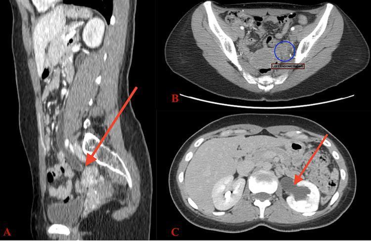
This composite diagnostic image features three contrast-enhanced CT views demonstrating severe obstructive uropathy. Image A (sagittal view) shows a significantly dilated left ureter (hydroureter) extending inferiorly toward a pelvic mass (indicated by a red arrow). Image B (axial pelvic view) highlights a spiculated, circular mass in the left adnexal region (circled), which is inseparable from the adjacent ureteral segment, suggesting intrinsic or extrinsic invasion. Image C (axial abdominal view) illustrates severe left hydronephrosis characterized by a markedly dilated renal pelvis (arrow) and thinning of the renal parenchyma compared to the normal right kidney. Notably, there is an absence of contrast flow within the dilated left collecting system, indicating complete obstruction. These findings are clinically consistent with ureteral endometriosis causing high-grade urinary tract obstruction. This material is suitable for medical education regarding radiological signs of hydronephrosis and differential diagnoses for ureteral masses in female patients.
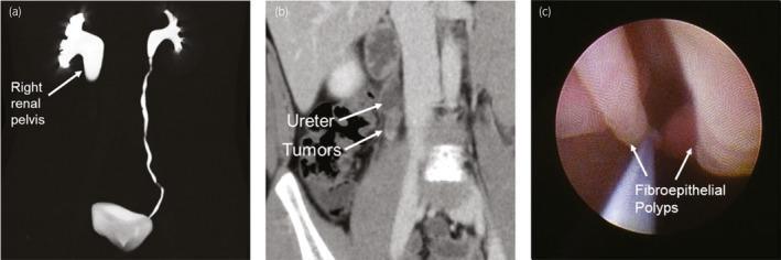
A multi-panel medical image illustrating the diagnosis of ureteral fibroepithelial polyps (UFEPs) causing obstruction. Panel (a) is a 3D reconstructed enhanced CT urogram demonstrating severe right-sided grade 3 hydronephrosis with a dilated renal pelvis and a sudden cutoff at the ureteropelvic junction (UPJ), while the left urinary tract remains normal. Panel (b) shows a coronal view of an enhanced CT scan identifying soft tissue masses (tumors) within the right proximal ureter at the level of the UPJ obstruction. Panel (c) provides a diagnostic ureteroscopy (URS) view showing multiple, smooth, pinkish, elongated finger-like masses projecting into the ureteral lumen, characteristic of benign fibroepithelial polyps. The image captures the diagnostic pathway from radiological identification of high-grade hydronephrosis to endoscopic visualization of the underlying etiology in a pediatric or adult urology context.
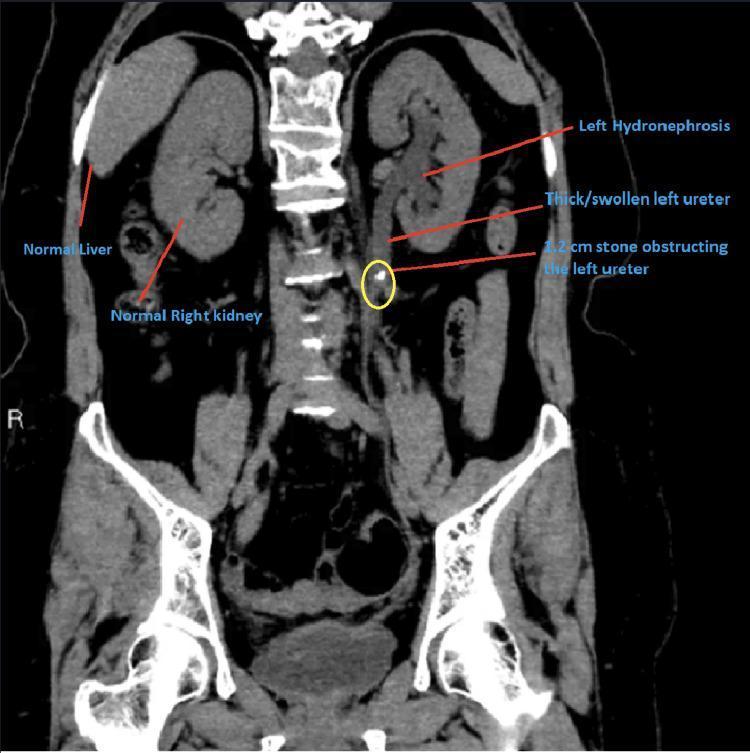
A diagnostic imaging study featuring a coronal CT scan of the abdomen and pelvis. The image highlights obstructive uropathy in the left urinary system. Key findings include a 1.2 cm hyperdense ureteral stone (highlighted by a yellow circle) located in the mid-to-proximal left ureter. Proximal to the obstruction, the left ureter exhibits significant dilation and wall thickening (hydroureter). The left kidney demonstrates moderate hydronephrosis, characterized by distention of the renal calyces and pelvis. In contrast, the right kidney, right ureter, and liver appear morphologically normal. Anatomical labels and text annotations clearly distinguish the pathological findings on the patient's left side from the normal anatomy on the right. This visual serves as a clear educational example of the upstream consequences of ureteral calculi, including secondary hydronephrosis and inflammatory ureteral changes, relevant for urology and emergency medicine curricula.
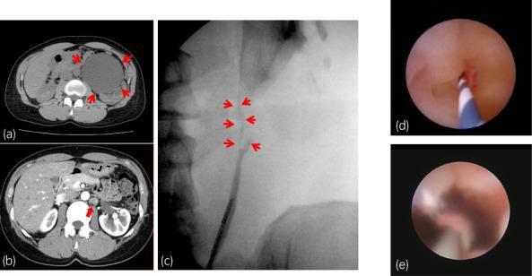
This composite figure illustrates the diagnosis and surgical outcome of ureteropelvic junction obstruction (UPJO) and subsequent stenosis. Panel (a) is an axial non-contrast CT scan showing severe left-sided hydronephrosis with a markedly dilated renal pelvis (red arrowheads). Panel (b) is a contrast-enhanced CT scan showing wall thickening at the UPJ and increased peri-ureteral fat stranding (red arrowhead), indicative of chronic inflammation or fibrosis. Panel (c) presents a retrograde pyelogram demonstrating a focal segment of anatomic ureteral stenosis (white arrowheads) where contrast flow is restricted. Panels (d) and (e) are ureteroscopic views. Image (d) shows the internal appearance of the high-grade ureteral stricture with a guide wire in place. Image (e) displays the same region following a redo pyeloplasty, showing a patent, widely open lumen with healthy-appearing mucosal lining, confirming successful surgical reconstruction and resolution of the obstruction.
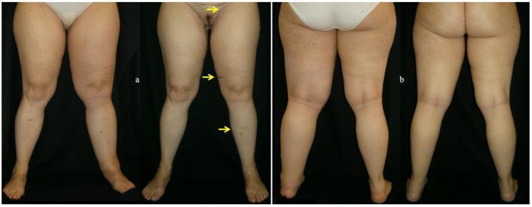
percutaneous nephrostomy ureteral stent placement
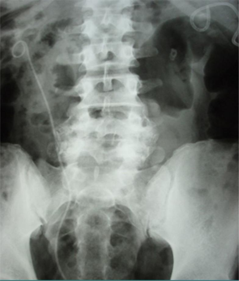
Anteroposterior KUB (kidneys, ureters, and bladder) radiograph of the abdomen and pelvis. The imaging demonstrates the lumbar spine, sacrum, and iliac crests with visible bowel gas patterns. Two distinct medical devices are present: a percutaneous nephrostomy (PCN) tube and a double-J ureteral stent. On the patient's left side, a radiopaque catheter is visible, originating from the flank and terminating in a pigtail-shaped coil in the upper left quadrant, consistent with a percutaneous nephrostomy placement for renal decompression. Concurrently, a double-J stent is visible on the patient's right side, with its proximal coil situated in the right renal pelvis and its distal end extending inferiorly through the ureter toward the bladder. This visual illustrates common urological interventions for managing urinary tract obstructions, such as those caused by urolithiasis, ureteral strictures, or malignancies. The radiograph is essential for confirming the correct anatomical positioning of these drainage devices relative to the renal and pelvic structures.
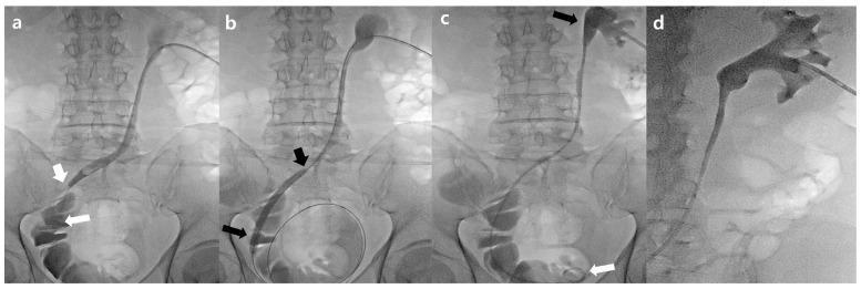
This composite of four fluoroscopic images (a-d) demonstrates the antegrade management of a uretero-ileal anastomosis stricture (UIAS) in a patient with a prior radical cystectomy and orthotopic neobladder. Image (a) is an antegrade nephrogram via an existing percutaneous nephrostomy (PCN) tube, showing a contrast-filled ureter with a severe segmental stenosis (approx. 2.4 cm) at the distal ureter/anastomosis site. Image (b) illustrates interventional balloon dilatation, featuring an inflated 6 mm balloon catheter at the point of stricture to restore patency. Image (c) shows the successful placement of a 6F double-J ureteral stent; the proximal pigtail is positioned in the renal pelvis (black arrow), and the distal pigtail is within the neobladder (white arrow). Image (d) is a post-procedural nephrogram confirming contrast flow and absence of significant blood clots (grade 1 retention). The series provides a visual guide for urological interventional procedures including nephrostomy access, balloon angioplasty of the urinary tract, and ureteral stenting for diversion-related complications.
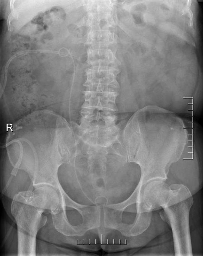
This diagnostic image is a postoperative anteroposterior (AP) Kidney, Ureter, and Bladder (KUB) radiograph of the abdomen and pelvis. The skeletal anatomy, including the lumbar vertebrae, pelvic girdle, and proximal femurs, is clearly visualized. Two radiopaque medical devices are visible in the right hemiabdomen: a nephrostomy tube and a double J (DJ) ureteral stent. The nephrostomy tube enters from the right flank, with its pigtail distal end positioned within the renal pelvis region. The double J stent is seen with its superior curl in the right kidney and its inferior curl residing in the pelvic cavity, corresponding to the anatomical location of the urinary bladder. An 'R' marker is positioned on the left side of the film, indicating the patient's right side. This image demonstrates successful placement of urinary diversion and drainage devices following a surgical intervention such as percutaneous nephrolithotomy (PCNL) or cyst unroofing. The bowel gas pattern appears normal, and there are no large radiopaque calculi visible in this postoperative view.
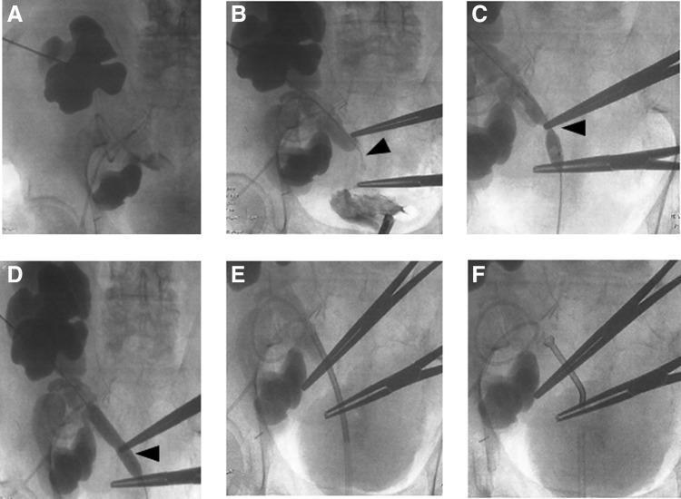
This composite of fluoroscopic images (A-F) illustrates an antegrade pyelogram and the interventional management of a ureteral stenosis in a 67-year-old patient with a double renal graft. Panel A demonstrates severe pelvicalyceal dilation (hydronephrosis) of the superior renal graft following percutaneous opacification. Panel B identifies the site of severe ureteral stenosis (arrowhead) at the level of the common iliac artery, with associated dilation of both the renal pelvis and proximal ureter. Panel C captures the use of a high-pressure balloon dilator over a 0.035-inch guidewire to treat the stricture. Panel D confirms the immediate post-dilation result, showing a wider ureteral lumen (arrowhead). Panel E shows the retrograde placement of an access sheath over the treated segment. Finally, Panel F visualizes the successful deployment and thermal expansion of a Memokath® 051 thermo-expandable metal stent, demonstrating restored ureteral patency. The series highlights critical steps in urological intervention, including nephrostomy access, balloon dilatation, and permanent stenting for obstructive uropathy in a complex transplant anatomy.
cervical cancer brachytherapy applicator
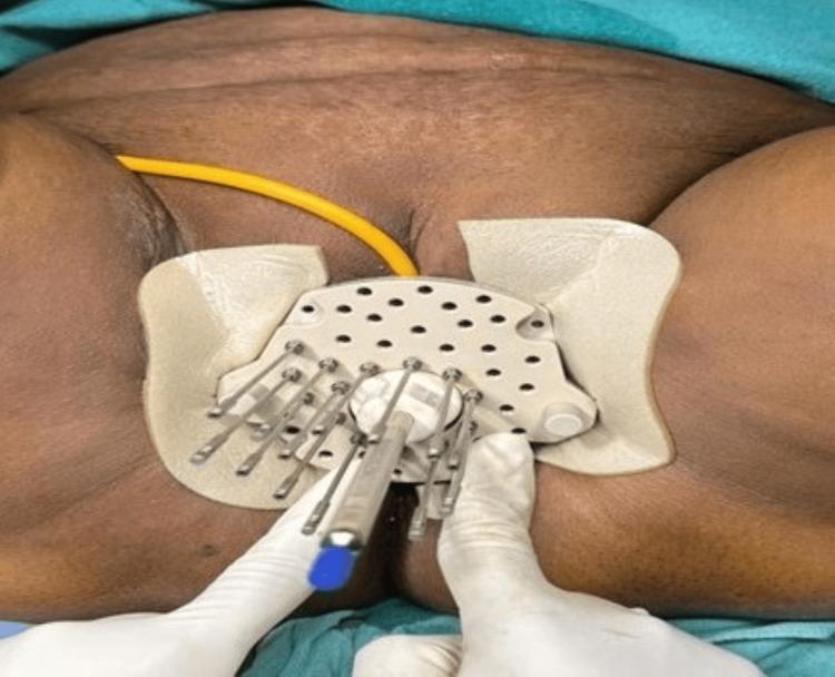
Clinical photograph of a patient undergoing high-dose-rate (HDR) interstitial brachytherapy for recurrent cervical cancer. The image shows a Kelowna template applicator secured over the perineal/vaginal region. Multiple thin, metallic interstitial needles are inserted through the circular grid of the applicator into the underlying tissue, arranged in a radial configuration to target the vaginal vault and paracervical regions. A Foley catheter (yellow) is visible, indicating bladder drainage during the procedure. The foreground shows a healthcare provider's gloved hands manipulating a central applicator component. This clinical setup demonstrates the surgical application of template-based interstitial brachytherapy, which allows for precise three-dimensional dose distribution in patients where conventional intracavitary brachytherapy is insufficient due to tumor anatomy or recurrence.
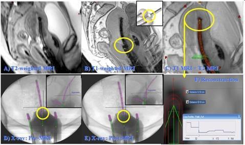
This composite figure illustrates the multi-modality imaging reconstruction of a titanium Tandem and Ovoid (T&O) brachytherapy applicator for cervical cancer treatment planning. (a) A sagittal T2-weighted MRI shows anatomical detail of the uterus and pelvic structures with 3 mm slice thickness. (b) A sagittal T1-weighted MRI highlights the applicator's cervical marker (flange) with high contrast, essential for position verification. (c) A fused T1 and T2 MRI image overlays the high-contrast applicator details onto the anatomical background for precise localization. (d) and (e) display orthogonal X-ray projections acquired prior to and following the MRI scan, respectively; yellow circles identify the flange position to calculate movement (Δd) and ensure reconstruction accuracy. (f) Provides a detailed view of the reconstructed tandem tip on MR, including an intensity profile used to account for artifacts and catheter thickness. This series demonstrates the clinical workflow for validating 3D MRI-based high-dose-rate (HDR) brachytherapy planning using multiple imaging modalities to minimize uncertainty in applicator reconstruction.
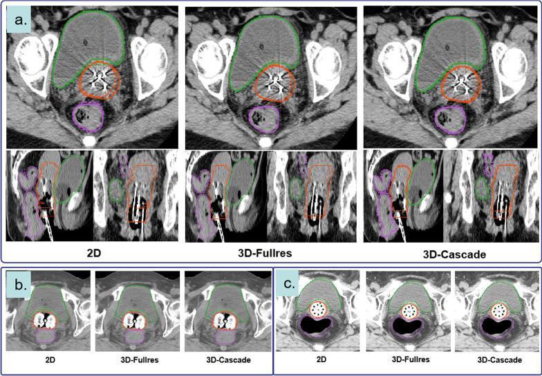
This diagnostic image set displays CT-based segmentation results for cervical cancer brachytherapy planning across three deep learning architectures: 2D, 3D-Fullres, and 3D-Cascade. The visualization compares manual ground-truth contours (solid lines) with auto-segmentation results (dashed lines) for critical structures: the bladder (green), rectum (purple), and High-Risk Clinical Target Volume (HRCTV, orange). The images are organized into three clinical scenarios based on the brachytherapy applicator used: (a) Needles and Tandem applicator, shown in axial, sagittal, and coronal planes; (b) Ovoid applicator in axial views; and (c) Vaginal multi-channel applicator in axial views. High-density metallic artifacts from the applicators are visible within the HRCTV. The comparison demonstrates the high level of spatial agreement between the automated AI models and expert manual segmentation for Organs at Risk (OARs) and target volumes, illustrating the clinical utility of nnU-Net frameworks in radiotherapy contouring workflows.
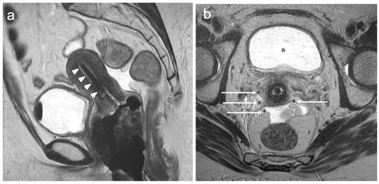
This diagnostic imaging composite displays T2-weighted magnetic resonance images (T2WI) of the female pelvis, illustrating the placement of brachytherapy equipment for the treatment of cervical cancer. Image (a) is a sagittal T2WI showing a dark, linear intracavitary brachytherapy applicator correctly positioned within the endometrial cavity, extending from the uterine fundus toward the cervix (marked by white arrowheads). Image (b) is an axial T2WI at the level of the cervix, demonstrating multiple hypointense, punctate signals representing parametrial interstitial needles (marked by white arrows). These needles are distributed within the parametrial tissues lateral to the cervix to deliver targeted radiation. The images highlight the use of MRI for real-time or post-procedural verification of applicator and needle positioning to ensure optimal dose distribution while sparing adjacent organs at risk, such as the bladder and rectum. This clinical material is intended for advanced medical education in gynecologic oncology and radiation oncology, focusing on locally advanced cervical cancer management.
pelvic arterial embolization hemorrhage control
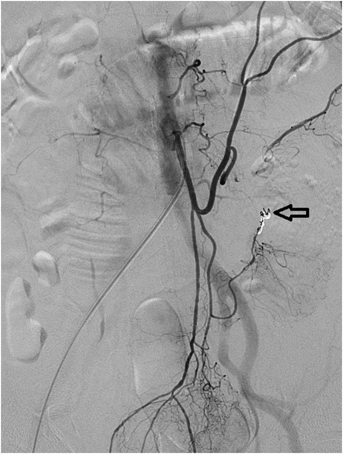
A digital subtraction angiogram (DSA) of the abdominal and pelvic vasculature, specifically targeting the inferior mesenteric artery (IMA) and its distal branches. The image displays the arterial tree as dark, contrast-filled branching structures against a grayscale background. An arrow points to a distinct area of intervention at the arcade between the left colic artery and the sigmoid artery. This area contains a radiopaque, high-density cluster of spring-like metallic coils and amorphous gel foam cubes, indicating a successful transarterial embolization (TAE) of a previously identified pseudoaneurysm. The embolization material forms a dense, occlusive mass to prevent further extravasation. The surrounding pelvic arterial anatomy, including the iliac vessels and sigmoid branches, remains visible with normal contrast filling. This diagnostic image demonstrates the post-procedural appearance of an emergency interventional radiology procedure for gastrointestinal hemorrhage control.
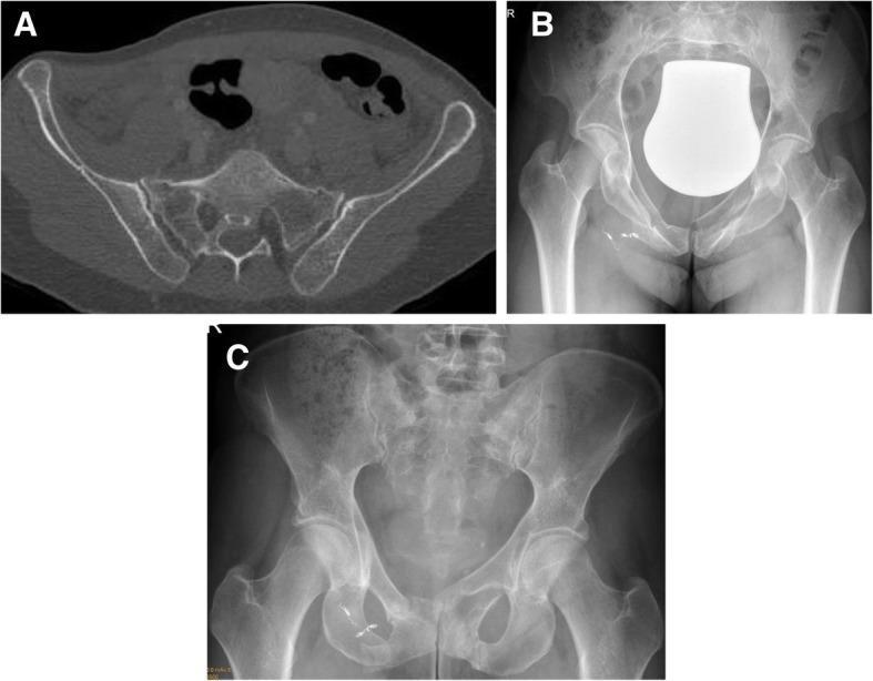
This composite clinical image illustrates the progression and non-operative management of a lateral compression (Type B2) pelvic fracture. Panel A is an axial CT scan of the pelvis showing bilateral sacral fractures involving the sacral alae, a characteristic finding in lateral compression injuries. Panel B is an anteroposterior (AP) pelvic radiograph demonstrating fractures of the superior and inferior pubic rami. Notable interventions include multiple radiopaque embolization coils in the right pelvic region, likely used to control arterial hemorrhage, and a large, bell-shaped radiopaque bladder shield. Panel C is a follow-up AP radiograph taken 6 weeks later, showing evidence of bone consolidation and interval healing of the pubic rami fractures with persistent embolization coils and no surgical hardware. This sequence demonstrates successful non-operative therapy where structural stability is achieved through natural remodeling and conservative management.
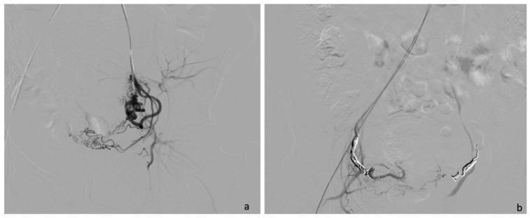
This diagnostic image pair displays Digital Subtraction Angiography (DSA) of the pelvic region, specifically focused on a uterine artery embolization (UAE) procedure. Panel (a) illustrates a pre-embolization selective angiogram of the uterine artery. It shows a dense, complex network of hypervascular branching structures and tortuous vessels consistent with the increased blood supply required for a gestational sac in a cesarean scar pregnancy (CSP). Contrast medium clearly delineates the arterial anatomy and distal capillary blush. Panel (b) shows the final post-procedural control angiography. The previous vascular network is no longer opacified, indicating a successful cessation of blood flow. Highly radiopaque, coiled metallic embolization agents (Interlock or Nester coils) are visible within the arterial lumens, serving as the mechanical obstruction. This comparison demonstrates the therapeutic devascularization of the affected uterine segment to prevent hemorrhage during subsequent surgical evacuation. The image is a critical educational tool for interventional radiology and obstetrics, highlighting the role of minimally invasive techniques in managing complex ectopic pregnancies.
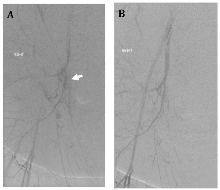
This diagnostic image set consists of two panels (A and B) showing catheter-based digital subtraction angiography (DSA) of the right internal iliac artery in a 36-year-old female patient with placenta accreta and postpartum hemorrhage. Panel A (Pre-embolization): The angiogram demonstrates the right pelvic vasculature with a focus on the internal iliac artery. A white arrow points to a distinct area of hypervascularity characterized by a dense cluster of multiple irregular, tortuous, and small-caliber vessels, consistent with pathological neovascularization or extravasation in the setting of retained placenta. Panel B (Post-embolization): Following the administration of Gelfoam into the anterior division of the internal iliac artery, a repeat angiogram shows successful devascularization of the previously identified abnormal vessels. There is a notable absence of the irregular vascular cluster and any signs of active extravasation, while the primary proximal arterial branches remain patent. This comparison illustrates the use of uterine artery embolization (UAE) as an emergency interventional radiology procedure for hemodynamic stabilization and hemorrhage control.
radical hysterectomy cervical cancer surgery diagram
rectovaginal fistula repair Martius flap
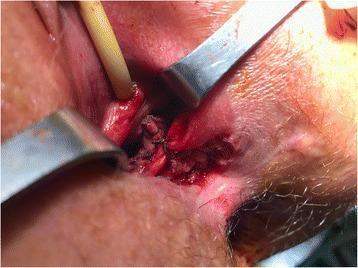
This clinical photograph captures a surgical field during a complex gynecological and proctological reconstruction for Crohn's disease-related fistulae. The image illustrates the 'final set-up' of a multi-step procedure involving the repair of a rectovaginal fistula and a longitudinal vaginal septum. Key anatomical features visible include the perineovaginal tract, which has been 'laid open' to expose underlying structures. Central to the operative site is the bulbocavernous muscle, carefully isolated and transposed as a Martius flap to reinforce the fistula repair. The vaginal mucosa is shown closed with meticulously placed interrupted absorbable sutures following the sectioning of the longitudinal vaginal septum. Surgical retractors are positioned to provide visualization of the deep pelvic tissues and the reinforced rectovaginal septum. This image serves as an educational resource for pelvic floor reconstruction, demonstrating tissue transposition and the management of congenital anomalies (septate vagina) in the context of chronic inflammatory bowel disease complications.
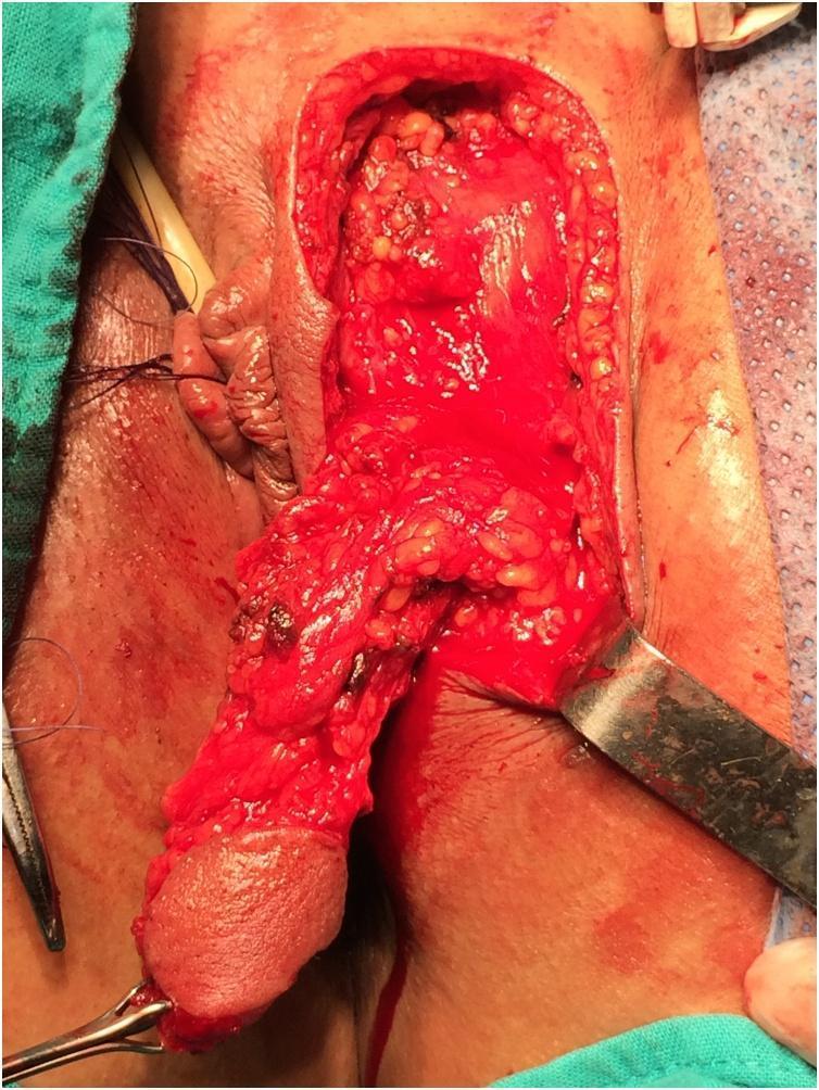
This clinical photograph captures an intraoperative view of a pedicled labial skin flap (Martius flap) being harvested for reconstructive surgery, likely for rectovaginal fistula repair. The image shows the inner aspect of the left labium majus where an elongated skin flap, approximately 4–5 cm in length, has been surgically dissected. The flap is pedicled, maintaining its vascular supply through a broad base (meso), allowing for mobilization without tension into the vaginal or rectal area. The distal end of the flap displays a smooth epithelial surface, while the underlying deep surface is granular, showing exposed subcutaneous adipose tissue and minor surgical bleeding. The donor site reveals deep perineal tissues, including muscle and fat, with a metal surgical retractor positioned to the right to maintain exposure. Sutures and surgical instruments are visible at the periphery, and anatomical landmarks like the clitoris are partially visible within the operative field. This visual demonstrates the surgical anatomy and mobilization phase of a pedicled fat/skin graft in gynecological or colorectal reconstructive procedures.
mkdir -p /tmp/workspace/cervical-fistula-ppt/assets && cd /tmp/workspace/cervical-fistula-ppt/assets && python /tmp/skills/shared/scripts/fetch_images.py \ "https://cdn.orris.care/cdss_images/pmc_clinical_VQA_19de324835218dfafb2ba0db000d30be5d5af0b4611cac9c1937e9c96f8cdafc.jpg" \ "https://cdn.orris.care/cdss_images/pmc_clinical_VQA_16ae4202cad875b971e4d65e44e7520bdb68847e1faa5400fc3899435ce1df69.jpg" \ "https://cdn.orris.care/cdss_images/pmc_clinical_VQA_3b3e11b86ab01d1f688be8c29f789ee718dcd5384c787d79d7febc7cddb95457.jpg" \ "https://cdn.orris.care/cdss_images/pmc_clinical_VQA_000fc1c134c15cad7c7a26b7502bb98c356c94c3203136d5aa18d9512b7a40ba.jpg" \ "https://cdn.orris.care/cdss_images/pmc_clinical_VQA_4d6980aeae1fd06804245eea975eeb07a30b3f33ba2cd32be39c26c7a6b53cb0.jpg" \ "https://cdn.orris.care/cdss_images/pmc_clinical_VQA_58eb9fafbaebf2d003bbd4c26d66f3a8e0f6109b8fc967b8f207375c41badf88.jpg" \ "https://cdn.orris.care/cdss_images/pmc_clinical_VQA_4532500310f486a7588ffe9f079a04c0c3190179026e33f684c66120e88148dd.jpg" \ > images.json 2>err.log; echo DONE; wc -c images.json; python -c "import json; d=json.load(open('images.json')); [print(x['url'][:60], 'OK' if x['base64'] else x['error']) for x in d]"