
Renal pain in detail for POM assignment
Reading File
Reading File
Searching the Web
renal colic clinical features referred pain diagnosis red flags guideline
Searching PubMed
renal colic AND diagnosis
Finding Sources
Finding Sources
Finding Sources
Reading File
Reading File
Reading File
Finding Sources
Reading File
Reading File
Reading File
Finding Sources
Reading File
Finding Sources
Searching Images
anatomical diagram radiation of ureteric colic pain flank groin testicle labium
Renal Pain
Definition
Renal pain is pain arising from the kidney, usually due to sudden distension of the renal capsule, renal pelvis, or collecting system. It is classically a dull, constant ache in the loin or costovertebral angle.
It must be differentiated from ureteric colic. Although often called “renal colic,” this is more accurately pain caused by acute ureteric obstruction, most commonly by a ureteric calculus. It is severe and may be colicky or continuously intense with waxing and waning severity.
Bailey and Love's Short Practice of Surgery, p. 2057; Smith and Tanagho's General Urology, p. 281.
Bailey and Love's Short Practice of Surgery, p. 2057; Smith and Tanagho's General Urology, p. 281.
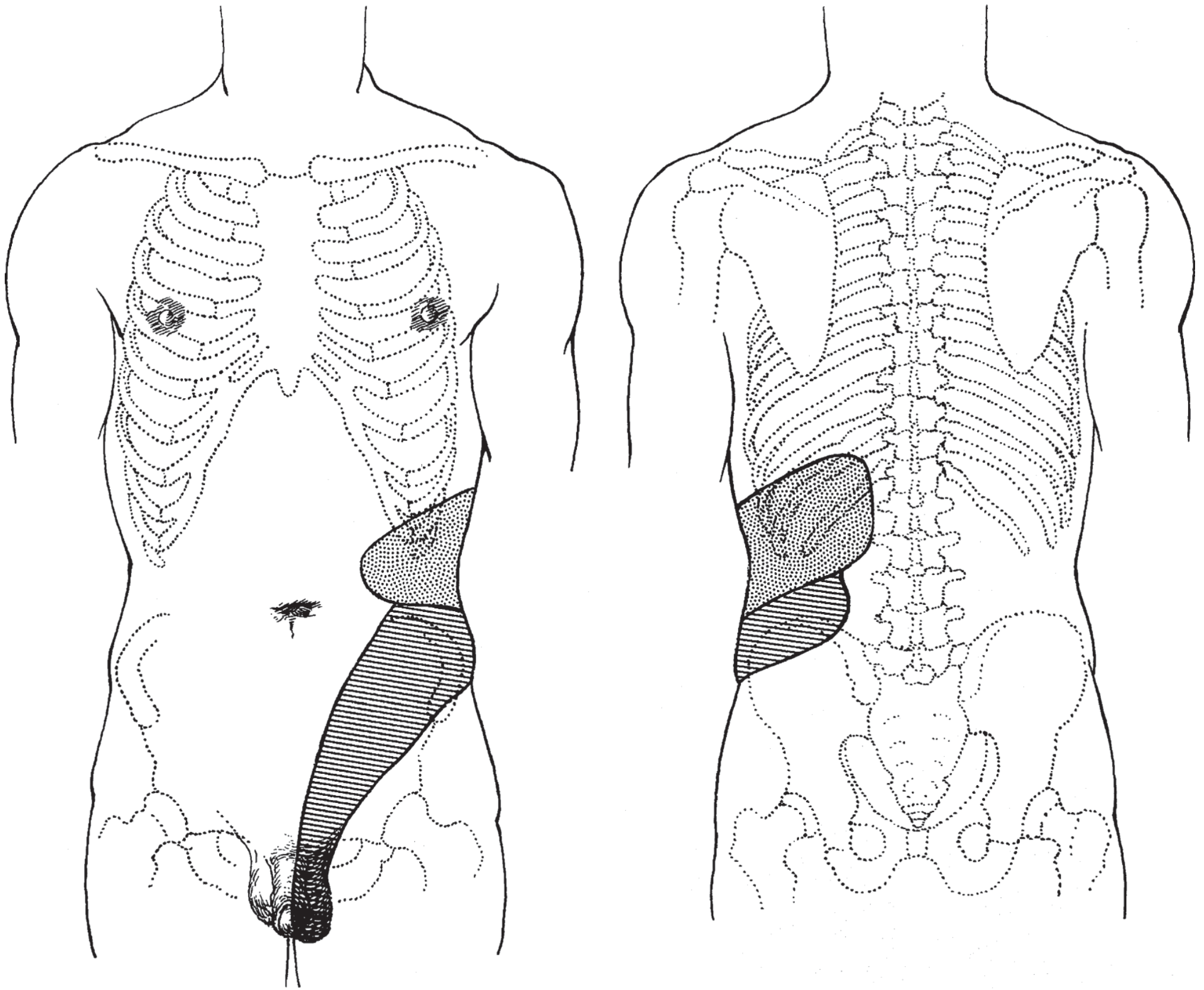
Mechanism of pain
The kidney parenchyma itself is relatively insensitive. Pain occurs when:
-
Renal capsule is stretched
Due to edema, inflammation, hemorrhage, or acute obstruction causing enlargement of the kidney. This produces typical constant renal pain. -
Renal pelvis and ureter are distended
Acute obstruction raises intraluminal pressure, stretches the collecting system, and stimulates visceral pain fibers. -
Ureteric smooth muscle undergoes spasm and hyperperistalsis
When a stone obstructs the ureter, powerful contractions attempt to push it onward. This produces severe ureteric colic. -
Inflammation and mucosal irritation occur
Edema and local inflammation around a calculus can add to pain and urinary symptoms.
Smith and Tanagho's General Urology, pp. 281-282.
Types of renal tract pain
| Feature | True renal pain | Ureteric colic |
|---|---|---|
| Main cause | Distension of renal capsule | Acute obstruction/stretching of renal pelvis or ureter |
| Character | Dull, aching, constant, gnawing | Severe, spasmodic, waxing and waning, sometimes continuously severe |
| Site | Loin/costovertebral angle, below 12th rib | Starts in flank or costovertebral angle and follows ureter downward |
| Radiation | Subcostal region, umbilicus, lower abdomen | Groin, lower abdomen, scrotum/testicle in men, labium/vulva in women |
| Patient behavior | May lie still due to discomfort | Restless, unable to find a comfortable position |
| Common examples | Acute pyelonephritis, acute hydronephrosis, renal infarction | Ureteric calculus, blood clot, sloughed renal papilla |
Smith and Tanagho's General Urology, pp. 45, 281-282; Bailey and Love's Short Practice of Surgery, pp. 2057-2058.
Site and radiation
True renal pain
Typical pain is felt in the costovertebral angle, lateral to the vertebral column and just below the 12th rib. It may spread anteriorly along the subcostal area toward the umbilicus or lower abdominal quadrant.
Ureteric colic
The radiation helps estimate the level of ureteric obstruction:
- Upper ureter or ureteropelvic junction: flank pain, often radiating to lower abdomen and ipsilateral testicle or labium.
- Mid-ureter: pain moves to lower abdominal quadrant. On the right it may mimic appendicitis, and on the left it may mimic diverticulitis.
- Lower ureter: pain radiates to groin, scrotum, vulva, or bladder region.
- Ureterovesical junction: frequency, urgency, dysuria, and suprapubic discomfort may dominate because of irritation around the bladder trigone.
The upper ureter and testis share segmental innervation, mainly T11-T12, explaining referred testicular pain.
Smith and Tanagho's General Urology, pp. 45-46, 281-282; National Kidney Foundation Primer on Kidney Diseases, p. 469.
Smith and Tanagho's General Urology, pp. 45-46, 281-282; National Kidney Foundation Primer on Kidney Diseases, p. 469.
Causes of renal pain
A. Causes of constant renal pain
-
Acute pyelonephritis
Renal inflammation and edema distend the capsule. Fever, chills, dysuria, pyuria, and costovertebral-angle tenderness may occur. -
Acute urinary obstruction with hydronephrosis
Sudden back pressure distends the renal pelvis and capsule. -
Renal or perinephric abscess
-
Renal trauma or subcapsular hematoma
-
Renal infarction or renal vein thrombosis
-
Hemorrhage into a renal cyst, especially in polycystic kidney disease.
-
Rapid enlargement of renal tumor or cyst
-
Acute glomerulonephritis, rarely, if capsular distension is significant.
Chronic kidney disease, chronic hydronephrosis, chronic pyelonephritis, renal tuberculosis, staghorn calculus, and many renal cancers may remain painless because slow expansion does not cause sudden capsular stretching.
Smith and Tanagho's General Urology, p. 45.
Smith and Tanagho's General Urology, p. 45.
B. Causes of ureteric colic
- Ureteric calculus, the commonest cause.
- Blood clot in the ureter, for example after trauma, tumor, or surgery.
- Sloughed renal papilla, such as in papillary necrosis.
- Ureteric stricture.
- Urothelial tumor causing obstruction.
- Rarely, obstruction due to fungal ball or other intraluminal material.
Bailey and Love's Short Practice of Surgery, p. 2058; National Kidney Foundation Primer on Kidney Diseases, p. 469.
Clinical features of acute ureteric colic
- Sudden onset of severe unilateral flank pain.
- Pain starts in the loin and typically travels downward and forward toward the groin.
- Pain does not necessarily completely disappear between waves. It often waxes and wanes.
- The patient is restless, constantly changing position, and unable to lie comfortably.
- Nausea and vomiting are common.
- Microscopic or gross hematuria may occur.
- Frequency, urgency, dysuria, and strangury occur with a stone near the ureterovesical junction.
- Costovertebral-angle tenderness may be present.
- Fever, chills, pyuria, hypotension, or toxicity suggest associated infection and require urgent assessment.
A stone may be present incidentally in the kidney, so finding a renal stone does not by itself prove that it is the cause of abdominal pain.
National Kidney Foundation Primer on Kidney Diseases, pp. 469-470.
National Kidney Foundation Primer on Kidney Diseases, pp. 469-470.
Examination
General examination
- Patient with colic is distressed and restless.
- Record pulse, blood pressure, temperature, hydration status, and urine output.
- Fever and tachycardia raise concern for infection or sepsis.
Abdominal examination
- Tenderness in the loin or costovertebral angle.
- Guarding may occur but marked peritonism suggests another diagnosis.
- Examine for abdominal mass, distended bladder, or pulsatile abdominal mass.
Systemic examination
- Examine genitalia, especially in males with scrotal pain, to exclude testicular torsion.
- Pelvic examination may be required in women with lower abdominal pain to exclude ectopic pregnancy or ovarian pathology.
Investigations
1. Urine tests
- Urinalysis for red blood cells, white blood cells, nitrites, protein, and crystals.
- Urine microscopy and culture if infection is possible.
- Hematuria supports but does not prove stone disease.
- Absence of hematuria does not exclude a stone, particularly in complete ureteric obstruction.
2. Blood tests
- Complete blood count: leukocytosis may be due to stress or infection.
- Serum creatinine, urea, and electrolytes: assess renal function and obstruction.
- C-reactive protein and blood culture if fever or sepsis is suspected.
- Pregnancy test in women of reproductive age before imaging and drug selection.
3. Imaging
- Ultrasound: identifies hydronephrosis, renal calculi, and some proximal or distal ureteric stones. It is preferred initially in pregnancy and when radiation should be avoided.
- Non-contrast low-dose CT of kidney, ureter, and bladder: most informative test for acute renal colic. It identifies the site, size, and density of stones and may reveal hydronephrosis, hydroureter, renal enlargement, and perinephric stranding.
- Plain KUB radiograph: limited in acute diagnosis but can help monitor selected known radiopaque stones.
- MRI: may be useful during pregnancy when ultrasound is inconclusive, though it is less practical for stone detection than CT.
National Kidney Foundation Primer on Kidney Diseases, p. 470; Bailey and Love's Short Practice of Surgery, pp. 714-730.
Differential diagnosis
Conditions that can mimic renal or ureteric pain include:
- Acute pyelonephritis
- Appendicitis
- Acute cholecystitis
- Diverticulitis
- Biliary colic
- Peptic ulcer disease
- Pancreatitis
- Abdominal aortic aneurysm or aortic dissection
- Herpes zoster, including pain before the rash appears
- Musculoskeletal back pain or radiculopathy
- Testicular torsion, epididymo-orchitis
- Ectopic pregnancy, ovarian torsion, pelvic inflammatory disease
- Acute cystitis
- Renal tumor, renal infarction, or perinephric abscess
National Kidney Foundation Primer on Kidney Diseases, p. 469.
Principles of management
Immediate management
- Assess for sepsis, shock, renal failure, solitary kidney, bilateral obstruction, or anuria.
- Give analgesia. NSAIDs are usually preferred if there is no contraindication because they reduce pain and ureteric inflammation. Opioids may be used if pain remains uncontrolled.
- Treat nausea and vomiting with an antiemetic.
- Check urine output and renal function.
- Obtain urine culture and start antibiotics if infection is suspected.
- Confirm obstruction and stone characteristics with appropriate imaging.
Indications for urgent urological drainage
An obstructed urinary tract with infection is a urological emergency. Urgent drainage by ureteric stent or percutaneous nephrostomy is needed when obstruction is associated with:
- Fever, pyuria, systemic sepsis, or pyonephrosis
- Anuria
- Acute kidney injury or rising creatinine
- Bilateral obstruction
- Obstruction in a solitary functioning kidney
- Uncontrolled pain or vomiting despite treatment
This is often summarized as “pus under pressure.”
National Kidney Foundation Primer on Kidney Diseases, pp. 469-470.
National Kidney Foundation Primer on Kidney Diseases, pp. 469-470.
Key points for viva or short note
- True renal pain is constant, dull, and felt in the loin due to renal capsular distension.
- “Renal colic” is usually more accurately called ureteric colic.
- Ureteric colic is caused by acute ureteric obstruction, most commonly a stone.
- Pain radiates loin to groin, and may extend to the testicle, scrotum, labium, or vulva.
- The colicky patient is restless, whereas patients with peritoneal inflammation generally prefer to lie still.
- Fever with obstructed ureteric stone indicates possible infected obstruction and needs urgent decompression.
- Non-contrast CT KUB is the most informative imaging test in many nonpregnant adults, while ultrasound is useful when radiation avoidance is important.
This is a shared conversation. Sign in to Orris to start your own chat.