
Make short and crisp notes with relevant points on apoptosis for md dermatology exam
apoptosis skin keratinocyte
apoptosis intrinsic and extrinsic pathway diagram caspase
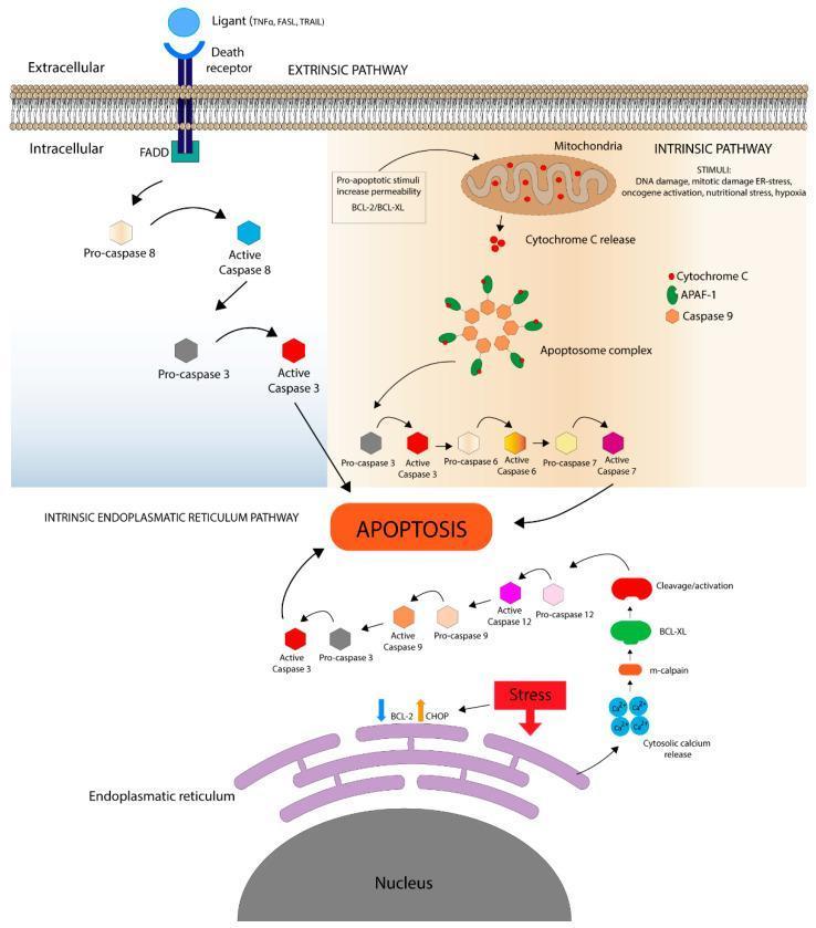
A comprehensive pathophysiology diagram illustrating the three primary signaling pathways of apoptosis: the extrinsic pathway, the intrinsic mitochondrial pathway, and the intrinsic endoplasmic reticulum (ER) pathway. The Extrinsic Pathway (top left) depicts extracellular ligands (TNF̑, FASL, TRAIL) binding to death receptors, recruiting the FADD adaptor protein to activate the Caspase 8 to Caspase 3 cascade. The Intrinsic Pathway (top right) shows cellular stressors (DNA damage, hypoxia, ER-stress) acting on the mitochondria to trigger Cytochrome C release. This facilitates the formation of the Apoptosome complex (Cytochrome C, APAF-1, and Caspase 9), which activates executioner caspases 3, 6, and 7. The Intrinsic ER Pathway (bottom) demonstrates ER stress leading to cytosolic calcium release, m-calpain activation, and a Caspase 12/9/3 cascade, alongside CHOP/BCL-2 regulation. All three pathways converge on the final stage of programmed cell death (Apoptosis), shown in central focus above the cell nucleus. The diagram uses standard biochemical nomenclature and color-coded symbols to represent pro-caspases versus active caspases, serving as a high-level educational resource for cell biology and molecular pathology.
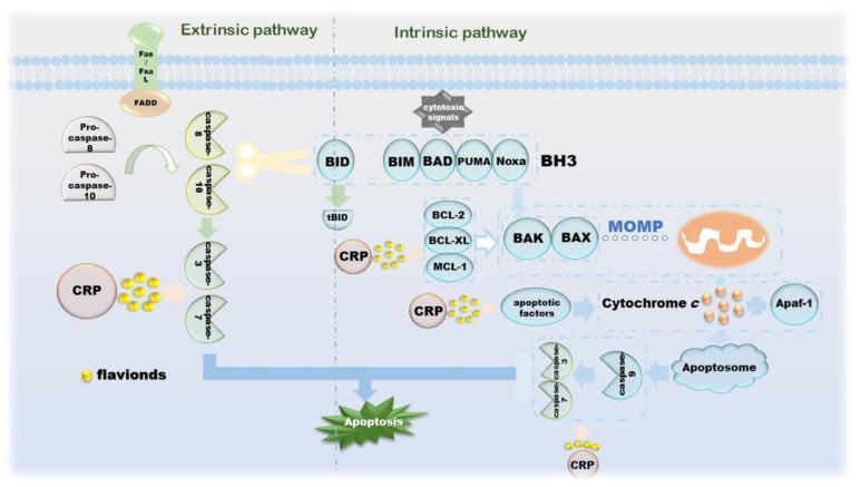
This pathophysiology diagram illustrates the extrinsic and intrinsic apoptotic signaling pathways and the modulatory role of Citri Reticulatae Pericarpium (CRP) and its flavonoids in cancer cell death. The extrinsic pathway (left) shows Fas/FasL binding to FADD, triggering the activation of Pro-caspase-8 and Pro-caspase-10 into active caspase-8 and caspase-10, which subsequently activate executioner caspases-3 and -7. The intrinsic pathway (right) is initiated by cytotoxic signals acting on BH3-only proteins (BID, BIM, BAD, PUMA, Noxa). These proteins modulate anti-apoptotic factors (BCL-2, BCL-XL, MCL-1) and pro-apoptotic BAK/BAX, leading to Mitochondrial Outer Membrane Permeabilization (MOMP). This results in Cytochrome c release, which binds to Apaf-1 to form the apoptosome, activating caspase-9 and executioner caspases. CRP and flavonoids are shown interacting at multiple nodes: promoting caspase activation in the extrinsic pathway, influencing tBID and mitochondrial factors in the intrinsic pathway, and ultimately enhancing the progression toward cellular apoptosis. The diagram serves as an educational summary of how natural compounds can target apoptotic dysregulation in oncology.
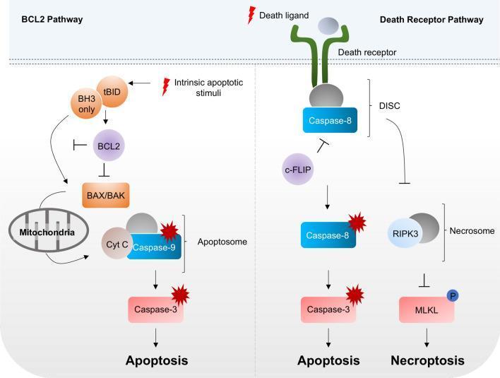
This pathophysiology diagram illustrates the signaling pathways of programmed cell death, specifically comparing BCL2-regulated apoptosis, death receptor-mediated apoptosis, and necroptosis. The left panel shows the intrinsic BCL2 pathway where intrinsic apoptotic stimuli activate BH3-only proteins and tBID, which inhibit BCL2. This relieves the inhibition on BAX/BAK, leading to mitochondrial outer membrane permeabilization, release of Cytochrome C (Cyt C), and formation of the apoptosome with Caspase-9, ultimately activating Caspase-3 to induce apoptosis. The right panel depicts the extrinsic Death Receptor Pathway. Binding of a death ligand to its receptor forms the Death-Inducing Signaling Complex (DISC), activating Caspase-8. This pathway bifurcates based on Caspase-8 activity: full activation leads to Caspase-3 cleavage and apoptosis, while c-FLIP inhibition of Caspase-8 can modulate the response. If Caspase-8 is lost or inhibited, the necrosome (comprising RIPK3) forms, leading to MLKL phosphorylation and execution of necroptosis. The schematic highlights key molecular targets for understanding cell death regulation in human biology and vascular diseases.
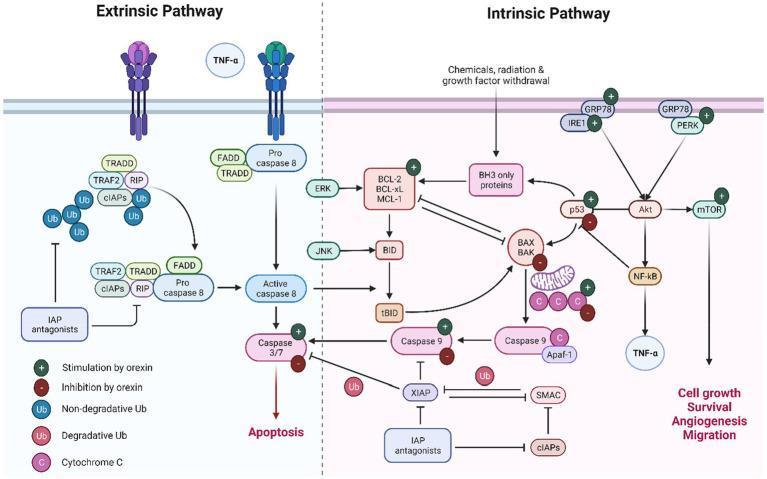
A comprehensive pathophysiology diagram illustrating the extrinsic and intrinsic pathways of apoptosis, with additional focus on the regulatory influence of orexin. The extrinsic pathway section details the binding of TNF-α to death receptors, leading to the recruitment of TRADD, FADD, and the formation of a signaling complex involving TRAF2, RIP, and cIAPs with non-degradative ubiquitin chains. This results in the activation of caspase-8 and subsequent execution by caspase-3/7. The intrinsic pathway section depicts triggers like radiation and growth factor withdrawal acting via BH3-only proteins to modulate the BCL-2 family (BCL-2, BCL-xL, MCL-1, BAX, and BAK). This triggers cytochrome C release from mitochondria, forming the apoptosome with Apaf-1 and caspase-9. The diagram uses green plus and red minus icons to indicate where orexin provides stimulation (e.g., GRP78, PERK, Caspase-9) or inhibition (e.g., p53, BAX, Caspase-3/7). Key signaling hubs like Akt, mTOR, and NF-κB are shown linking apoptosis to cell growth, survival, and migration.
keratinocyte apoptosis necrotic epidermis histology TUNEL
Apoptosis — Short Notes (MD Dermatology)
1. Definition
2. Apoptosis vs Necrosis (high-yield table)
| Feature | Apoptosis | Necrosis |
|---|---|---|
| Trigger | Physiologic or mild injury | Severe injury |
| Cell size | Shrinks | Swells |
| Membrane | Intact till late | Disrupted early |
| DNA | Fragmented (nucleosome-sized) | Random digestion |
| Inflammation | Absent | Present |
| Fate | Phagocytosed as apoptotic bodies | Lysed, contents leak out |
3. Morphology (H&E)
4. Two Core Pathways
- Triggered by DNA damage, ER stress, growth factor withdrawal, UV injury.
- BH3-only sensor proteins (BID, BIM, BAD, PUMA, Noxa) tilt the balance toward BAX/BAK oligomerization -> mitochondrial outer membrane permeabilization -> cytochrome c release.
- Cytochrome c + APAF-1 -> apoptosome -> activates caspase-9 -> executioner caspases 3, 6, 7.
- Regulated by Bcl-2 family: anti-apoptotic (Bcl-2, Bcl-XL, Mcl-1) vs pro-apoptotic (Bax, Bak, BH3-only).
- p53 is a key upstream activator (via Bax) after DNA damage - central to UV-induced keratinocyte apoptosis ("sunburn cells") - loss of p53 gives UV-damaged keratinocytes a survival advantage, permitting clonal expansion of mutant cells -> skin carcinogenesis (Fitzpatrick's Dermatology - TP53 section).
- Death receptors: Fas (CD95) and TNF receptor I, containing cytoplasmic "death domains."
- FasL (on activated cytotoxic T cells/NK cells) cross-links Fas -> adaptor FADD -> activates caspase-8 -> downstream caspase cascade.
- Also mediated by Granzyme B (released by CD8+ Tc cells), which directly activates caspase-3.
- Both pathways converge on executioner caspases -> nuclear/cytoplasmic protein degradation -> apoptotic morphology.
5. Clearance
6. Physiological Roles in Skin (exam relevant)
- Epidermal homeostasis (balances basal proliferation and desquamation).
- Catagen phase of hair cycle: apoptosis drives involution of the inferior hair follicle segment.
- Elimination of UV-damaged/self-reactive keratinocytes and lymphocytes.
- Note: terminal keratinocyte differentiation (cornification) is a distinct process from classic apoptosis, though it shares some features (caspase-14 mediated, not classic caspase-3/9 cascade) - Scott-Brown's Otorhinolaryngology.
7. Dermatology-Relevant Pathology (must-know for exam)
| Condition | Apoptosis-related mechanism |
|---|---|
| Sunburn / UV injury | p53-mediated intrinsic apoptosis of damaged keratinocytes ("sunburn cells") |
| SJS/TEN | Massive, rapid keratinocyte apoptosis in lesional skin (Fas-FasL and granzyme/perforin-mediated), overwhelming phagocytic clearance -> full-thickness epidermal necrosis (Dermatology, 2-Vol Set, p. 1989-92) |
| Lichen planus / lichenoid reactions | CD8+ Tc cell-mediated keratinocyte apoptosis (civatte/colloid bodies); driven by CD4-Th/Langerhans cell interaction |
| Graft-versus-host disease | Similar CD8+ Tc-mediated apoptotic keratinocyte injury |
| Mastocytosis | Failure of mast cell apoptosis leads to pathologic accumulation (demonstrated by TUNEL) |
| Autoimmune lymphoproliferative syndrome (ALPS) | Mutations in Fas (TNFRSF6)/FasL or caspase 8/10 -> defective lymphocyte apoptosis -> autoimmune cytopenias, lymphadenopathy |
| Cutaneous carcinogenesis (BCC/SCC/melanoma) | Apoptosis evasion via p53 loss, Bcl-2 overexpression -> survival of mutated keratinocytes |
| Photodynamic therapy | Therapeutically induces apoptosis in target (dysplastic/tumor) cells |
8. Colloid/Civatte bodies
Diagram for quick recall