
Facial nerve , from embryology to pathology Involving all the anatomy from scratch , syndromes associated , pathway ,everything .
Management of hypocalcemia after thyroidectomy , also describe what all are complications of intra and post op thyroidectomy And management
post-thyroidectomy hypocalcemia management complications 2024 guidelines
thyroidectomy hypocalcemia management
thyroidectomy complications diagram parathyroid recurrent laryngeal nerve
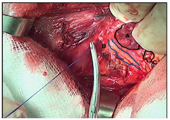
This clinical photograph displays an intraoperative view of a thyroidectomy during a recurrent laryngeal nerve (RLN) dissection, captured under 2.5x optical magnification. The surgical field highlights critical anatomical structures labeled as follows: (A) the reddish-brown, lobulated thyroid gland parenchyma; (B) the right-sided recurrent laryngeal nerve, identified as a slender, yellowish-white string-like structure outlined in blue; and (C) the inferior parathyroid gland, appearing as a small, beige-colored nodule in close proximity to the thyroid and nerve. Surgical instruments including a metallic hemostat and a blue suture thread are visible, facilitating tissue retraction and identification. White surgical gauze and metallic retractors define the periphery of the operative site. This image serves as an educational tool for endocrine surgery, illustrating the precise identification and preservation of the RLN and parathyroid glands to prevent postoperative complications such as vocal cord paralysis and hypocalcemia.
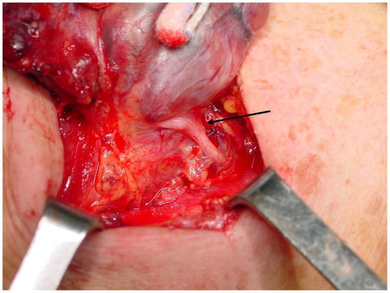
This clinical photograph captures an intraoperative view of a thyroidectomy, specifically illustrating the critical anatomical relationship between the recurrent laryngeal nerve (RLN) and the tubercle of Zuckerkandl. The surgical field is exposed using metal retractors, revealing the lateral lobe of the thyroid gland. A black arrow points to the recurrent laryngeal nerve, which appears as a distinct, thin, whitish cord-like structure. The nerve is seen coursing in close proximity to the tubercle of Zuckerkandl, a posterior projection of the thyroid lobe that serves as a vital surgical landmark. The surrounding field shows dissected adipose tissue, small local vessels, and minor surgical hemorrhage. This image demonstrates a common anatomical configuration where the nerve is positioned slightly inferior and deep to the tubercle, highlighting the necessity for careful dissection in this region to prevent iatrogenic nerve injury during thyroid or parathyroid surgery.
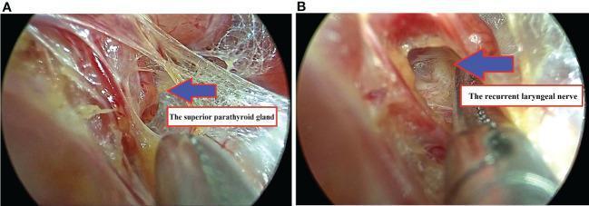
Dual-panel intraoperative endoscopic photographs demonstrating critical landmarks during transoral endoscopic thyroidectomy. Panel A provides a close-up view of the superior parathyroid gland, identified as a distinct, yellowish-tan ovoid structure nestled within perilaryngeal connective tissue and surgical dissection planes. Panel B illustrates the identification and exposure of the right recurrent laryngeal nerve (RLN), appearing as a slender, whitish-grey cord-like structure. A surgical instrument is visible in the foreground, highlighting the careful dissection required to preserve the nerve's integrity within the paratracheal groove. These images serve as educational references for endocrine surgery, emphasizing the importance of visual identification and meticulous preservation of the parathyroid glands and laryngeal nerves during thyroid or parathyroid procedures to avoid post-operative complications like hypocalcemia or vocal cord paralysis.
Thyroidectomy: Intraoperative & Postoperative Complications + Management of Post-Thyroidectomy Hypocalcemia
PART 1 - COMPLICATIONS OF THYROIDECTOMY
A. Intraoperative Complications
1. Recurrent Laryngeal Nerve (RLN) Injury
- Transection (sharp injury)
- Traction/stretch
- Thermal injury from energy devices
- Inadvertent ligation
- Unilateral injury: hoarseness, weak voice, aspiration risk
- Bilateral injury: stridor, aphonia, respiratory distress - may require immediate tracheostomy
2. Superior Laryngeal Nerve (SLN) - External Branch Injury
- Controls the cricothyroid muscle (voice pitch)
- Injury causes loss of high-pitched voice, early voice fatigue
- Often underdiagnosed
- Particularly relevant for singers or public speakers
3. Parathyroid Gland Devascularization / Inadvertent Removal
- All 4 parathyroid glands derive their blood supply from the inferior thyroid artery
- Dissection too far from the thyroid capsule risks devascularization
- Inferior parathyroids are more variable in location
- Prevention: Stay close to thyroid capsule; confirm parathyroid tissue with intraoperative PTH or biopsy; autotransplant into sternocleidomastoid muscle if viability is uncertain
4. Vascular Injury / Hemorrhage
- Injury to the inferior thyroid artery, superior thyroid artery, or their branches
- Rare but serious: injury to the carotid artery, internal jugular vein
- Management: Direct pressure, vascular control, call for vascular backup if needed
- Intraoperative hemorrhage can obscure the RLN and parathyroids
5. Tracheal / Esophageal Injury
- Rare, more common with large goiters, retrosternal extension, or reoperative surgery
- Tracheal injury: recognized by air leak - primary repair with absorbable sutures
- Esophageal injury: primary repair, wide drainage, and may need diversion
6. Thoracic Duct Injury (Left side)
- Can occur with left-sided central neck dissection
- Presents as chyle leak postoperatively
- Prevention: careful dissection in left paratracheal region
B. Postoperative Complications
1. Postoperative Hematoma / Hemorrhage (MOST URGENT)
- Occurs in ~1% of cases, typically within the first 4-6 hours
- Can cause airway compromise rapidly due to tracheal compression from expanding hematoma in the fixed neck compartment
- Signs: neck swelling, stridor, dyspnea, inability to swallow
- Management:
- Immediate bedside wound opening (if airway compromise)
- Return to operating room for exploration and hemostasis
- Every thyroid ward patient should have a suture removal kit at the bedside for emergency decompression
2. Hypocalcemia / Hypoparathyroidism (MOST COMMON)
- Most common complication of total thyroidectomy
- Transient: resolves within 6-12 months (~10% of cases)
- Permanent: persists beyond 6-12 months (~1-2% with experienced surgeons)
- Symptoms begin 24-72 hours postoperatively (when calcium levels nadir)
3. Recurrent Laryngeal Nerve Injury (Postoperative Recognition)
- Transient injury: neuropraxia from stretch/thermal effects; usually resolves within weeks to months
- Permanent injury: vocal cord paralysis
- All patients with voice change post-thyroidectomy must be referred to a voice specialist / ENT for laryngoscopy
- Management: voice therapy; for bilateral cord paralysis - arytenoidectomy or lateralization procedures; tracheostomy if acute
4. Seroma / Wound Complications
- Seroma: fluid collection in dead space; usually resolves spontaneously; drain if persistent
- Wound infection: uncommon (~0.3-0.5%); treat with antibiotics; open if abscess
- Keloid/hypertrophic scar: more common in predisposed individuals; manage with silicone sheets, steroid injection, or laser
5. Thyroid Storm
- Rare but life-threatening (mortality up to 25%)
- Triggered by: surgery on an inadequately prepared hyperthyroid patient, stress, acute illness
- Diagnosis: Burch-Wartofsky scoring system (temperature, CNS status, cardiovascular, GI/hepatic, precipitating factors)
- Management:
- PTU 200-250 mg every 4h (blocks synthesis and peripheral conversion) OR methimazole
- Potassium iodide 1h after antithyroid drugs (Wolff-Chaikoff effect)
- Beta-blockers: propranolol 60-80 mg every 4-6h (controls adrenergic symptoms)
- Hydrocortisone 300 mg IV loading, then 100 mg every 8h
- Cooling blankets, acetaminophen (avoid aspirin)
- Supportive ICU care
6. Hungry Bone Syndrome
- Specific to patients with pre-existing hyperthyroidism or hyperparathyroidism
- High bone turnover state: after surgery removes thyroid hormone excess, bones rapidly uptake calcium
- Severe, prolonged hypocalcemia despite normal PTH levels
- Electrolytes: low Ca, low phosphate, low Mg, high K
- Requires aggressive calcium and magnesium supplementation
7. Hypothyroidism
- Expected and intentional after total thyroidectomy
- Start levothyroxine at 1.5-1.7 mcg/kg/day (lower doses in elderly)
- TSH checked at 6-8 weeks, then every 1-2 months until stable, then annually
- For cancer: TSH suppression targets depend on risk stratification
8. Tracheomalacia
- Rare, associated with long-standing compressive goiters
- Weakened tracheal cartilage may collapse after goiter removal (loss of external support)
- Can cause acute airway obstruction at extubation
- Management: keep intubated, reintubate, consider tracheostomy or tracheal resection if severe
9. Chyle Leak
- Occurs with central or lateral neck dissection (left > right)
- Milky drainage, worsens with oral feeding
- Management: low-fat diet with medium-chain triglycerides; octreotide; if persistent >2 weeks, surgical re-exploration and ligation
10. Pneumothorax / Pneumomediastinum
- Rare, associated with retrosternal goiter, deep central neck dissection
- Recognized by chest pain, dyspnea, decreased breath sounds
- Management: chest tube if significant; observation if small
PART 2 - MANAGEMENT OF POST-THYROIDECTOMY HYPOCALCEMIA
Pathophysiology
- Inadvertently removed (confirmed on pathology)
- Devascularized (ischemic/necrotic)
- Stunned (transient dysfunction from manipulation)
- Decreased osteoclast-mediated bone calcium release
- Decreased renal calcium reabsorption
- Decreased 1-alpha-hydroxylase activity (less active vitamin D / calcitriol)
- Net result: hypocalcemia + hyperphosphatemia
Diagnosis and Monitoring
- PTH < 10 pg/mL at 1h post-op = high risk of hypocalcemia
- PTH-guided protocols reduce emergency readmissions and allow risk-stratified calcium supplementation (meta-analysis, PMID [38013484])
| Symptom | Notes |
|---|---|
| Perioral / acral paresthesias | Earliest |
| Chvostek sign | Tap facial nerve → ipsilateral twitch (also positive in 10% of normal people) |
| Trousseau sign | Inflate BP cuff above systolic x3 min → carpopedal spasm |
| Laryngospasm / bronchospasm | Medical emergency |
| Tetany | Ionized Ca < 4.3 mg/dL |
| Seizures | Advanced |
| Prolonged QT, arrhythmias | Cardiac |
| Papilledema | Raised ICP |
Management Protocol
Step 1 - Mild/Asymptomatic Hypocalcemia (Ca 7.5-8.5 mg/dL)
- Oral calcium carbonate: 1-2 g elemental calcium/day in divided doses (2-3 times daily)
- Take WITH food (requires gastric acid for absorption)
- Oral calcitriol: 0.25-0.5 mcg twice daily
- Bypasses the need for PTH-mediated renal 1-alpha-hydroxylation
- Reassess calcium in 24-48 hours
- Calcium citrate preferred in: patients on PPIs, prior gastric bypass, achlorhydria (does not require acid for absorption)
Step 2 - Moderate Symptomatic Hypocalcemia
- IV calcium gluconate: 10-20 mL of 10% solution (90-180 mg elemental Ca) over 10-20 minutes
- Follow with a continuous infusion: 60 mL of 10% calcium gluconate in 500 mL D5W at 1 mL/kg/hr
- Adjust every 4 hours based on serum calcium and symptoms
- Target: symptomatic relief and calcium 8.0-9.0 mg/dL
- Do NOT give calcium and bicarbonate through the same IV line (precipitates)
Step 3 - Severe/Life-Threatening (Tetany, Seizures, Laryngospasm)
- Calcium chloride 10 mL of 10% solution IV (faster-acting, does not need hepatic metabolism to release ionized calcium) - give via central line due to vein irritation risk
- Or calcium gluconate as emergency bolus (safer for peripheral veins)
- Infusion rate must not exceed 1.5 mEq/min to minimize cardiac arrhythmia risk
- Continuous cardiac monitoring
- If seizures persist: consider diphenhydantoin
Step 4 - Correct Concurrent Hypomagnesemia
- Hypomagnesemia impairs PTH secretion and causes PTH resistance at target organs
- Always check Mg in hypocalcemic patients
- If Mg < 1.8 mg/dL: IV magnesium chloride or magnesium sulfate
- Hypocalcemia will NOT correct if hypomagnesemia persists
Vitamin D Management
| Vitamin D Form | Indication |
|---|---|
| Calcitriol (1,25-OH2D) | First-line post-thyroidectomy (active form, bypasses PTH-dependent step) |
| Ergocalciferol (D2) | Long-term supplementation |
| Calcifediol (25-OH D) | Liver disease |
| Calcitriol preferred | Renal disease (impaired 1-alpha-hydroxylation) |
Long-term / Permanent Hypoparathyroidism
- Oral calcium carbonate: 2 g/day in divided doses + calcitriol 0.5-1.0 mcg/day
- Low-phosphate, low-oxalate diet
- Monitor for hypercalciuria (over-supplementation can cause renal calcium deposits)
- Target serum calcium: low-normal range (8.0-8.5 mg/dL) to avoid urinary calcium overload
- Recombinant human PTH (rhPTH 1-84 / Natpara): FDA-approved for chronic hypoparathyroidism; reduces calcium and calcitriol requirements; considered when conventional therapy is inadequate
- Parathyroid autograft: implant cryopreserved or fresh parathyroid tissue into sternocleidomastoid or forearm; functional recovery in ~50-60% of cases
PTH-Guided Protocol (Current Evidence)
- Low-risk patients (PTH > 15 pg/mL): oral calcium supplementation only; early discharge
- Intermediate risk (PTH 10-15): oral calcium + calcitriol; close monitoring
- High risk (PTH < 10): IV calcium as needed + oral calcium + calcitriol; delayed discharge
Quick Summary Table
| Complication | Timing | Management |
|---|---|---|
| Hematoma/airway compromise | 0-6h | Open wound, return to OR |
| RLN injury | Intraop / immediate | IONM, nerve visualization; rehab if persistent |
| Hypocalcemia (transient) | 24-72h | Oral Ca + calcitriol |
| Hypocalcemia (severe) | 24-72h | IV Ca gluconate infusion |
| Thyroid storm | Periop | PTU + KI + beta-blocker + steroids |
| Hungry bone syndrome | 24-72h | Aggressive Ca + Mg replacement |
| Hypothyroidism | Days-weeks | Levothyroxine replacement |
| Tracheomalacia | At extubation | Keep intubated, may need tracheostomy |
| Chyle leak | 2-5 days | Low-fat diet, octreotide, re-explore if >2 weeks |
| Seroma/infection | Days-weeks | Aspiration, antibiotics |