


Pic with circle is 27th June, another is today
plantar wart verruca plantaris sole foot
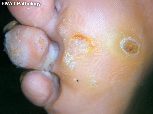
Clinical photography of a plantar foot displaying a verruca plantaris (plantar wart). Modality is a high‑quality gross image used in dermatology documentation. Precise location is the plantar surface of the foot (sole), with a focal, endophytic lesion that is rough and hyperkeratotic. Visual features include a circular to oval papule with a central crater or indent, irregular surface texture, and surrounding callus‑like thickening. Punctate black dots representing thrombosed capillaries may be seen within the lesion, while surrounding epidermis shows white scaling and fissuring. The lesion is typically mildly elevated above the surrounding skin and may be tender with pressure; it often coexists with other warts on the same sole. The clinical impression is verruca plantaris caused by human papillomavirus infection, with characteristic epidermal hyperplasia, papillomatosis, and hyperkeratosis evident on gross inspection. Differential diagnoses include callus/corn, plantar keratoderma, and foreign body reaction; histology would show koilocytosis and viral cytopathic effects if examined. This image supports clinical decision making, differential diagnosis, and treatment planning (topical salicylic acid, cryotherapy, or laser), and serves as an educational reference for students, residents, and clinicians evaluating plantar lesions. Clinical correlation includes assessing for additional lesions and patient history of HPV exposure; follow-up imaging is not required.
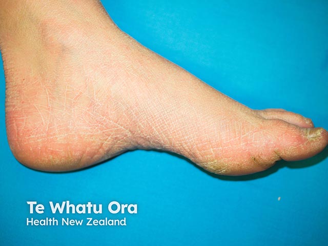
This is a macroscopic clinical photograph of the plantar aspect of the foot, captured for dermatology/foot-health assessment. Imaging modality: Clinical photography; technique: high-resolution color image of the sole, direct view, with standard diffuse lighting and no magnification. Anatomical location: Plantar surface of the hindfoot and midfoot, primarily the heel region, extending to the medial plantar arch. Visual features: a red-orange to pink-tan hyperkeratotic plaque with a thick, rough surface and fissuring; surrounding hypochromic to xerotic skin; margins are relatively ill-defined but the lesion remains clearly localized to the plantar skin. The lesion appears as a diffuse keratotic patch rather than a single discrete nodule, with variable thickness across the plantar surface. Pathology (clinically): evidence of epidermal hyperplasia with thickened stratum corneum (hyperkeratosis) and surface fissures, typical of friction-induced plantar keratopathy; no signs of vesiculation, purulence, or acute inflammation evident. Diagnostic significance: characteristic of friction-related plantar hyperkeratosis or callus; differential considerations include plantar corn, plantar wart (verruca plantaris), and diffuse plantar keratoderma; correlate with symptoms and history. Clinical correlation: evaluate footwear, gait, pressure points, activity level; screen for diabetes risk; may respond to keratolytics, debridement, padding, and orthotic optimization; useful for monitoring response to therapy and for educational and research material.
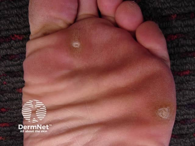
Clinical photography of the plantar surface of the foot demonstrating a classic hard corn (clavus) due to repetitive pressure. The lesion is a small, well‑circumscribed, hyperkeratotic papule with a central keratin plug, surrounded by peripheral epidermal thickening (callus) on the weight‑bearing sole. The surrounding skin is erythematous to slightly inflamed under pressure. The core appears pale yellow to white and slightly translucent; the lesion projects above the adjacent skin, producing a painful sensation with direct compression. This appearance distinguishes clavus from a plantar wart or simple callus by the presence of a dense keratinized core and a more pointed cone shape toward the dermis. The lesion is typically located at pressure points near the metatarsal heads or along the distal toe margins, often due to ill‑fitting footwear or abnormal gait. Etiology is chronic friction and pressure; pathophysiology involves localized epidermal hyperplasia and keratinocyte hypertrophy. Management focuses on debulking the hyperkeratosis (paring), application of keratolytics (salicylic acid), protective footwear, and orthotic correction; addressing footwear fit reduces recurrence risk. Differential diagnoses include plantar callus, plantar wart (verruca plantaris), and foreign body granuloma. Clinically correlates with pain on palpation and history of footwear‑related irritation. This image is educational for dermatology, podiatry, primary care, and medical education in skin pathology.
Comparison: June 27 vs. August 9 (Today)
What both images show
June 27 (circled image)
- A raised, slightly reddened/flushed circular area, roughly 2-3 cm in diameter
- Several tiny dark pinpoint dots scattered within the lesion - these are thrombosed (clotted) capillaries, a hallmark sign of a plantar wart
- Surrounding skin lines (dermatoglyphics) appear interrupted/distorted, which is characteristic of warts and distinguishes them from calluses
- The area appears somewhat inflamed or at an active/symptomatic stage
August 9 - Today
- The same area now shows a smaller, more discrete, well-circumscribed bump - roughly 5-7 mm
- It has a thickened outer border with a slightly depressed, darker central core
- The lesion looks more consolidated and keratotic (hardened) compared to the broader, flushed appearance in June
- The skin lines are still interrupted around it
- Less surrounding redness - the broader flush has reduced
What has changed over ~6 weeks
| Feature | June 27 | August 9 |
|---|---|---|
| Size/spread | Larger, broader reddened zone | Smaller, more focal/discrete |
| Appearance | Diffuse flush with multiple black dots | Compact, hardened dome with central core |
| Inflammation | More inflamed/red | Less surrounding inflammation |
| Keratosis | Less defined | More defined, thicker |
Most likely diagnosis: Plantar Wart (Verruca Plantaris)
- Interrupted skin lines - a key distinguishing sign from calluses/corns (which preserve skin lines)
- Thrombosed capillary dots (black pinpoints) visible in the June photo
- Location on the weight-bearing sole
- Circular, endophytic lesion with hyperkeratotic surface
What to do
- See a podiatrist or dermatologist for confirmation - a trained clinician can pare down the surface to look for the black dots beneath, which settles the diagnosis immediately
- Avoid cutting or picking at it yourself, as this spreads HPV to surrounding skin
- Over-the-counter salicylic acid (e.g., Bazuka gel, Wartner patches) can be applied nightly after soaking and filing - this is the standard first-line self-treatment for plantar warts
- If it persists, a doctor can offer cryotherapy (freezing), Candida antigen injection (now a first-line approach for stubborn warts - [Pfenninger & Fowler's Procedures, p. 4167]), or other in-office treatments
- Surgical excision on the sole is generally avoided as the resulting scar can itself be painful (like a pebble in a shoe)
Disclaimer: This is an AI-assisted visual assessment for informational purposes only - not a medical diagnosis. Please consult a healthcare professional for proper evaluation and treatment.
No when I take a bath in hot shower this fully turn white... And one small bup white arises may be right now the colour because I walk bare foot in my house no pain while sitting only pain when it gets pressed while walking
plantar wart white maceration after water soaking
Your Symptoms - What They Mean
| Symptom | Significance |
|---|---|
| Turns fully white in hot shower | Classic "maceration" sign - thickened keratin (dead skin) over a wart absorbs water and turns white. This happens more dramatically with warts than with normal skin because the keratin layer is abnormally thick. |
| Small white bump visible | The wart's core - the hyperkeratotic centre becomes very visible when wet |
| No pain at rest/sitting | Typical of plantar warts - they are endophytic (grow inward), so compression is needed to cause pain |
| Pain only when pressed/walking | Direct pressure on the wart's body is what causes the pain - exactly how plantar warts behave |
| No pain barefoot at rest | If it were a corn (heloma), you'd typically have more constant tenderness around it |
Diagnosis: Plantar Wart (Verruca Plantaris) - High Confidence
What You Should Do (Step by Step)
- Soak the foot in warm water for 10-15 minutes (you're already doing this in the shower - good)
- After soaking, while the skin is soft and white, gently file the top with a pumice stone or nail file - this removes dead white skin and lets treatment penetrate
- Apply salicylic acid gel/liquid (available at any pharmacy - brands like Bazuka, Compound W, or Dr. Scholl's) directly on the lesion, let it dry, and cover with a plaster
- Repeat every night - consistency is key; it usually takes 6-12 weeks
- Do not share the pumice stone/file with others (warts are contagious via HPV)
- Don't try to cut or dig it out - this spreads the virus into the surrounding skin
- Avoid walking barefoot in shared areas (swimming pools, gym floors) to prevent spreading it to others or picking up more strains
- The new satellite bump grows or more appear
- No improvement after 8-10 weeks of salicylic acid
- The pain worsens significantly
- Any doubt about the diagnosis
This is informational guidance only - not a clinical diagnosis. Please see a podiatrist or dermatologist for confirmation.
Suggest indian brand salicylic acid patchs and give instructions how to apply it and everything with mrp
salicylic acid wart removal patches India brand MRP price 2025 2026
Dr Scholl's Bazuka wart patch India pharmacy price plantar wart
salicylic acid wart corn remover patches available India pharmacy 1mg netmeds price rupees
https://www.1mg.com/otc/salactin-salicylic-lactic-acid-paint…
https://www.1mg.com/search/all?name=salicylic+acid+wart+patc…
Indian Products for Plantar Wart Treatment
Important note first
- Salicylic acid paints/solutions (most effective, most used in India)
- Corn/callus plasters with salicylic acid (work well for plantar warts too)
Best Indian Products (Easily Available at Chemist / Online)
Option 1 - BEST CHOICE
- Contains: Salicylic Acid 16.7% + Lactic Acid 16.7%
- Form: Paint/solution with applicator brush (10 ml bottle)
- MRP: ~₹495-512 for 10 ml (currently ~₹495 on 1mg)
- Available: 1mg, local chemists, Netmeds, PharmEasy
- Rating: 4.3/5 with 928 ratings - very popular in India
- This is the #1 recommended option for plantar warts in India
Option 2
- Contains: Salicylic Acid 16.7% + Lactic Acid 16.7%
- Form: Solution with applicator (15 ml)
- MRP: ~₹180-220
- Available: Most chemist shops, 1mg, Netmeds
Option 3
- Contains: Salicylic Acid 40% in a medicated plaster/pad
- Brands: Carnation, Dr. Foot, Scholl (imported, sometimes available)
- MRP: ~₹80-150 for a pack of 6-9 plasters
- Available: Big chemist shops, Apollo Pharmacy, MedPlus
- 40% SA in plaster form is stronger - good for a well-established plantar wart like yours
Option 4 (Prescription-strength, ask your chemist)
- Generic medicated plaster sold at compounding pharmacies
- Ask chemist for "Salicylic Acid 40% Plaster" or "Monoplus Plaster"
- MRP: ~₹50-100 per strip
- You cut it to the exact size of your wart
Step-by-Step Instructions (For Salactin Paint or Duofilm)
For Plaster/Patch Method (Corn Plasters 40% SA)
- Soak and file as above (Steps 1 & 2)
- Cut the plaster to the exact size of your wart (don't make it bigger - it will burn surrounding skin)
- Stick it directly on the wart
- Leave it on for 48 hours (change every 2 days)
- When you remove it, the skin will be white/soft - file it again
- Apply a fresh plaster
What to Expect
| Week | What you'll see |
|---|---|
| Week 1-2 | Wart turns more white/soft, skin peeling |
| Week 3-4 | Top layer starts coming off in layers |
| Week 6-8 | Wart shrinks, black dots disappear |
| Week 8-12 | Wart fully gone and normal skin replaces it |
Important DOs and DON'Ts
✅ DO protect surrounding healthy skin with petroleum jelly (Vaseline) before applying the paint
✅ DO be consistent - skipping days slows everything down
✅ DO wear footwear inside the house to reduce pressure on the wart while it heals
❌ DON'T use on diabetic feet without doctor's advice
❌ DON'T apply more than one layer thinking it works faster - it just burns the skin
❌ DON'T stop early even if it looks like it's gone - complete 2 weeks after it appears healed
If No Improvement in 8 Weeks
- Cryotherapy (liquid nitrogen) - ₹500-1500 per session, usually 2-4 sessions needed
- Cantharidin application (done in clinic only)
- Candida antigen injection - very effective, minimal scarring
This is informational guidance only. If you have diabetes or poor circulation, consult a doctor before using any salicylic acid product.