
Indications for oparative management in clavicle fracture
clavicle fracture operative fixation plate
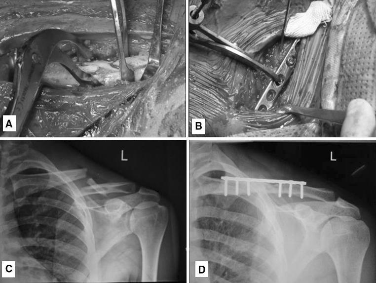
This composite educational image illustrates the surgical management of a midshaft clavicle fracture through plate osteosynthesis. Panels A and B are intra-operative clinical photographs. Panel A shows the fracture reduction phase, where surgical instruments (reduction clamps and retractors) are used to align the cortical bone fragments. Panel B demonstrates the fixation phase, showing a 3.5 mm dynamic compression plate (DCP) being contoured and secured to the superior aspect of the clavicle. Panels C and D are anteroposterior (AP) radiographs of the left shoulder. Radiograph C displays the preoperative displaced midshaft clavicle fracture with clear discontinuity and superior displacement of the medial fragment. Radiograph D is a 6-month post-operative follow-up showing successful fracture union and consolidation. The internal fixation hardware, consisting of a superiorly placed plate and six cortical screws, remains in situ with restored anatomical alignment. This sequence highlights the transition from acute injury to clinical recovery via open reduction and internal fixation (ORIF).
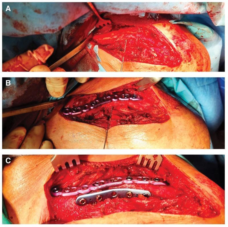
This sequence of three clinical intraoperative photographs (A, B, C) documents the progression of an open reduction and internal fixation (ORIF) of a midshaft clavicle fracture using a dual-plating technique.
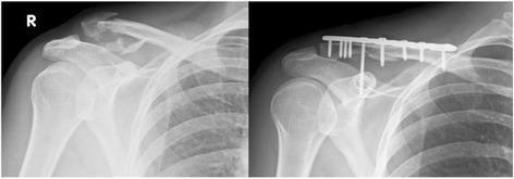
Two anteroposterior (AP) radiographs of the right shoulder demonstrate a comparison between a pre-operative and post-operative state of a lateral clavicle fracture. The pre-operative image (left) shows a displaced, fragmented fracture of the distal (lateral) third of the clavicle, consistent with a Neer Type IIB classification, which indicates coracoclavicular ligament involvement and inherent instability. The post-operative image (right) displays the results of open reduction and internal fixation (ORIF). Visible hardware includes a locking compression plate (LCP) positioned on the superior aspect of the clavicle, secured by multiple cortical and locking screws. A long coracoclavicular (CC) screw is notably inserted through the plate, traversing from the clavicle into the coracoid process of the scapula to provide additional stability and address ligamentous disruption. The radiographs highlight key orthopedic concepts in shoulder trauma management, specifically the stabilization of unstable distal clavicle fractures to prevent non-union and restore joint mechanics.
Indications for Operative Management of Clavicle Fractures
Absolute Indications
- Open fracture - exposed bone with breach of skin
- Neurovascular injury requiring repair - vascular injury (e.g., subclavian artery) or progressive neurologic deficit involving the brachial plexus
- Skin tenting - where the fracture fragment threatens to perforate the skin (risk of converting to open fracture)
Relative Indications
Fracture-Specific Factors
| Factor | Threshold |
|---|---|
| Displacement | > 2 cm |
| Shortening | > 2 cm |
| Comminution | Increasing / segmental |
| Segmental fracture | Inherent instability |
| Scapular malposition | Winging on initial examination |
Associated Injuries
- Ipsilateral upper extremity fractures (floating shoulder)
- Multiple ipsilateral upper rib fractures (destabilize the shoulder girdle)
- "Floating shoulder" - combined clavicle + glenoid neck fracture
- Ipsilateral upper extremity injuries that require early arm use
Patient Factors
- Polytrauma requiring early upper extremity weight-bearing
- High functional demand (overhead workers, athletes)
- Elite sports or self-employed professionals requiring rapid return to function
- Multiple risk factors for nonunion converging (female sex + displacement + comminution + age)
Special Situations
Timing of Surgery
Key Clinical Note
- Rockwood and Green's Fractures in Adults, 10th ed. (2025), Chapter 34 - Clavicle Fractures, pp. 1208-1209
- Miller's Review of Orthopaedics, 9th ed. - Clavicle Fracture section
- Rosen's Emergency Medicine - Clavicle chapter, p. 647