
draw a schematic diagram of erythrocyte membrane showing the Protein Structure (ankyrin , band 3 )
Detailed scientific schematic diagram of erythrocyte (red blood cell) membrane protein structure. Show a cross-section of the lipid bilayer membrane with the following labeled components clearly: Integral membrane proteins: - Band 3 protein (anion exchanger, large transmembrane protein spanning the bilayer multiple times, shown as a large barrel/cylinder crossing the membrane, labeled "Band 3 (Anion Exchanger)") - Glycophorin A (single-pass transmembrane protein with carbohydrate chains on extracellular side) Peripheral/cytoskeletal proteins on the cytoplasmic (inner) side: - Ankyrin (shown connecting Band 3 to spectrin, labeled clearly) - Spectrin (alpha and beta heterodimer, shown as long flexible filaments forming a meshwork beneath the membrane) - Actin (short filaments at junctional complexes) - Protein 4.1 (at junctional complex connecting spectrin to glycophorin) - Protein 4.2 (near ankyrin-Band 3 complex) - Adducin (at actin junctions) Lipid bilayer: - Phospholipid bilayer clearly shown with hydrophilic heads (circles) and hydrophobic tails (lines) - Outer leaflet labeled "Extracellular space" with carbohydrate (glycocalyx) chains on glycoproteins - Inner leaflet labeled "Cytoplasm / Intracellular space" - Cholesterol molecules interspersed in the bilayer Connections clearly shown: - Ankyrin bridges Band 3 (transmembrane) to Beta-spectrin (cytoskeletal) - Protein 4.1 links spectrin-actin junction to glycophorin A - The spectrin network forms a 2D lattice under the membrane Use a clean white background, bright distinct colors for each protein type, bold clear labels with arrows pointing to each structure, anatomical diagram style, educational medical illustration quality.
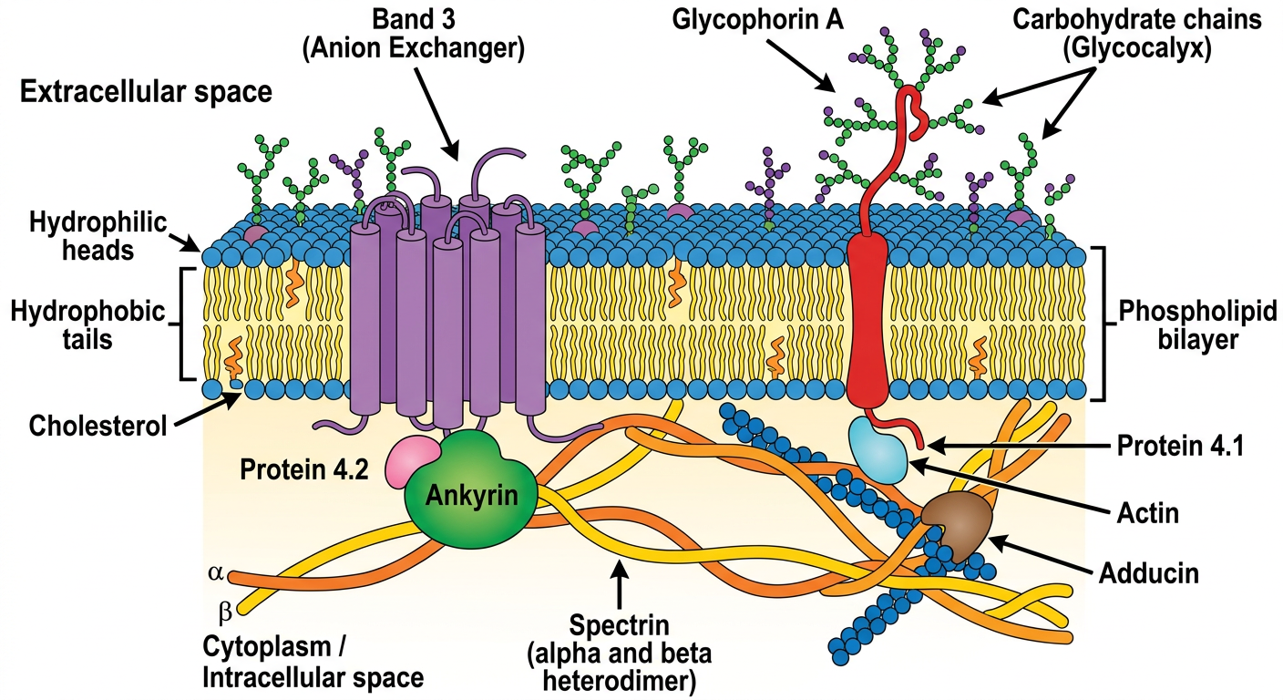
- Band 3 (Anion Exchanger 1 / AE1) - the most abundant RBC membrane protein; a multipass transmembrane protein that exchanges Cl⁻/HCO₃⁻ and serves as the main anchoring point for the cytoskeleton
- Glycophorin A - single-pass transmembrane protein carrying sialic acid residues (contributes to the negative surface charge of RBCs)
- Ankyrin - the critical linker protein that attaches Band 3 on the membrane to Beta-spectrin below; loss of ankyrin causes hereditary spherocytosis
- Spectrin (α/β heterodimers) - forms the flexible meshwork scaffold just beneath the membrane; provides RBC deformability
- Protein 4.1 - links the spectrin-actin junctional complex to Glycophorin A
- Protein 4.2 - stabilizes the ankyrin-Band 3 interaction
- Actin - short protofilaments that form the junctional nodes of the spectrin lattice
- Adducin - caps actin at the junctional complexes
- Defects in spectrin or ankyrin → Hereditary Spherocytosis
- Defects in Band 3 → Hereditary Stomatocytosis or Southeast Asian Ovalocytosis
- The Band 3 / Ankyrin / Spectrin vertical connection is the primary anchor holding the lipid bilayer to the cytoskeleton