
Stretch and inverse strech in physiology
Reading File
Searching sources...
Finding Sources
Finding Sources
Now let me read the key sections from Ganong's and Guyton:
Reading File
Reading File
Now let me read the inverse stretch reflex section from Ganong's and also fetch relevant images:
Reading File
Reading File
Now let me read the Golgi tendon organ / inverse stretch reflex sections and fetch the key diagram:
Reading File
Reading File
Loading Image
I now have all the information needed to give a thorough answer. Let me also fetch the Golgi tendon organ diagram:
Loading Image
Here is a complete, well-sourced explanation of both reflexes.
Stretch Reflex and Inverse Stretch Reflex
1. Stretch Reflex (Myotatic Reflex)
Definition
When a skeletal muscle with an intact nerve supply is stretched, it contracts. This is the stretch reflex (also called the myotatic reflex). The stimulus is stretch of the muscle; the response is contraction of that same muscle. - Ganong's Review of Medical Physiology, 26th Edition
Receptor - The Muscle Spindle
The sense organ is the muscle spindle, a small fusiform structure within the fleshy belly of the muscle. Each spindle contains:
- Intrafusal fibers (nuclear bag + nuclear chain) - in parallel with the regular extrafusal fibers
- Type Ia (primary) afferents - large, fast myelinated fibers wrapping the nuclear bag region; respond to both the rate of change in length (dynamic) and steady-state length (static)
- Type II (secondary) afferents - from nuclear chain fibers; mainly a static response
- γ-motor neuron (fusimotor) supply - controls the sensitivity of the spindle
Neural Pathway (Monosynaptic)
- Muscle stretched → spindle nuclear bag region deformed
- Ia fiber fires → enters spinal cord via dorsal root
- Directly synapses (glutamate) on alpha motor neurons in the ventral horn of the same segment
- Alpha motor neuron fires → extrafusal muscle contracts, opposing the stretch
This is the only truly monosynaptic reflex in the body. - Guyton & Hall Textbook of Medical Physiology
Dynamic vs. Static Components
| Component | Stimulus | Mediator | Effect |
|---|---|---|---|
| Dynamic (phasic) | Sudden rapid stretch | Ia fibers (primary endings) | Instantaneous strong contraction; over in fraction of a second |
| Static (tonic) | Sustained stretch | Ia + II fibers (primary + secondary) | Prolonged, weaker contraction maintaining muscle length |
The dynamic response opposes sudden changes in muscle length. The static response keeps contraction intensity constant over time, resisting slow positional drift. - Guyton & Hall
Associated Features
Reciprocal innervation: Ia fibers also send a collateral to an inhibitory interneuron (Ia inhibitory interneuron) that synapses on the antagonist muscle's motor neurons. This causes simultaneous relaxation of the antagonist - the agonist contracts while the antagonist yields.
Damping/smoothing function: The spindle apparatus smooths out jerk-like, intermittent signals from the CNS into smooth, graded contraction. If spindle sensory nerves are cut, contraction becomes oscillatory and unsmooth.
γ-motor neuron coactivation: When the brain sends signals to contract a muscle, α and γ motor neurons are coactivated. γ activation shortens the intrafusal fiber poles, keeping the central nuclear bag taut even as the extrafusal fiber shortens - this prevents "unloading" of the spindle and maintains its sensitivity throughout the contraction.
Clinical Example: Knee Jerk (DTR)
Tapping the patellar tendon instantaneously stretches the quadriceps → activates Ia fibers → monosynaptic reflex → quadriceps contracts (knee jerks forward). Graded 0-5+. Absent DTRs suggest LMN lesion (peripheral neuropathy); exaggerated DTRs suggest UMN lesion with loss of descending inhibition. - Ganong's
2. Inverse Stretch Reflex (Clasp-Knife Reflex / Golgi Tendon Reflex)
Definition
Up to a point, the harder a muscle is stretched, the stronger the reflex contraction. However, when tension becomes great enough, contraction suddenly ceases and the muscle relaxes. This relaxation in response to strong stretch is the inverse stretch reflex. - Ganong's Review of Medical Physiology, 26th Edition
It is "inverse" because it is opposite to the stretch reflex - strong stretch causes relaxation rather than contraction.
Receptor - The Golgi Tendon Organ (GTO)
Located in the tendon, in series with the muscle fibers (unlike spindles, which are in parallel). Each GTO consists of:
- A netlike collection of knobby nerve endings among tendon fascicles
- About 3-25 (Ganong's) to 10-15 (Guyton's) muscle fibers per organ
- Innervated by Ib myelinated fibers (large, rapidly conducting, ~16 µm diameter)
Because GTOs are in series, they are activated by both passive stretch and active contraction of the muscle. This is the key difference from the muscle spindle - the GTO responds to tension (force), not just length.
Neural Pathway (Disynaptic - Inhibitory)
- Excessive muscle tension → GTO deformed → Ib fiber fires
- Ib fiber enters spinal cord → synapses on an inhibitory interneuron (releases glycine)
- Inhibitory interneuron hyperpolarizes the alpha motor neurons of the same muscle (IPSP)
- With strong enough stretch, IPSPs are so great that motor neurons stop discharging → muscle relaxes suddenly
The Ib fibers also make excitatory connections with motor neurons supplying the antagonist muscles - the opposite of what the stretch reflex does. - Ganong's, Guyton's
The Golgi tendon reflex is disynaptic (Ib fiber → inhibitory interneuron → motor neuron).

Why "Clasp-Knife"?
In spasticity, passively flexing an extremity meets increasing resistance (stretch reflex in the extensor), then at a critical point, resistance suddenly gives way like closing a penknife - the moment of give is the inverse stretch reflex cutting in.
Functions of the GTO Reflex
- Protective - prevents muscle tearing or tendon avulsion when tension is excessive ("lengthening reaction" in Guyton's terminology)
- Force regulation - provides negative feedback to maintain appropriate tension
- Load equalization - fibers under excess tension are inhibited; under-loaded fibers are disinhibited, spreading load evenly across the muscle
- Fine motor control - GTOs act as transducers to signal force to the cerebellum via spinocerebellar tracts, contributing to smooth coordination
3. Comparison at a Glance
| Feature | Stretch Reflex | Inverse Stretch Reflex |
|---|---|---|
| Also called | Myotatic reflex | Golgi tendon / clasp-knife reflex |
| Receptor | Muscle spindle (intrafusal fibers) | Golgi tendon organ |
| Location | In parallel with extrafusal fibers | In series with muscle fibers |
| Afferent fiber | Ia (type II also for static) | Ib |
| Synapse type | Monosynaptic | Disynaptic (via inhibitory interneuron) |
| Neurotransmitter | Glutamate (excitatory) | Glycine (inhibitory, via interneuron) |
| Response | Muscle contraction | Muscle relaxation |
| Stimulus detected | Muscle length / rate of change | Muscle tension (stretch + contraction) |
| Effect on antagonist | Inhibits (reciprocal innervation) | Excites |
| Purpose | Resist unexpected length changes; maintain posture | Protect against excessive tension; regulate force |
4. Combined Diagram (Ganong's Figure 12-3)
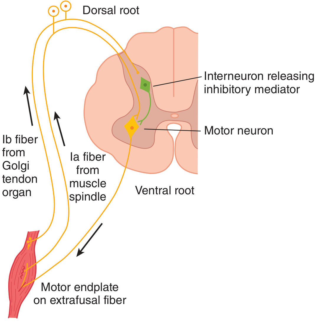
Ia fibers from the spindle directly excite the motor neuron (stretch reflex). Ib fibers from the GTO excite an inhibitory interneuron that hyperpolarizes the same motor neuron (inverse stretch reflex). With strong stretch, the IPSP dominates and the motor neuron goes silent. - Ganong's Review of Medical Physiology, 26th Edition
5. Clinical Correlations
- Hyper-reflexia / clonus: Exaggerated stretch reflex due to loss of descending inhibition (UMN lesion). Clonus is a rhythmic oscillation of the stretch reflex-inverse stretch reflex sequence, where the muscle repeatedly contracts and relaxes when under sustained stretch. Sustained clonus (≥5 beats) is always abnormal.
- Hyporeflexia / areflexia: LMN lesion, peripheral neuropathy, or disruption anywhere in the reflex arc.
- Clasp-knife phenomenon: A hallmark of spasticity - initial resistance from the stretch reflex, then sudden give from the inverse stretch reflex.
- Physiologic tremor (~10 Hz): Partly a result of the spindle feedback loop; the velocity sensitivity of Ia fibers normally damps this. Tremor amplifies in anxiety, fatigue, or drug toxicity.
Sources:
- Ganong's Review of Medical Physiology, 26th Edition, pp. 238-241 (stretch reflex, inverse stretch reflex, Golgi tendon organ, clonus)
- Guyton & Hall Textbook of Medical Physiology, pp. 679-682 (myotatic reflex, dynamic/static stretch reflexes, Golgi tendon organ, lengthening reaction)
Dorsal ganglia
Searching sources...
Finding Sources
Finding Sources
Reading File
Reading File
Reading File
Reading File
Loading Image
Loading Image
Finding Sources
I now have comprehensive information from multiple sources. Here is the full answer:
Dorsal Root Ganglia (DRG)
Definition and Location
The dorsal root ganglion (DRG), also called the spinal ganglion, is a cluster of sensory neuron cell bodies located on the dorsal root of each spinal nerve, just outside and adjacent to the spinal cord. There is one DRG at each spinal level on each side, giving 31 pairs in humans (8 cervical, 12 thoracic, 5 lumbar, 5 sacral, 1 coccygeal).
DRG neurons originate from the neural crest during embryological development. - Eric Kandel, Principles of Neural Science, 6th Edition
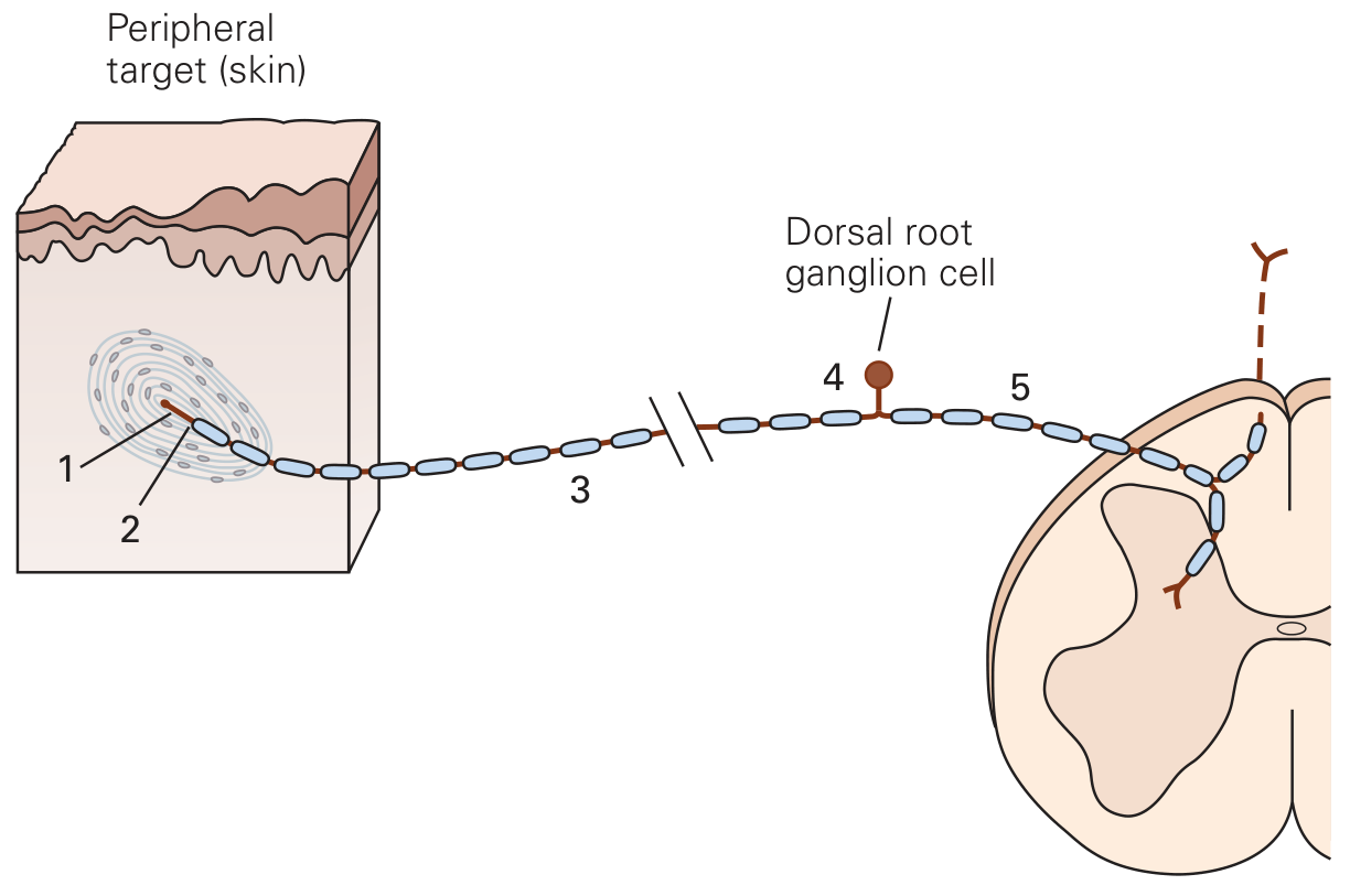
Cell Type: Pseudounipolar Neurons
DRG neurons are pseudounipolar (also called unipolar). Developmentally they begin as bipolar neurons, but the axon and dendrite migrate together and fuse into a single process that exits the cell body, then divides in a T-shape into two branches:
- Peripheral branch - travels to the periphery (skin, muscle, joints, viscera) where its terminal contains specialized sensory receptors
- Central branch - travels into the spinal cord (or brainstem for cranial nerve equivalents) to synapse on second-order neurons
The cell body itself sits "off to the side" of this through-line - it monitors the signal but does not participate in transmission synaptically. There are no synapses within the DRG itself - this distinguishes it sharply from autonomic ganglia, which do contain synaptic connections. - Histology: A Text and Atlas, Wheater's
Five Functional Zones of a DRG Neuron
Each DRG neuron has five distinct functional zones:
| Zone | Location | Function |
|---|---|---|
| 1. Receptive zone | Distal peripheral terminal | Contains specialized receptor channels (mechano-, thermo-, chemoreceptors) that convert stimulus energy into a receptor potential |
| 2. Spike generation site | Initial axon segment / first node of Ranvier | Voltage-gated Na⁺/K⁺ channels convert receptor potential into action potentials |
| 3. Peripheral nerve fiber | Between receptor and cell body | Conducts action potentials to the DRG cell body |
| 4. DRG cell body (soma) | In the ganglion | Contains nucleus; site of protein synthesis; expresses sensory receptor proteins |
| 5. Central branch | Spinal/cranial nerve into cord | Connects DRG neuron to ipsilateral spinal cord or brainstem; forms first synapse in somatosensory pathways |
- Kandel, Principles of Neural Science, 6th Edition
Histology of the DRG
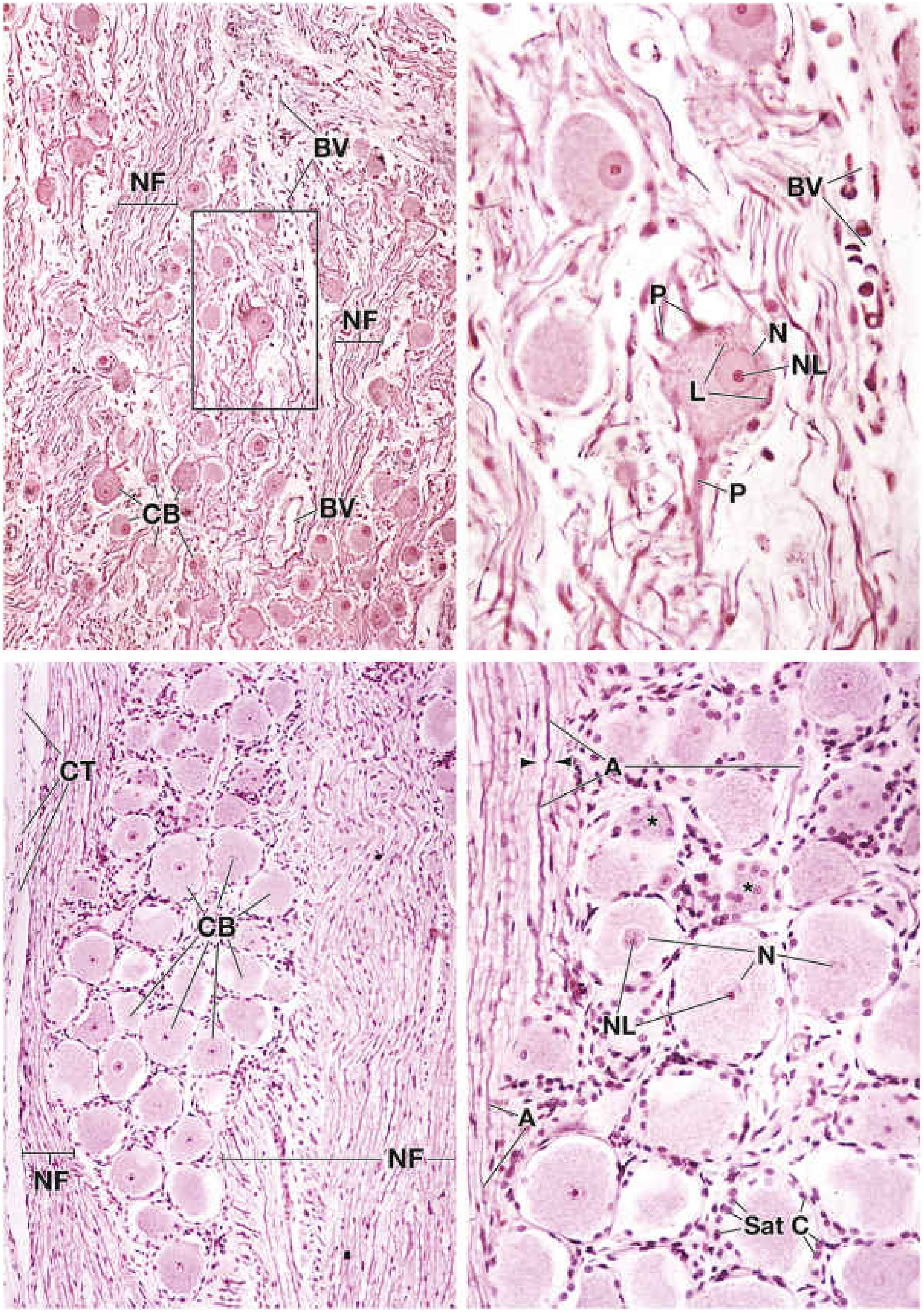
On H&E staining, the DRG shows:
-
Large neuronal cell bodies (CB) arranged in closely packed clusters, with large pale-staining spherical nuclei (N), prominent nucleoli (NL), and Nissl bodies (rough ER/ribosomes)
-
Satellite cells (Sat C) - small flattened glial cells that completely surround each neuron cell body (analogous to Schwann cells in the peripheral nerve). They are continuous with the Schwann cell investment of the axon. They regulate the ionic microenvironment of the neuron
-
Bundles of nerve fibers (NF) passing between cell clusters - the axons and their myelin sheaths, with centrally placed axons (A) surrounded by the space where myelin was dissolved in processing (arrowheads = neurilemma)
-
Connective tissue (CT) capsule at the periphery
-
Blood vessels (BV) throughout
-
Lipofuscin (L) - yellow-brown age pigment visible in older neurons
-
Histology: A Text and Atlas with Correlated Cell and Molecular Biology
Diversity of DRG Neurons
DRG neurons are heterogeneous - they differ in:
| Property | Large DRG neurons | Small DRG neurons |
|---|---|---|
| Cell body size | Large | Small |
| Axon | Large myelinated (Aα, Aβ) | Thinly myelinated (Aδ) or unmyelinated (C) |
| Conduction velocity | Fast | Slow |
| Function | Touch, proprioception, pressure | Temperature, pain, itch, chemical irritants |
| Molecular markers | Npy2r, Parvalbumin (PV) | CGRP, IB4 lectin |
The large DRG neurons transmit non-noxious sensations; the small ones transmit pain and temperature (nociceptive/thermoreceptive). - Kandel, Principles of Neural Science
Central Projections - Body Map
The central axons of DRG neurons are topographically organized. They enter the spinal cord via the dorsal root and terminate in a pattern that maps the body surface - adjacent body regions are represented in adjacent spinal segments. This forms the basis of dermatomes. Fibers carrying touch/pressure synapse in the dorsal column nuclei (gracile and cuneate nuclei); pain/temperature fibers synapse in the dorsal horn (Rexed laminae I, II, and V). - Kandel, Principles of Neural Science
DRG vs. Autonomic (Sympathetic) Ganglia - Key Differences
| Feature | Dorsal Root Ganglion | Autonomic Ganglion |
|---|---|---|
| Neuron type | Pseudounipolar (sensory) | Multipolar (motor/autonomic) |
| Synapses in ganglion | None | Present (pre→post ganglionic) |
| Function | Somatic and visceral sensation | Autonomic efferent output |
| Location | Dorsal root, just outside cord | Paravertebral chain or prevertebral plexus |
| Glial support | Satellite cells | Satellite cells |
- Histology: A Text and Atlas
Clinical Relevance
Dorsal root ganglionopathies (sensory neuronopathies) present as pure sensory syndromes because the DRG is the first-order sensory neuron. Causes include:
| Onset | Causes |
|---|---|
| Acute | Guillain-Barré (AIDP), varicella zoster (herpes zoster), Lyme disease |
| Subacute/Chronic | Paraneoplastic (anti-Hu antibodies, associated with lung/ovarian cancer - causes inflammation and destruction of DRG neurons); Sjögren's syndrome; vitamin B12 deficiency; cisplatin/taxol toxicity; diabetes |
The paraneoplastic form is notable: inflammation of the DRG (dorsal root ganglionitis) produces a sensory ataxia + areflexia with large fiber loss. Because it targets the cell body rather than the axon, it produces a non-length-dependent sensory loss (the whole dermatome is affected, not just the distal limbs).
Herpes zoster (shingles): Varicella zoster virus lies latent in DRG neurons and reactivates to cause pain and vesicular eruption in a dermatomal distribution - the pain results from direct viral inflammation and destruction of DRG neurons.
DRG stimulation is used therapeutically as a form of neuromodulation for complex regional pain syndrome (CRPS) and chronic lower limb pain, as an alternative to conventional spinal cord stimulation. - Kaplan & Sadock's Comprehensive Textbook of Psychiatry; Barash Clinical Anesthesia
Sources:
- Kandel, Principles of Neural Science, 6th Edition, pp. 456-458
- Histology: A Text and Atlas with Correlated Cell and Molecular Biology, pp. 1027-1029
- Kaplan & Sadock's Comprehensive Textbook of Psychiatry, p. 2009
- Barash, Cullen & Stoelting's Clinical Anesthesia, 9th Edition
This is a shared conversation. Sign in to Orris to start your own chat.