
Abscess
What aspect of abscess are you interested in?
abscess pathology pus formation histology
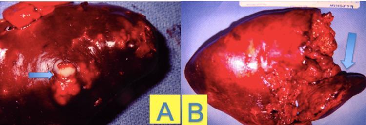
Gross pathology specimens of a human spleen demonstrating focal and diffuse features of a splenic abscess. Panel A displays a side-view of the dark-red, inflamed splenic capsule with a localized area of purulent exudate (pus formation), indicated by a blue arrow. The pus appears as a pale, off-white, viscous material extruding from a disrupted focal point on the diaphragmatic surface. Panel B shows the splenic parenchyma in a late stage of inflammation, characterized by a predominantly dark red and irregular surface. A blue arrow in Panel B points to a region of severe tissue disruption, consistent with necrosis and parenchymal liquefaction, where the normal splenic architecture is lost and replaced by amorphous, friable tissue. This comparison illustrates the progression from localized abscess formation to widespread inflammatory necrosis within the organ, relevant for surgical pathology and infectious disease education.
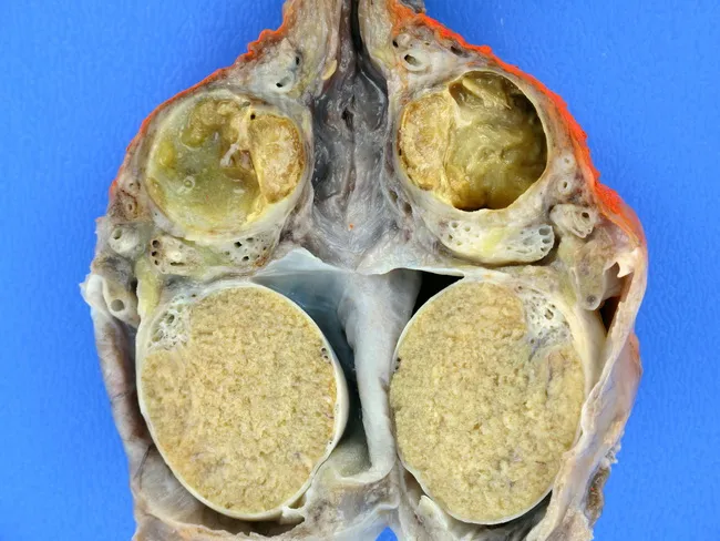
Gross pathology photograph of the scrotal contents after dissection shows pronounced inflammatory changes centered in the epididymis with secondary abscess formation. The epididymal walls are thickened and friable, separating a pale yellow to tan pus-filled cavity from surrounding tissue. The abscess cavities appear irregular and are lined by inflammatory granulation tissue. The adjacent testicular parenchyma is largely unremarkable or only minimally involved. In this case, numerous foamy histiocytes populate the walls of the abscess, consistent with xanthogranulomatous epididymo-orchitis. The foamy macrophages are lipid-laden and may be accompanied by multinucleated giant cells, lymphocytes, and plasma cells; cholesterol clefts and necrotic debris may be present in the inflammatory loci. The overall pattern is chronic inflammation with focal granulomatous response. Clinically, this entity can simulate an infiltrating neoplasm and may present with scrotal swelling, pain, or mass. Pathologic significance lies in recognizing xanthogranulomatous inflammation as a distinct chronic infectious/inflammatory process rather than malignancy; microbiologic correlation is helpful; treatment typically involves antibiotic therapy and surgical drainage or epididymectomy, depending on extent. Differential diagnoses include tuberculous epididymo-orchitis, malakoplakia, chronic bacterial epididymitis, and less commonly granulomatous vasculitis. This specimen provides educational utility for gross-pathology and diagnostic pathology teaching, illustrating how histiocyte-rich, foamy macrophage inflammation can shape scrotal disease.
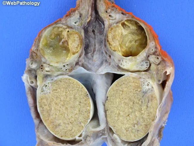
Comprehensive Description: This is a gross pathology photograph of a male scrotal specimen showing xanthogranulomatous epididymo-orchitis with abscess formation. The inflammatory process is predominantly centered in the epididymis with secondary involvement of adjacent testicular tissue. The abscess walls are thickened and lined by numerous foamy histiocytes, lipid-laden macrophages, and chronic inflammatory cells. On cut sections, the epididymal ducts and surrounding stroma display pale, granulomatous nodules intermixed with necrotic debris and sparse lymphocytes. The overall appearance is a granulomatous, macrophage-rich inflammatory reaction consistent with xanthogranulomatous inflammation, which can mimic neoplasm clinically. In this case, bilateral scrotal contents are involved, though the most conspicuous changes reside in the epididymal tissue; testicular parenchyma is relatively preserved. The image illustrates the histologically distinctive foamy macrophage infiltrate within the abscess walls and surrounding granulomatous response. Clinically, this entity represents a chronic infectious or inflammatory process that may require surgical drainage or excision and antibiotic therapy. Differential considerations include tuberculous epididymo-orchitis, malakoplakia, other granulomatous infections, and rarely epididymal/neoplastic processes. This image is educational for medical students, pathologists, urologists, and radiologists to recognize xanthogranulomatous epididymo-orchitis and its abscess morphology. The accompanying histology demonstrates macrophage-rich inflammatory tissue with lipid-laden cytoplasm and occasional multinucleated giant cells for educational reference.
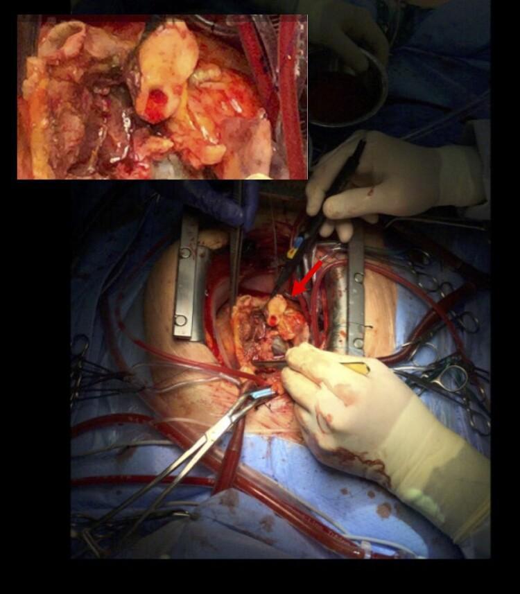
This clinical photograph captures an intraoperative view of an open cardiac surgical site during a complex cardiothoracic procedure. The image depicts a sternotomy with the heart exposed and a surgical field maintained by a metal rib retractor. An inset in the upper-left corner provides a magnified view of the primary pathology: a significant aortic root abscess. This abscess is characterized by localized, yellowish-white purulent material and necrotic tissue extending from the aortic valve area toward the left main truncus. A red arrow indicates the specific involvement of the circumflex artery, which is encased in a sheath of pus. The surgical environment includes essential equipment such as cardiopulmonary bypass cannulae (red tubing carrying blood), an electrocautery device held by a surgeon, and various surgical clamps and forceps. The educational focus is on the visual manifestation of infective endocarditis complications, specifically the formation of perivalvular abscesses and the surgical anatomy of the aortic root.
types of abscess skin subcutaneous brain liver lung diagram
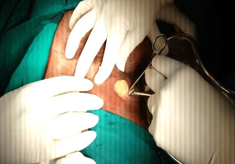
This clinical photograph captures a surgical incision and drainage (I&D) procedure of a subcutaneous abscess. The image focuses on a small, transverse incision made in the right hypochondriac region of the abdomen. Thick, opaque, yellowish purulent material (pus) is visible emerging from the opening, indicating an active infectious process. Several hands clad in sterile white surgical gloves are positioned around the site; one set of hands applies pressure to the surrounding skin to facilitate drainage, while another hand holds a surgical instrument, likely a hemostat or forceps, to keep the incision edges open. The surgical field is prepared with green sterile drapes, and the surrounding skin appears erythematous and taut. This visual demonstrates the management of a liver abscess with parietal extension, illustrating key steps in bedside or operating room drainage of localized collections. It serves as an educational example for general surgery and infectious disease specialties, highlighting the physical characteristics of purulent exudate and basic surgical technique.
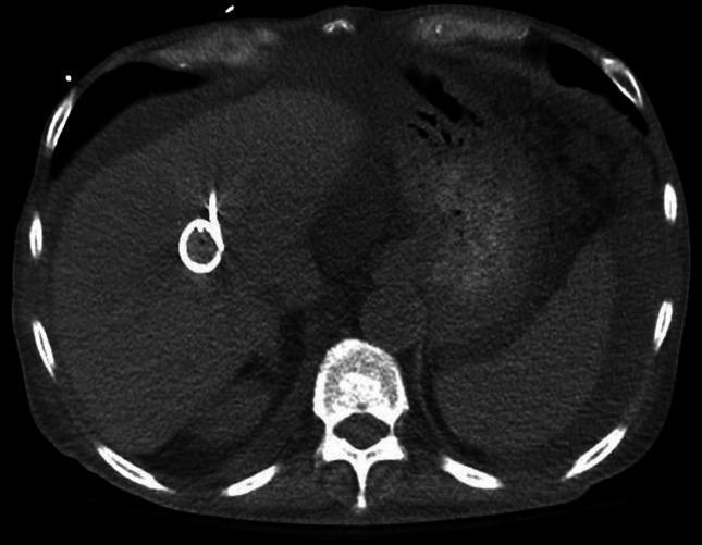
This diagnostic image is an axial computed tomography (CT) scan of the upper abdomen, specifically focused on the liver. The scan reveals a large, ill-defined, hypodense lesion within the right lobe of the liver, consistent with a hepatic abscess. Centered within this fluid collection is a highly radiopaque, pigtail-shaped structure, identifying a percutaneous drainage catheter (drain) placed for therapeutic intervention. The surrounding liver parenchyma appears otherwise unremarkable in density. Posteriorly, the vertebral body and neural arch are visible as hyperdense (bright white) bony structures. The rib cage is partially visualized at the periphery of the abdominal cavity. Subcutaneous fat is seen as a low-density (dark gray) layer beneath the skin. This image serves as a clinical example of radiographic monitoring for image-guided drainage of complex intra-abdominal infections, such as those caused by vancomycin-resistant Enterococcus (VRE) or other multidrug-resistant organisms.
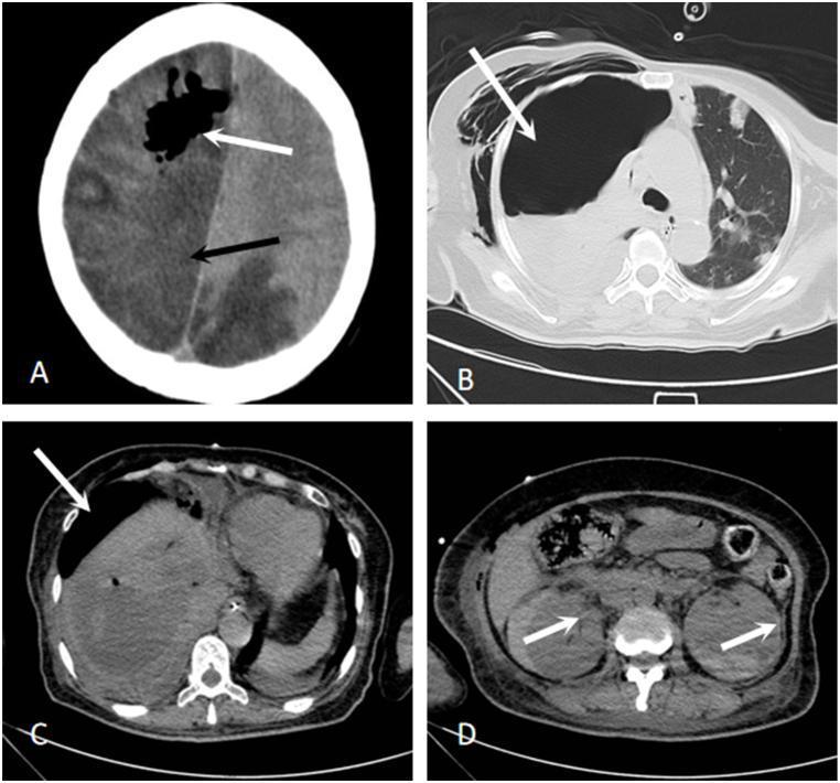
A series of four axial CT images (A–D) demonstrating multi-organ complications in a clinical case. Image A (Brain CT): Shows extensive regions of low density across the right hemisphere and left posterior lobes, consistent with massive infarction. Intraparenchymal gas pockets (white arrow) and a significant midline shift to the left indicate pneumocephalus and cerebral herniation. Image B (Chest CT): Displays a large right-sided pneumothorax (white arrow) with underlying lung atelectasis and consolidation, accompanied by subcutaneous emphysema in the soft tissues of the chest wall. Image C (Abdominal CT): Reveals a large hepatic lesion with an air-fluid level and a visible drainage tube (white arrow), characteristic of a liver abscess under treatment. Image D (Abdominal CT): Shows the bilateral kidneys with internal areas of altered density and scattered gas (white arrows), suggesting emphysematous or hemorrhagic foci. This sequence illustrates a severe systemic infectious process, such as invasive Klebsiella pneumoniae syndrome, leading to metastatic infections and emphysematous changes in multiple organ systems.
Abscess - General Overview
Definition
Classical Clinical Features
| Sign | Latin | Manifestation |
|---|---|---|
| Heat | Calor | Warm overlying skin |
| Redness | Rubor | Erythema |
| Pain | Dolor | Tenderness, throbbing |
| Swelling | Tumor | Fluctuant mass |
Composition of Pus
- Dead and dying neutrophils (predominant) - killed by bacterial toxins
- Tissue debris and liquefied necrotic material
- Living and dead bacteria
- Proteinaceous fluid (inflammatory exudate)
- Cellular breakdown products (DNA, lipids, enzymes)
Pathophysiology
- A puncture wound, surgical incision, bacteraemia, or contiguous spread introduces pyogenic organisms into tissue.
- Bacteria (commonly Staphylococcus aureus) release toxins and enzymes (e.g., coagulase, hyaluronidase, leukocidins).
- Neutrophils are recruited en masse and attempt to phagocytose bacteria.
- Both bacteria and neutrophils die, releasing proteolytic enzymes that liquefy the tissue.
- Central liquefactive necrosis creates the pus-filled cavity.
- Surrounding tissue shows an acute inflammatory exudate with fibrin, oedema, and inflammatory cells.
- A fibrinous wall (pyogenic membrane) forms around the cavity, attempting to contain the infection.
- Granulation tissue (macrophages, fibroblasts, new blood vessels) develops around this.
- This leads to collagen deposition - the fibrous capsule.
- As pressure increases, the abscess tracks along planes of least resistance, typically toward the skin surface ("points").
- It may burst spontaneously or require drainage.
- If incompletely treated, a chronic abscess or antiboma (partially sterilized by antibiotics) may form.
Microbiology
- Staphylococcus aureus (most common, including MRSA)
- Streptococcus pyogenes
- Mixed flora - gram-negative enteric organisms (E. coli, Klebsiella) and anaerobes (Bacteroides fragilis)
- Mycobacterium tuberculosis (cold abscess - no heat, no redness)
- Actinomyces israelii (actinomycotic abscess with sulfur granules)
- Mixed aerobic/anaerobic oral flora (Streptococcus, Fusobacterium, Prevotella)
Types of Abscess
By Location
| Type | Common Site | Key Features |
|---|---|---|
| Cutaneous / Subcutaneous | Skin and soft tissue | Furuncle (single follicle), carbuncle (multiple), hidradenitis |
| Peritonsillar | Around the tonsil | Muffled voice, trismus, uvular deviation |
| Dental / Periapical | Root of a tooth | Tooth pain, facial swelling, fistula to skin |
| Intraabdominal | Post-op, perforated viscus | Deep-seated fever, ileus |
| Perianal / Perirectal | Around the anus | Anorectal pain, may lead to fistula-in-ano |
| Liver (hepatic) | Liver parenchyma | Pyogenic (E. coli) or amoebic (E. histolytica) |
| Lung | Pulmonary parenchyma | Cavity with air-fluid level on CXR, foul sputum |
| Brain | Cerebral parenchyma | Headache, focal deficits, raised ICP |
| Spinal epidural | Epidural space | Back pain, neurological deficit |
| Bartholin's | Bartholin gland | Vulvar swelling, dyspareunia |
| Pilonidal | Natal cleft | Young men, midline pit, hair nidus |
| Psoas | Psoas muscle | Fever, hip/flank pain, TB or Crohn's |
| Breast | Breast tissue | Lactational (S. aureus) or non-lactational |
By Chronicity
| Type | Features |
|---|---|
| Acute abscess | Classic hot, red, tender, fluctuant; short history |
| Chronic abscess | Minimal heat/redness; lymphocytes and plasma cells; tissue sequestration; may calcify; associated with TB or actinomycosis |
| Cold abscess | No inflammatory signs (no calor/rubor); typical of TB; tracks along fascial planes |
| Antiboma | Partly sterilised by antibiotics; sterile pus within fibrous capsule; may still need drainage |
Natural History and Complications
- Resolve - spontaneously rupture and drain
- Spread - track along fascial planes to involve adjacent structures
- Become chronic - fibrous walling off with chronicity
- Form a sinus - a blind-ended track from the abscess to an epithelial surface
- Form a fistula - an abnormal communication between two epithelial surfaces
- Cause septicaemia/bacteraemia - haematogenous spread leading to metastatic abscesses
- Calcify - dystrophic calcification in old, sterile abscess cavities
On Imaging
Key Principles of Management
- Incision and Drainage (I&D) - the definitive treatment. "Ubi pus, ibi evacua" (Where there is pus, evacuate it).
- Antibiotics alone are insufficient - they cannot penetrate the avascular pus-filled cavity adequately.
- Healing by secondary intention - the cavity is left open to drain; granulation fills from the base.
- Antibiotics are indicated when: the cavity is surgically closed after drainage, there is surrounding cellulitis, the patient is immunocompromised, or there is systemic sepsis.
- MRSA coverage (trimethoprim-sulfamethoxazole or clindamycin) should be considered for community-acquired skin abscesses in high-prevalence settings.