
Cerebellum cerebrum and spinal cord histology viva theory
Reading File
This is a broad visual/histology topic. I'll run parallel searches for all three structures.
Finding Sources
Finding Sources
Finding Sources
Finding Sources
Finding Sources
Finding Sources
Good. Now let me search specifically for histology content using paragraph search, and also read the most relevant histology sections.
Finding Sources
Finding Sources
Finding Sources
Excellent results. Now let me read the key sections in parallel - the Histology textbook for cerebellum, Guyton for cerebral cortex, and the spinal cord anatomy sections.
Reading File
Reading File
Reading File
Now let me read the spinal cord and cerebral cortex layer details, plus fetch key histology images.
Reading File
Reading File
Loading Image
Excellent content gathered. Now let me fetch the cerebral cortex layers diagram and spinal cord cross-section image.
Loading Image
Loading Image
Loading Image
I have rich, high-quality content from all three structures. Now I'll compose the comprehensive viva/theory answer.
Histology of the Cerebellum, Cerebrum, and Spinal Cord
I. CEREBELLUM
Gross Overview
The cerebellum lies in the posterior fossa, dorsal to the pons and medulla. Its cortex is folded into narrow leaf-like folds called folia (analogous to gyri of the cerebrum). Beneath the cortex is white matter and three pairs of deep cerebellar nuclei (dentate, emboliform/globose, fastigial).
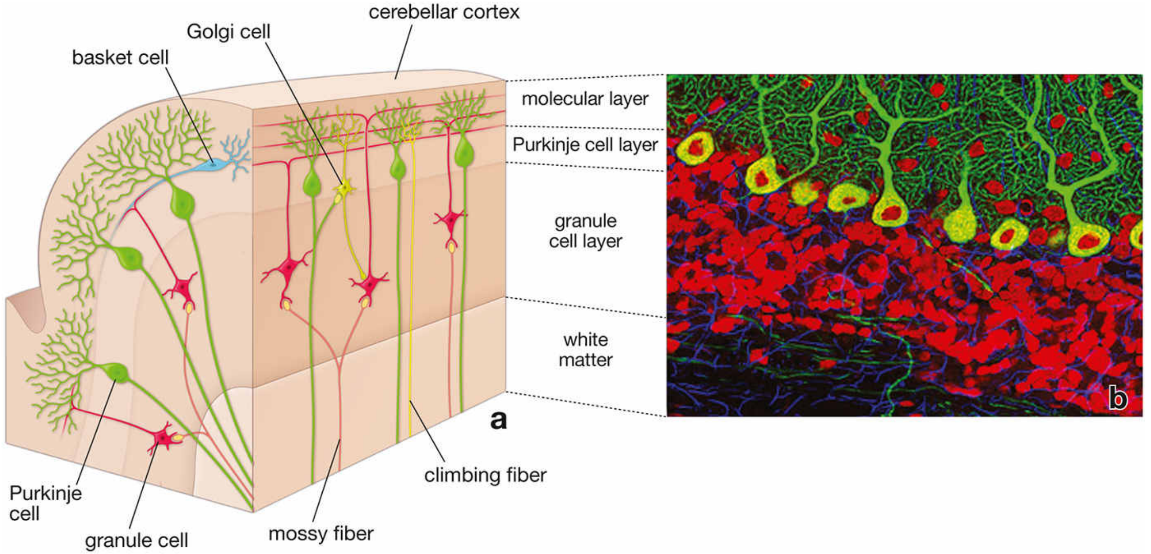
Histological Layers of the Cerebellar Cortex
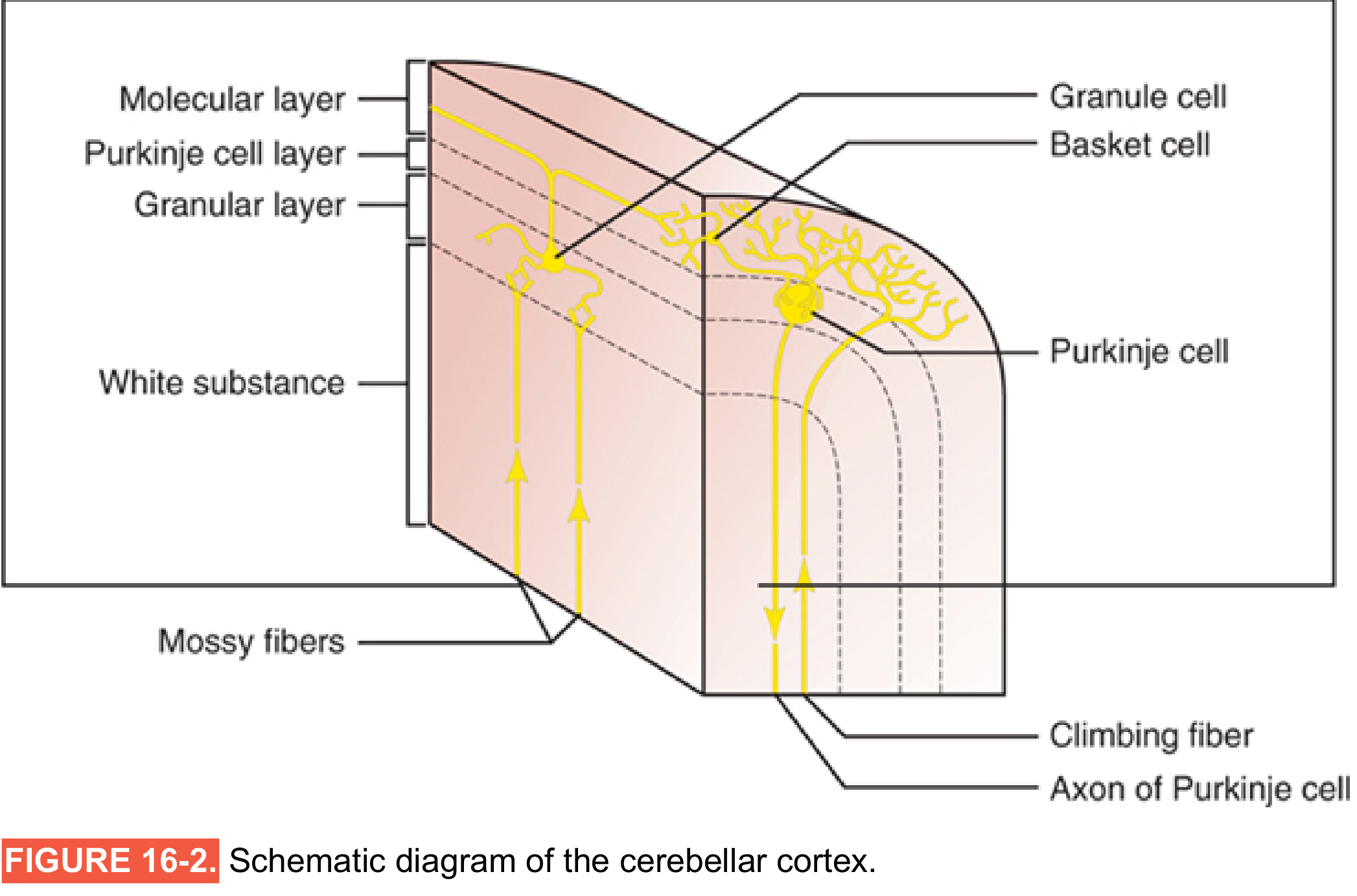
The cerebellar cortex has 3 layers (outer to inner):
| Layer | Position | Cell Types | Key Features |
|---|---|---|---|
| 1. Molecular layer | Outermost | Outer stellate cells, inner basket cells | Cell-sparse; rich in unmyelinated axons and dendrites; receives parallel fibers (axons of granule cells) |
| 2. Purkinje cell layer | Middle | Purkinje cells (single row) | Only output neurons of cerebellar cortex; large flask-shaped cells; rich dendritic arborization in molecular layer; axons project to deep cerebellar nuclei |
| 3. Granular cell layer | Innermost (adjacent to white matter) | Granule cells, Golgi cells | Most densely packed neurons in the CNS; granule cell axons ascend and bifurcate as parallel fibers in molecular layer |
Five Cell Types Summary
| Cell | Layer | Neurotransmitter | Role |
|---|---|---|---|
| Stellate cell | Molecular | GABA (inhibitory) | Inhibits Purkinje cell dendrites |
| Basket cell | Molecular | GABA (inhibitory) | Forms "basket" around Purkinje cell somata; inhibits Purkinje cells |
| Purkinje cell | Purkinje | GABA (inhibitory) | Sole output of cerebellar cortex; inhibits deep nuclei |
| Granule cell | Granular | Glutamate (excitatory) | Most numerous CNS neuron; sends parallel fibers to excite Purkinje cells |
| Golgi cell | Granular | GABA (inhibitory) | Inhibits granule cells (feedback inhibition) |
Afferent Fiber Systems
- Climbing fibers: From inferior olivary nucleus; each fiber synapses with ONE Purkinje cell (powerful, one-to-one)
- Mossy fibers: All other afferents (spinocerebellar, pontocerebellar, etc.); divergent - one fiber excites thousands of Purkinje cells via granule cells
Special Histological Feature - Negri Bodies
In rabies virus infection, eosinophilic intracytoplasmic inclusions called Negri bodies (2-10 µm) are classically found in Purkinje cells of the cerebellar cortex and pyramidal cells of the hippocampus. They represent viral replication compartments.
II. CEREBRUM (Cerebral Cortex)
General Structure
The cerebral cortex is a thin layer (2-5 mm thick) covering all convolutions of the cerebrum. Total area ~0.25 m². Contains >80 billion neurons.
Three Main Neuron Types
- Granular (stellate) cells - short axons, function as interneurons; excitatory (glutamate) or inhibitory (GABA); predominate in sensory cortex
- Pyramidal cells - named for their shape; large, give rise to long output fibers including the corticospinal tract; project to spinal cord, basal ganglia, brainstem
- Fusiform (polymorphic) cells - spindle-shaped; mostly in deeper layers; project mainly to thalamus
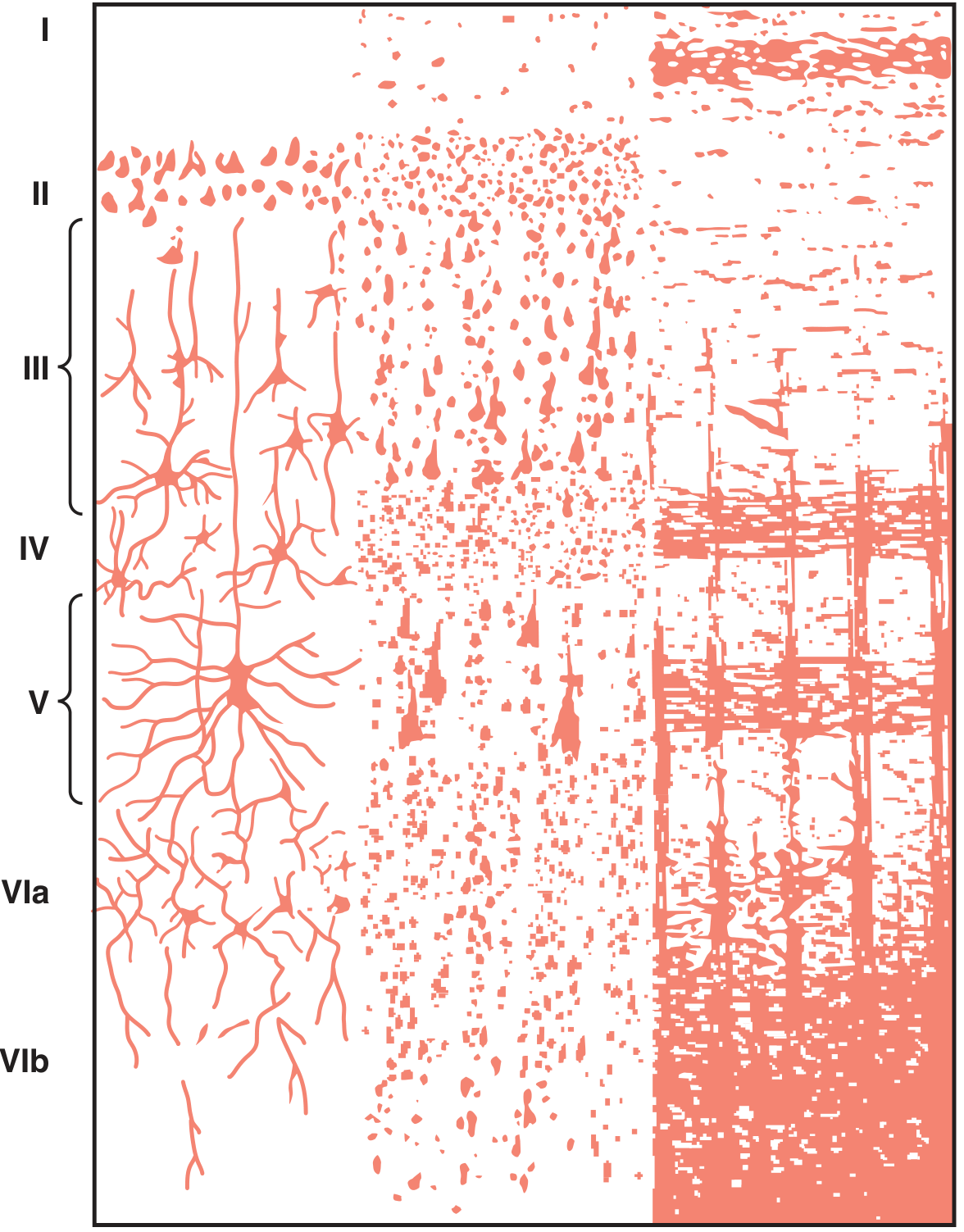
Six Layers of the Neocortex (I - VI, surface to deep)
| Layer | Name | Main Cell Type | Key Connections/Function |
|---|---|---|---|
| I | Molecular (plexiform) layer | Few neurons; mostly dendrites and axons | Receives diffuse nonspecific input from brainstem; controls general cortical excitability |
| II | External granular layer | Small pyramidal + stellate cells | Commissural fibers; sends axons via corpus callosum to contralateral cortex |
| III | External pyramidal layer | Medium pyramidal cells | Cortico-cortical and commissural connections; sends axons to opposite hemisphere via corpus callosum |
| IV | Internal granular layer | Granule (stellate) cells | PRIMARY SENSORY INPUT LAYER - receives thalamic (specific sensory) afferents; well-developed in primary sensory cortex (koniocortex); absent in motor cortex |
| V | Internal pyramidal (ganglionic) layer | Large pyramidal cells (Betz cells in motor cortex) | Output to spinal cord, brainstem, basal ganglia; gives rise to corticospinal tract |
| VI | Multiform (fusiform) layer | Fusiform/polymorphic cells | Projects to thalamus (corticothalamic fibers); modulates thalamic gating |
Special Cortical Types
- Koniocortex (granular cortex): Layer IV very thick; granule cells dominant; seen in primary sensory areas (S1, V1, A1) - very little layer V
- Agranular cortex: Layer IV absent/rudimentary; pyramidal cells dominant; seen in primary motor cortex (M1)
- Heterotypic cortex: One layer is disproportionately prominent
- Homotypic cortex: All 6 layers roughly equal; seen in association areas
Columnar Organization
Neurons are organized into vertical columns (~0.3-0.5 mm diameter, ~10,000 neurons/column), each responding to one specific sensory modality or serving one motor function. This is a fundamental principle of cortical organization.
III. SPINAL CORD
Gross Cross-Sectional Structure
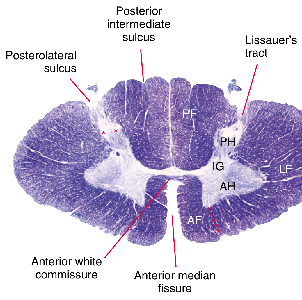
A cross-section reveals:
- Inner H-shaped (butterfly-shaped) gray matter - neuronal cell bodies
- Outer white matter - myelinated axons organized into funiculi (columns)
Gray Matter Horns
| Horn | Contains | Function |
|---|---|---|
| Anterior (ventral) horn | Cell bodies of lower motor neurons (alpha and gamma motor neurons) | Motor output to skeletal muscle |
| Posterior (dorsal) horn | Cell bodies receiving sensory input from periphery | Sensory relay; contains substantia gelatinosa (SG) |
| Lateral horn | Preganglionic sympathetic neurons | Present only from T1 to L2 (sympathetic); parasympathetic at S2-S4 |
White Matter Funiculi (Columns)
| Column | Contents | Modality |
|---|---|---|
| Posterior funiculus | Dorsal columns (fasciculus gracilis + cuneatus) | Fine touch, vibration, conscious proprioception |
| Anterior funiculus | Anterior corticospinal tract, tectospinal, vestibulospinal | Mainly motor |
| Lateral funiculus | Lateral corticospinal tract, spinothalamic tract, spinocerebellar tracts | Mixed sensory + motor |
Rexed's Laminae
The gray matter is further divided into 10 laminae (I-X) based on cytoarchitecture:
| Lamina | Location | Key Note |
|---|---|---|
| I | Marginal zone (dorsal tip of posterior horn) | Pain and temperature relay |
| II | Substantia gelatinosa | Pain modulation; rich in enkephalin interneurons |
| III-IV | Nucleus proprius | Touch, pressure relay |
| V-VI | Neck/base of posterior horn | Convergence of cutaneous + proprioceptive input |
| VII | Intermediate gray | Clarke's column (C8-L3) - spinocerebellar relay; lateral horn (T1-L2) |
| VIII | Ventral horn (commissural) | Motor interneurons |
| IX | Motor columns (anterior horn) | Alpha and gamma motor neurons |
| X | Around central canal | Commissural neurons |
Key Histological Features of Spinal Cord
- Anterior horn cells (motor neurons): Large multipolar neurons; rich in Nissl substance (rough ER); have prominent nucleolus - these are the cells destroyed in poliomyelitis and motor neuron disease (ALS)
- Substantia gelatinosa (lamina II): Small cells, gelatinous appearance; site of action of opioid analgesics (gate control theory)
- Clarke's column (nucleus dorsalis, lamina VII): Found C8-L3; relay station for unconscious proprioception to cerebellum via spinocerebellar tract
- Lissauer's tract: Thin pale zone at the tip of the dorsal horn; carries pain and temperature fibers a few segments up/down before synapsing
Quick Viva Summary Table
| Feature | Cerebellum | Cerebrum | Spinal Cord |
|---|---|---|---|
| Number of cortical layers | 3 | 6 | N/A (gray/white matter + laminae) |
| Key output neuron | Purkinje cell (GABA, inhibitory) | Large pyramidal cell (Layer V) | Alpha motor neuron (Anterior horn) |
| Special cell marker | Purkinje cell (Negri bodies in rabies) | Betz cells (layer V of motor cortex) | Anterior horn cells (polio target) |
| Primary input layer | Granular layer (mossy/climbing fibers) | Layer IV (thalamic input) | Dorsal horn / posterior funiculus |
| Key neurotransmitter | GABA (Purkinje, basket, stellate, Golgi) | Glutamate (pyramidal) + GABA (interneurons) | Glutamate, Glycine, GABA, Substance P |
Sources: Localization in Clinical Neurology 8e, p.961-962 | Histology: A Text and Atlas (Ross), p.996-998 | Guyton & Hall Medical Physiology, pp.600, 716 | Gray's Anatomy for Students, p.1326
This is a shared conversation. Sign in to Orris to start your own chat.