
I want you to give me a clear cut explaination about the embryological development of CVS incuding devt of portal vein , IVC ,arch of aorta with beautiful diagrams
Reading File
Now let me search the medical library for cardiovascular embryology content in parallel.
Finding Sources
Finding Sources
Finding Sources
Finding Sources
Finding Sources
Finding Sources
Excellent - Langman's Medical Embryology has the CVS content. Let me read those sections now.
Reading File
Reading File
Great finds. Now let me read the core CVS embryology content from Langman's and the venous development from THIEME in parallel.
Reading File
Reading File
Excellent - I now have very rich content from both Langman's and THIEME. Let me fetch the key diagrams while reading more Langman's content on IVC and heart tube formation.
Reading File
Loading Image
Loading Image
Beautiful diagrams retrieved. Now let me get a few more critical diagrams - the IVC development, portal vein/vitelline vein diagram, and heart tube formation.
Loading Image
Loading Image
Loading Image
Excellent - all the key diagrams are retrieved. Let me also get the heart tube looping and aortic arches diagrams from Langman's.
Reading File
Loading Image
I now have all the key content and diagrams. Let me also fetch one more important diagram - the heart tube looping - and read the early heart tube formation section.
Reading File
I now have comprehensive content and all the key diagrams. Let me now compile the full, detailed answer.
Embryological Development of the Cardiovascular System (CVS)
Sources: Langman's Medical Embryology; General Anatomy and Musculoskeletal System, THIEME Atlas
OVERVIEW: The Three Venous Inputs in Week 4
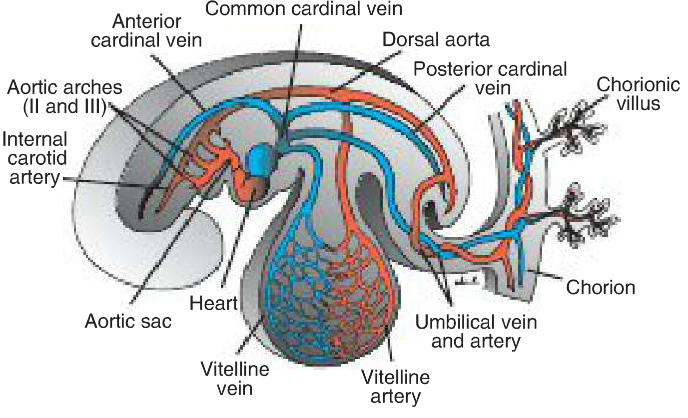
By the end of week 4, three paired venous systems drain into the sinus venosus of the primitive heart:
| Venous System | Origin | Function |
|---|---|---|
| Vitelline veins (omphalomesenteric) | Yolk sac | Carry nutrient-rich blood from gut |
| Umbilical veins | Chorionic villi | Carry oxygenated blood from placenta |
| Cardinal veins | Embryo body | Drain systemic body of the embryo |
PART 1: EARLY HEART TUBE FORMATION
Week 3 - Cardiogenic Crescent
- Angioblastic cells in the splanchnic mesoderm form a horseshoe-shaped cardiogenic plate (crescent) in front of the prechordal plate.
- Two endothelial strands (lateral heart-forming regions) fuse at the midline to form a single primitive heart tube by days 21-22.
- The heart begins beating around day 21-22 - the first organ to function.
Week 4 - Cardiac Looping (D-loop)
The straight heart tube consists of (craniocaudal):
- Truncus arteriosus (outflow)
- Bulbus cordis (future right ventricle outflow)
- Primitive ventricle (future left ventricle)
- Primitive atrium
- Sinus venosus (inflow - receives all three venous pairs)
The tube undergoes rightward D-looping - the bulboventricular region bends to the right and caudally, bringing the future ventricles to their definitive position. This looping is under control of NODAL signaling (left-right axis). Failure produces dextrocardia or situs inversus.
Septation (Weeks 4-8)
- Atrial septation: Septum primum descends, leaving ostium primum at bottom. As primum fuses with endocardial cushions, cell death creates ostium secundum in the upper part. Then septum secundum grows to the right of primum, leaving the foramen ovale - this closes at birth by pressure change.
- AV canal: Four endocardial cushions fuse to form the superior and inferior cushions, dividing the canal into right (tricuspid) and left (mitral) AV orifices.
- Ventricular septation: The thick muscular interventricular septum grows up from the floor. The remaining membranous portion is closed by contributions from the inferior AV cushion plus the right and left conus swellings.
- Conotruncal septation: The spiral aorticopulmonary septum (contributed by neural crest cells) divides the truncus arteriosus into the ascending aorta and pulmonary trunk. Because it spirals ~180°, the pulmonary trunk winds around the aorta.
PART 2: DEVELOPMENT OF THE ARCH OF THE AORTA
The Six Aortic Arches - Overview
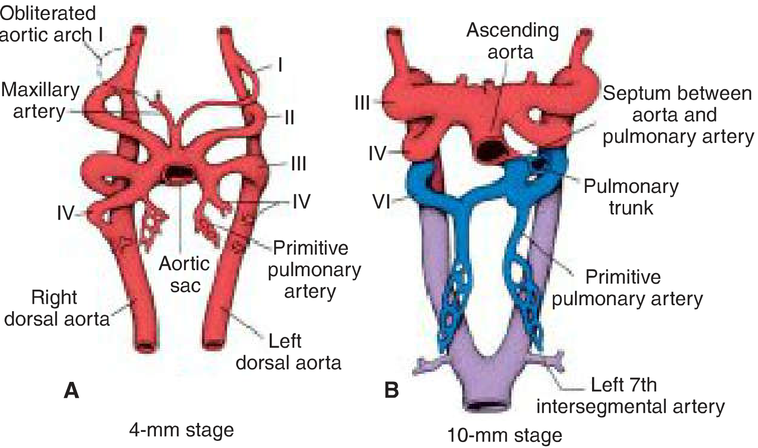
Each of the five pharyngeal arches (1st, 2nd, 3rd, 4th, and 6th - the 5th never fully forms) contains an aortic arch artery connecting the:
- Ventral aortic roots (aortic sac) - feeding from the heart
- Paired dorsal aortae - receiving blood and fusing below to form the single descending aorta
The arches do NOT all exist simultaneously - they appear and regress in a craniocaudal sequence.
Fate of Each Aortic Arch
| Arch | Left | Right |
|---|---|---|
| 1st | Regresses (tiny remnant = maxillary artery) | Regresses |
| 2nd | Regresses (remnant = stapedial & hyoid arteries) | Regresses |
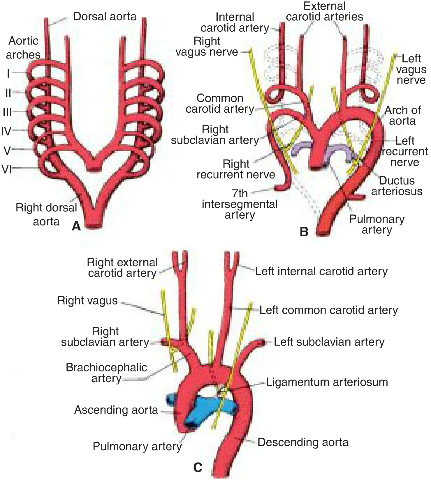
| 3rd | Common carotid + proximal internal carotid | Common carotid + proximal internal carotid |
| 4th | Arch of aorta (between L. common carotid & L. subclavian) | Proximal right subclavian artery |
| 5th | Never completely forms | Never completely forms |
| 6th | Proximal: left pulmonary artery; Distal: ductus arteriosus | Proximal: right pulmonary artery; Distal: REGRESSES |
Key additional contributions:
- The external carotid is a sprout from the 3rd arch
- The remainder of internal carotid beyond the 3rd arch comes from the cranial dorsal aorta
- The left subclavian arises from the 7th intersegmental artery (not an aortic arch)
- The brachiocephalic trunk is formed from the right 4th arch + right dorsal aorta + right 7th intersegmental
The Recurrent Laryngeal Nerves - A Classic Clue
The recurrent laryngeal nerves hook around the 6th arch arteries on both sides. Because the right distal 6th arch regresses, the right nerve migrates upward and now hooks around the right subclavian artery (derived from right 4th arch). On the left, the ductus arteriosus persists (as ligamentum arteriosum), so the left recurrent laryngeal nerve hooks around the ligamentum arteriosum - explaining why it descends into the thorax.
Common Anomalies
| Anomaly | Embryological Basis |
|---|---|
| Aberrant right subclavian artery | Right 4th arch + right dorsal aorta obliterate; right subclavian arises from left dorsal aorta, crosses behind esophagus (dysphagia lusoria) |
| Double aortic arch | Persistence of distal right dorsal aorta; forms vascular ring around trachea and esophagus |
| Right-sided aortic arch | Left 4th arch + left dorsal aorta obliterate; right side forms the arch |
| Coarctation of aorta | Abnormal migration of smooth muscle cells from ductus arteriosus into juxtaductal aorta |
| Patent ductus arteriosus (PDA) | Failure of 6th arch distal segment (ductus arteriosus) to close postnatally |
PART 3: DEVELOPMENT OF THE PORTAL VEIN
Vitelline (Omphalomesenteric) Veins - The Origin
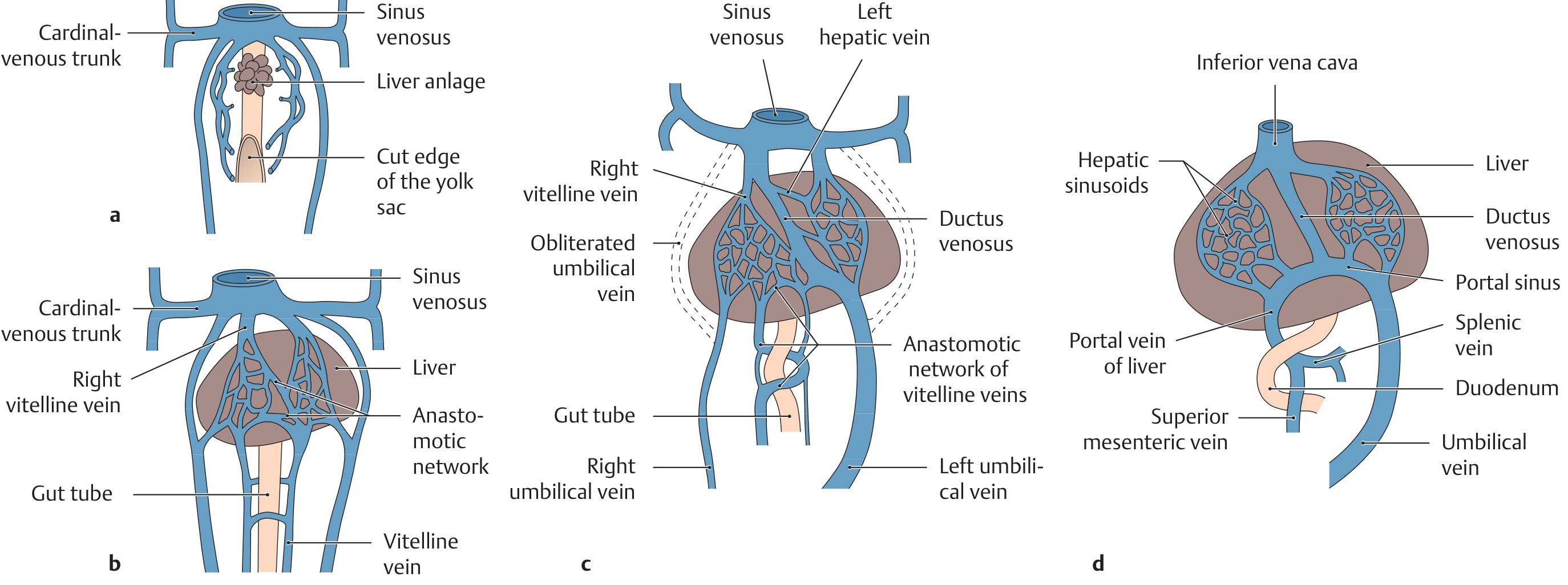
The two vitelline veins initially run alongside the gut tube and drain the yolk sac into the sinus venosus. Before reaching the heart, they:
- Form a venous plexus around the duodenum
- Penetrate the septum transversum, where liver cords grow into and surround them
- Break up into the hepatic sinusoids (first hepatic sinusoids)
Step-by-Step Portal Vein Formation
- Week 4: Both vitelline veins are symmetric and enter the sinus venosus.
- Week 5: The vitelline veins form three anastomotic channels around the duodenum - one ventral and two dorsal. The liver sinusoids disrupt the distal portions of both veins.
- Week 6-8: Through a complex series of obliterations and anastomoses, the distal portion of the RIGHT vitelline vein becomes the portal vein. It receives:
- The superior mesenteric vein (formerly right vitelline)
- The splenic vein
- The inferior mesenteric vein
- The proximal right vitelline vein (between liver and sinus venosus) becomes the hepatic veins and posthepatic IVC segment.
- The left vitelline vein largely regresses.
The Umbilical Veins and Ductus Venosus
- Initially, two umbilical veins bring oxygenated placental blood alongside the liver.
- The right umbilical vein regresses completely by month 2.
- The left umbilical vein persists and connects with the hepatic sinusoids.
- A shunt - the ductus venosus - develops inside the liver, bypassing the sinusoids and connecting the left umbilical vein directly to the proximal right vitelline trunk (future hepatic/IVC segment), allowing most blood to bypass liver metabolism and reach the sinus venosus.
- At birth, the left umbilical vein becomes the ligamentum teres hepatis (round ligament of liver), and the ductus venosus becomes the ligamentum venosum.
PART 4: DEVELOPMENT OF THE INFERIOR VENA CAVA (IVC)
The IVC is built from four distinct embryonic segments, each from a different venous source:
The Four Segments of the IVC
| Segment | Embryonic Source | Adult IVC Region |
|---|---|---|
| Hepatic | Right vitelline vein (proximal) + hepatic sinusoids | Above renal veins (suprahepatic) |
| Prerenal (suprarenal) | Right subcardinal vein | Between liver and renal veins |
| Renal | Subcardinal-supracardinal anastomosis | At renal vein level |
| Postrenal (sacrocardinal) | Right sacrocardinal vein | Below renal veins to iliac bifurcation |
The Cardinal Vein Story
Week 4: Only three venous pairs exist:
- Anterior cardinal veins - drain the head
- Posterior cardinal veins - drain the body
- Common cardinal veins (ducts of Cuvier) - join ant. + post. cardinals and enter sinus venosus
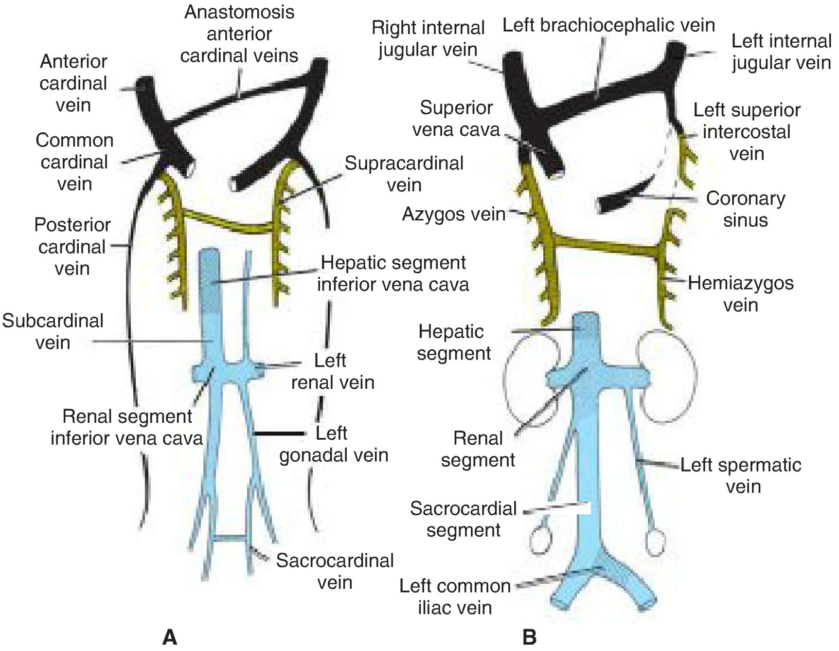
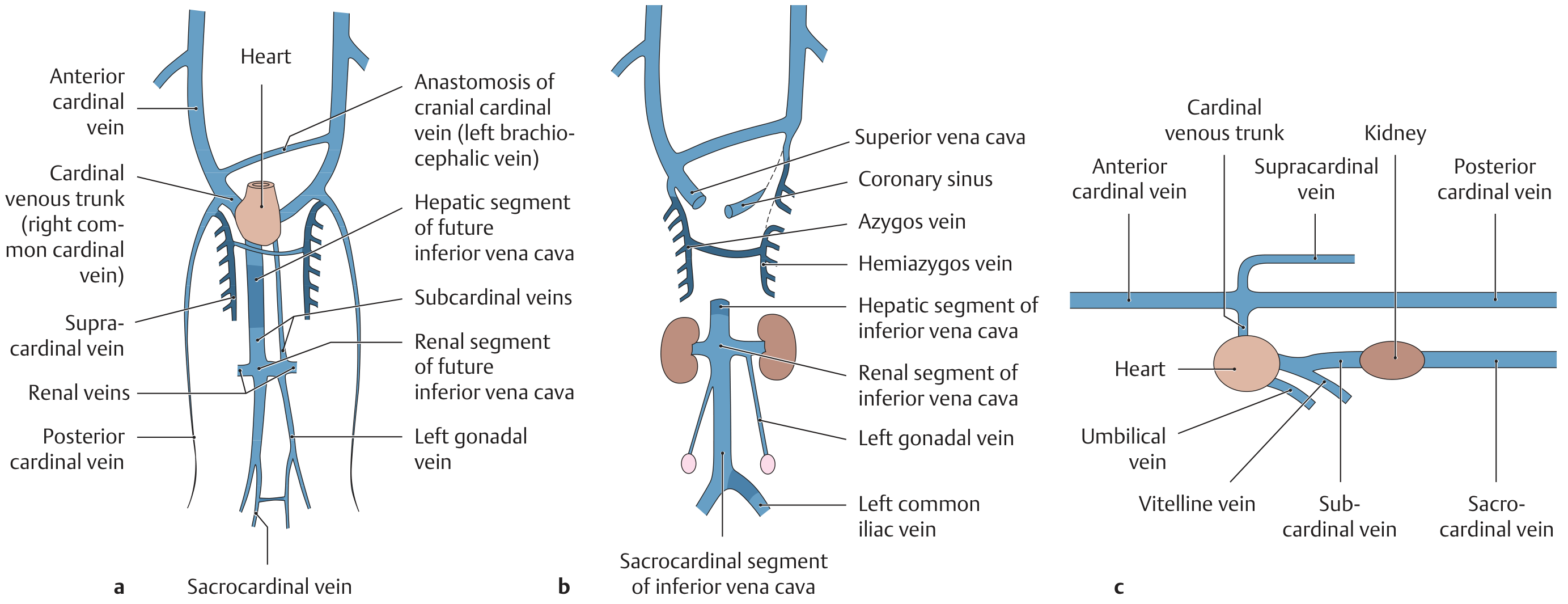
Weeks 5-7: Three new venous systems form to replace the posterior cardinals:
- Subcardinal veins - drain the kidneys (medial to posterior cardinals)
- Supracardinal veins - drain body wall via intercostal veins (lateral to posterior cardinals), future azygos system
- Sacrocardinal veins - drain the lower limbs
How Left-to-Right Channeling Builds the IVC
The key principle is that anastomoses form between left and right vessels, then the left side regresses, channeling all blood rightward:
- Subcardinal anastomosis → becomes left renal vein → left subcardinal disappears; right subcardinal becomes the IVC renal segment
- Sacrocardinal anastomosis → becomes left common iliac vein; right sacrocardinal becomes sacrocardinal IVC segment
- Anterior cardinal anastomosis → becomes left brachiocephalic vein → left anterior cardinal largely disappears; right side builds SVC
IVC Anomalies
| Anomaly | Basis |
|---|---|
| Double IVC | Failure of left sacrocardinal vein to regress |
| Left-sided IVC | Right sacrocardinal regresses; left persists |
| Azygos continuation of IVC | Failure of right subcardinal-hepatic anastomosis; blood bypasses liver and drains via azygos → SVC. Associated with asplenia/polysplenia syndromes |
PART 5: DEVELOPMENT OF THE SUPERIOR VENA CAVA (SVC)
The SVC is simpler - formed entirely from the right anterior and right common cardinal veins:
- The anastomosis between the two anterior cardinal veins (left and right) becomes the left brachiocephalic vein
- Once this anastomosis forms, blood from the left head/arm drains across to the right
- The left common cardinal vein regresses into the coronary sinus (which drains the heart's venous blood into the right atrium)
- The right common cardinal vein + right anterior cardinal become the SVC
- The anterior cardinal veins form the internal jugular veins
SUMMARY TABLE: Key Embryonic Vessels and Their Adult Derivatives
| Embryonic Vessel | Adult Derivative |
|---|---|
| Left 4th aortic arch | Arch of aorta |
| Right 4th aortic arch | Proximal right subclavian |
| 3rd aortic arch (bilateral) | Common carotid + proximal internal carotid |
| Left 6th arch (distal) | Ductus arteriosus → ligamentum arteriosum |
| Left 6th arch (proximal) | Left pulmonary artery |
| Right 6th arch (proximal) | Right pulmonary artery |
| 7th left intersegmental artery | Left subclavian artery |
| Distal right vitelline vein | Portal vein |
| Proximal right vitelline vein | Hepatic veins + hepatic segment of IVC |
| Right subcardinal vein | Renal segment of IVC |
| Right sacrocardinal vein | Sacrocardinal segment of IVC |
| Subcardinal-sacrocardinal join | Complete IVC |
| Right common cardinal + right ant. cardinal | SVC |
| Anastomosis of ant. cardinal veins | Left brachiocephalic vein |
| Left common cardinal vein | Coronary sinus |
| Ductus venosus | Ligamentum venosum |
| Left umbilical vein | Ligamentum teres hepatis |
MNEMONIC HELP
Aortic arch derivatives: "3 is C for Carotid, 4 is A for Aorta/Arch, 6 is P for Pulmonary"
IVC segments (top to bottom): "Happy Renal Surgeon" = Hepatic + (p)Rerenal + Renal + Sacrocardinal
Neural crest cells contribute to the conotruncal septum (aorticopulmonary septum), pharyngeal arch arteries, and the recurrent laryngeal hook - their migration failure causes DiGeorge syndrome (22q11 deletion) with interrupted aortic arch, conotruncal defects, and absent thymus/parathyroids.
References: Langman's Medical Embryology, 14th ed., pp. 226-240 | THIEME Atlas of Anatomy: General Anatomy and Musculoskeletal System, pp. 30-31
This is a shared conversation. Sign in to Orris to start your own chat.