
Clincial and examination finding CA lung
lung cancer chest X-ray clinical findings
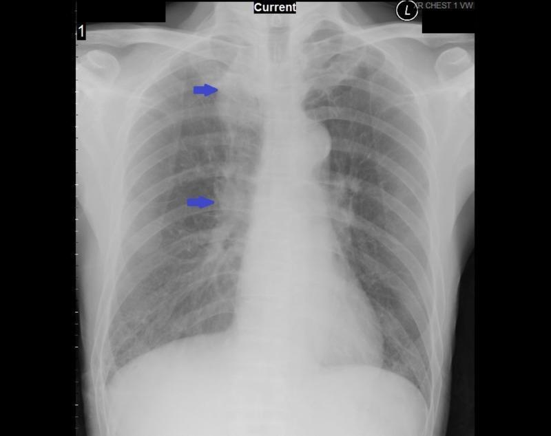
This diagnostic image is a posterior-anterior (PA) chest X-ray demonstrating several key clinical findings in a patient with a history of lung cancer. The primary feature includes postoperative changes in the right lung, specifically a reduction in lung volume consistent with a prior right upper lobe lobectomy. There is evidence of pulmonary fibrosis, visualized as patchy, increased interstitial opacities and thickening, which is more pronounced in the right lung field. Blue arrows point toward the mediastinal region, highlighting borderline enlarged lower mediastinal lymph nodes situated adjacent to the distal esophagus. The right hemithorax shows architectural distortion, while the left lung field appears relatively clearer but with subtle reticular markings. The skeletal structures, including the clavicles and scapulae, are partially visible at the periphery. This radiograph serves as an educational example of assessing chronic parenchymal changes and hilar/mediastinal lymphadenopathy in a post-surgical oncological context.
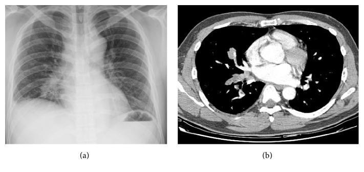
This composite clinical image presents diagnostic findings in a 41-year-old male with lung cancer and dyspnea. Image (a) is a posterior-anterior chest X-ray showing an ill-defined, heterogeneous area of increased opacity in the right lower lung field, suggestive of a parenchymal lesion. Image (b) is an axial contrast-enhanced CT scan of the thorax at the level of the pulmonary veins. The CT demonstrates high-density, irregular lesions within the right superior and right inferior pulmonary veins. These findings represent tumor-associated thrombotic lesions or direct tumor extension, which appear to distend the affected vessels and extend into the adjacent lung parenchyma. The visual evidence illustrates pulmonary vein obstruction syndrome (PVOS) as a complication of lung adenocarcinoma. The images are categorized as clinical imaging and x-ray radiography, suitable for intermediate-level medical education regarding oncological thoracic complications and vascular involvement in lung cancer.
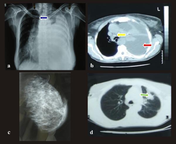
This composite of clinical imaging illustrates diagnostic findings in a case of primary lung adenocarcinoma with breast metastasis. (a) Anteroposterior chest x-ray reveals a massive left-sided pleural effusion causing near-complete opacification of the left hemithorax and a significant mediastinal shift toward the right (blue arrow). (b) Axial chest CT scan (mediastinal window) shows a massive pleural effusion (red arrow) resulting in compressive atelectasis of the left lung and severe contralateral displacement of the trachea and mediastinum; paratracheal lymphadenopathy is also noted (yellow arrow). (c) Digital mammography of the left breast demonstrates a diffuse, asymmetrical density in the subareolar and upper outer quadrant without discrete margins or microcalcifications, suggesting inflammatory or metastatic infiltration. (d) Axial chest CT (lung window) highlights a 3.5 x 4.5 cm irregular peripheral lesion in the left upper lobe (green arrow) with spiculation and extension into the surrounding parenchyma, making contact with the pleura. These images collectively demonstrate the radiological presentation of metastatic lung cancer involving the pleura and breast.
finger clubbing lung cancer paraneoplastic
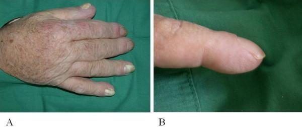
Two-panel clinical photograph illustrating manifestations of hypertrophic osteoarthropathy (HOA). Panel A presents a dorsal view of a right hand exhibiting generalized acral enlargement and diffuse edematous swelling. The fingers appear thickened, particularly at the distal aspects, and the nails show thickening and discoloration. Panel B provides a close-up lateral view of a finger demonstrating significant digital clubbing (Hippocratic fingers). Visible features include a loss of the normal Lovibond angle, a rounded bulbous deformity of the fingertip, and smooth, taut, shiny skin indicative of soft tissue hypertrophy and edema. Notable periungual erythema is present around the proximal nail fold. Clinically, these findings are highly suggestive of paraneoplastic syndrome, specifically secondary hypertrophic osteoarthropathy associated with underlying malignancies such as non-small cell lung carcinoma. The images serve as educational examples for recognizing secondary systemic signs of internal malignancy during physical examination.
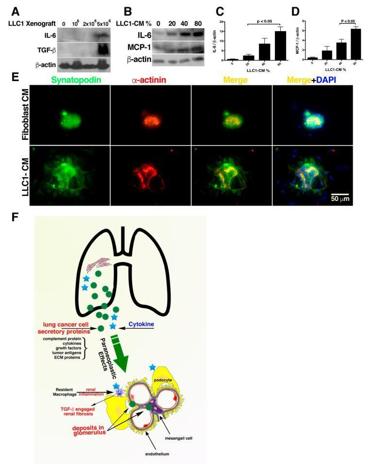
Educational medical composite depicting the paraneoplastic effects of lung cancer on renal pathophysiology. (A-B) Western blot analyses showing dose-dependent increases in inflammatory markers (IL-6, TGF-̢, and MCP-1) in both mouse kidney tissues and NRK-52E renal cells following exposure to LLC1 (Lewis Lung Carcinoma) xenografts or conditioned medium (LLC1-CM). (C-D) Bar graphs quantifying the significant (p < 0.05) increase in IL-6 and MCP-1 relative to ̢-actin loading controls. (E) Multi-channel immunofluorescence images of cultured glomeruli. Top row (Fibroblast-CM control) shows tightly clustered podocytes with high expression of Synaptopodin (green) and ̡-actinin (red). Bottom row (LLC1-CM) demonstrates loss of glomerular integrity, characterized by dispersed podocyte arrangement and disrupted microfilament bundling. (F) Pathophysiology diagram illustrating the proposed mechanism: lung cancer secretory proteins (cytokines, complement, growth factors) trigger paraneoplastic effects including resident macrophage activation, TGF-̢-engaged renal fibrosis, and glomerular deposits leading to glomerulopathy. This resource illustrates the remote systemic impact of tumor secretomes on kidney function.
Clinical and Examination Findings in Carcinoma Lung (CA Lung)
Overview
1. Symptoms from the Primary Tumor
Central/Endobronchial Tumors (e.g., Squamous Cell, SCLC)
- Cough (8-75%) - most common presenting symptom
- Hemoptysis (6-35%) - blood-streaked sputum; prompts urgent investigation
- Wheeze / stridor - due to airway narrowing
- Dyspnea (3-60%)
- Post-obstructive pneumonia - persistent pneumonia unresponsive to antibiotics is a red flag
Peripheral Tumors (e.g., Adenocarcinoma)
- Chest/pleuritic pain (20-49%) - pleural or chest wall involvement
- Lung abscess features - cavitating peripheral tumors
- Restrictive dyspnea - pleural effusion
| Symptom / Sign | Frequency |
|---|---|
| Cough | 8-75% |
| Weight loss | 0-68% |
| Dyspnea | 3-60% |
| Chest pain | 20-49% |
| Hemoptysis | 6-35% |
| Bone pain | 6-25% |
| Clubbing | 0-20% |
| Fever | 0-20% |
| Weakness | 0-10% |
| SVC obstruction | 0-4% |
| Dysphagia | 0-2% |
| Wheezing/stridor | 0-2% |
2. Regional Spread - Clinical Findings
| Structure Involved | Clinical Finding |
|---|---|
| Recurrent laryngeal nerve (left > right) | Hoarseness (up to 5% at presentation) |
| Phrenic nerve | Hemidiaphragm elevation, dyspnea |
| Sympathetic chain | Horner's syndrome: ptosis, miosis, enophthalmos, anhidrosis |
| SVC compression | SVC syndrome: facial/neck/arm swelling, dyspnea, dilated neck veins |
| Esophagus | Dysphagia |
| Pericardium/heart | Tamponade, arrhythmia, cardiac failure |
| Pleural space | Pleural effusion - pain, dyspnea, stony dull percussion |
| Trachea | Tracheal obstruction, mediastinal shift |
3. Pancoast (Superior Sulcus) Syndrome
- Shoulder pain radiating in the ulnar distribution of the arm (C8, T1, T2 nerve roots)
- Brachial plexopathy - upper limb weakness and paresthesia
- Horner's syndrome (coexists in most cases) - ptosis + miosis + anhidrosis
- Radiologic destruction of 1st and 2nd ribs
- Wasting of small muscles of the hand
4. Metastatic Disease - Examination Findings
| System | Findings |
|---|---|
| Lymph nodes | Supraclavicular/cervical lymphadenopathy (>1 cm) |
| Brain | Focal neurologic deficits, papilledema, headache, seizures, mental status changes |
| Bone | Bone tenderness, pathologic fractures, spinal cord compression |
| Liver | Hepatomegaly (>13 cm span), right upper quadrant pain, jaundice |
| Soft tissue | Subcutaneous masses |
| General | Weight loss >10 lb, cachexia, pallor (anaemia - Hct <40% in men, <35% in women) |
5. Paraneoplastic Syndromes
Endocrine/Metabolic
| Syndrome | Mediator | Associated Histology | Clinical Features |
|---|---|---|---|
| SIADH | ADH/vasopressin | SCLC (10-45%), squamous cell | Hyponatraemia, confusion, seizures |
| Ectopic Cushing's | ACTH | SCLC | Hypokalaemia, hypertension, hyperglycaemia, proximal myopathy |
| Hypercalcaemia | PTHrP | Squamous cell carcinoma | Polyuria, constipation, confusion, bone pain |
| Hypoglycaemia | IGF-2 | Various | Fasting hypoglycaemia |
| Carcinoid syndrome | Serotonin | Carcinoid tumour | Flushing, diarrhoea, wheeze |
Neurological (mainly SCLC)
- Lambert-Eaton Myasthenic Syndrome (LEMS) - proximal muscle weakness, depressed reflexes that improve with repetitive stimulation (antibody against presynaptic voltage-gated Ca²⁺ channels)
- Cerebellar degeneration - truncal ataxia, dysarthria
- Peripheral sensory neuropathy
- Encephalomyelitis - anti-Hu antibodies
Musculoskeletal
- Hypertrophic pulmonary osteoarthropathy (HPOA) - periosteal new bone formation in distal long bones; painful wrists/ankles
- Digital clubbing (0-20%) - loss of Lovibond angle, sponginess of nail bed
Haematological
- Migratory thrombophlebitis (Trousseau's syndrome) - more common with adenocarcinoma
- Anaemia of chronic disease
- DIC (disseminated intravascular coagulation)
Dermatological
- Acanthosis nigricans
- Dermatomyositis / polymyositis
- Erythema gyratum repens
6. Physical Examination - System-by-System Summary
| System | Signs to Look For |
|---|---|
| General | Cachexia, pallor, jaundice, clubbing |
| Hands | Clubbing, HPOA tenderness, wasting (Pancoast), flapping tremor (CO₂ retention) |
| Eyes | Ptosis, miosis (Horner's), papilledema |
| Face/Neck | Facial/neck oedema (SVC syndrome), dilated neck veins, cervical/supraclavicular lymphadenopathy, hoarseness on speaking |
| Chest | Reduced expansion, tracheal deviation, dullness to percussion (collapse, effusion), bronchial breathing, absent breath sounds (effusion), wheeze |
| Abdomen | Hepatomegaly (metastases) |
| Neurology | Focal deficits, ataxia, proximal myopathy, reduced reflexes |
| Musculoskeletal | Bone tenderness, joint swelling (HPOA) |
7. Radiological Clues on CXR
Key Clinical Pearls
- A persistent cough with hemoptysis in a smoker age >40 = lung cancer until proven otherwise, even with a normal CXR.
- New hoarseness + lung mass = left recurrent laryngeal nerve palsy from mediastinal involvement.
- Hypercalcaemia without bone metastases = PTHrP secretion from squamous cell carcinoma.
- Hyponatraemia (especially in SCLC) = SIADH until proven otherwise.
- Clubbing + periosteal pain = HPOA - a paraneoplastic marker of NSCLC.
- Pancoast + Horner's = apical (superior sulcus) tumour with brachial plexus and sympathetic chain invasion.
- SVC syndrome = lung cancer in ~75% of cases (mainly SCLC and right-sided squamous).
Physical examination findings
superior vena cava syndrome lung cancer facial swelling
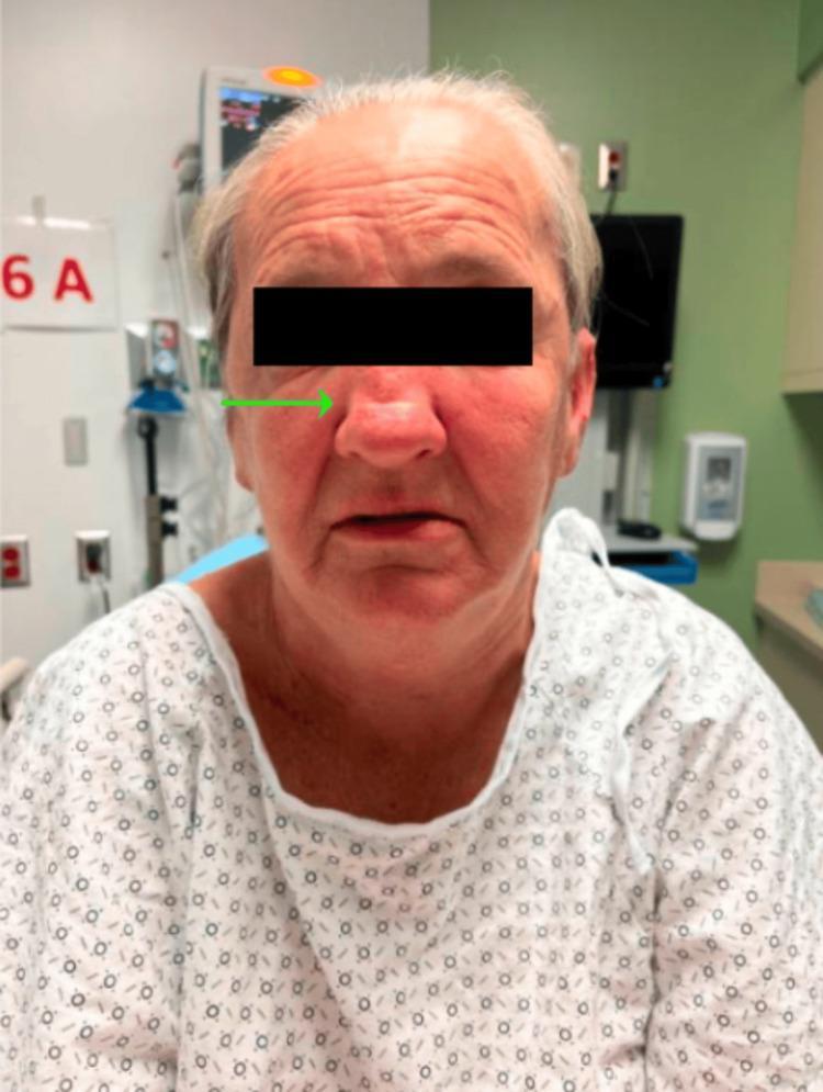
Clinical photograph of an adult female patient in a hospital setting demonstrating classic physical signs associated with Superior Vena Cava (SVC) Syndrome. The image shows significant, diffuse facial edema (facial plethora) and generalized erythema, particularly prominent in the periorbital and midfacial regions. The swelling appears slightly asymmetric, with greater involvement of the patient's left side. A green arrow highlights a distinct saddle nose deformity, characterized by a collapse of the nasal bridge. The patient exhibits a mildly anxious expression and is seated in a clinical environment, wearing a standard patterned hospital gown. This visual presentation illustrates the physical manifestations of venous congestion and obstruction often caused by mediastinal masses or thoracic malignancies, such as small-cell lung cancer (SCLC). The image is an educational example of oncologic or vascular emergencies and the importance of physical examination in diagnosing obstructive venous syndromes.
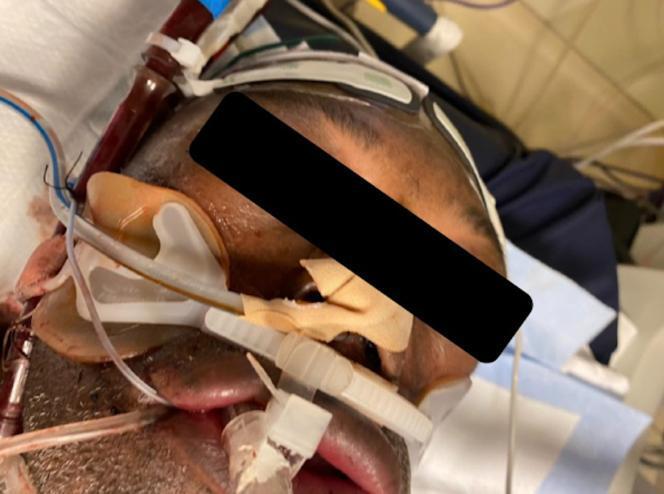
This clinical photograph illustrates a patient presenting with severe facial and neck edema, a classic hallmark of Superior Vena Cava (SVC) Syndrome. The patient exhibits massive swelling (anasarca-like) of the periorbital, cheek, and lip regions, resulting in a significantly turgid and distorted facial appearance. The neck also shows pronounced soft tissue swelling. Several critical care interventions are visible: an endotracheal tube is secured with a commercial holder for respiratory support, and a large-bore percutaneous cannula, likely part of a dual-lumen right ventricular assist device (RVAD), is inserted into the neck. There is visible blood within the proximal tubing of the cervical access. The image demonstrates the dramatic clinical presentation of venous congestion and high central venous pressure (CVP) following iatrogenic SVC obstruction. This visual serves as an educational example of device-related vascular complications in intensive care settings, highlighting the immediate physical manifestations of impaired venous return from the head and upper extremities.
Horner syndrome ptosis miosis lung cancer
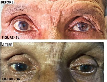
Two-panel clinical photograph displaying the 'Before' and 'After' ophthalmic findings in a patient with left-sided Horner's syndrome. The 'Before' image (Figure 3a) demonstrates classic clinical signs including left upper eyelid ptosis (drooping) and left-sided miosis (constricted pupil) compared to the right eye. The 'After' image (Figure 3b) illustrates the clinical response following three cycles of chemotherapy for an underlying superior sulcus tumor (Pancoast tumor). There is visible improvement in the left ocular presentation, characterized by the elevation of the left upper eyelid, reduction in ptosis severity, and a comparative increase in left pupil diameter (resolution of miosis). The image highlights the ocular manifestations of sympathetic nerve pathway disruption and its subsequent recovery during systemic treatment of the primary thoracic malignancy. Educational focus includes the recognition of Horner's syndrome triad (ptosis, miosis, and anhidrosis) and its clinical association with apical lung lesions.
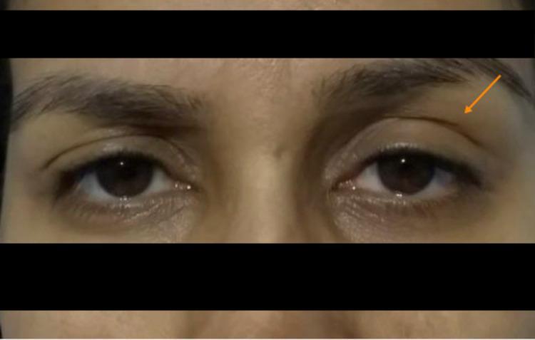
This clinical photograph displays a frontal view of a patient's eyes, highlighting diagnostic signs of Horner's syndrome. The image demonstrates a clear asymmetry between the left and right ocular regions. The left eye exhibits visible ptosis (drooping of the upper eyelid), indicated by an orange arrow, where the eyelid margin sits lower on the iris compared to the contralateral side. Furthermore, there is evidence of miosis (pupillary constriction) in the left eye, making the pupil appear significantly smaller than the right. The right eye appears normal with a baseline pupil size and expected eyelid position. Clinically, this presentation is characteristic of sympathetic nerve pathway disruption, which in this context followed the administration of neuraxial analgesia. The image serves as a high-quality educational example for identifying the ocular components of a partial Horner's syndrome in a clinical setting.
digital clubbing finger nails
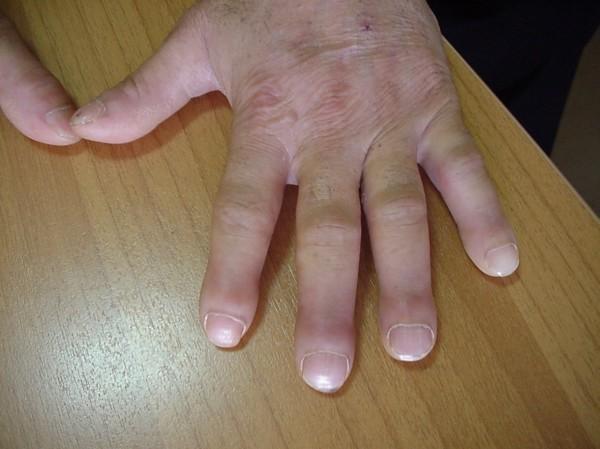
This clinical photograph displays a dorsal view of a human hand resting on a flat surface, illustrating classic features of digital clubbing. The distal phalanges of all visible fingers show significant bulbous enlargement and swelling, resulting in a 'drumstick' appearance. The nails exhibit increased longitudinal and transverse curvature (convexity), becoming rounded and broad. There is a visible loss of the normal angle between the nail plate and the proximal nail fold (Lovibond's angle), with the proximal nail fold appearing thickened and shiny. The skin over the hand is wrinkled, and an old linear scar is noted on the dorsal web space between the thumb and index finger. These findings are highly characteristic of digital clubbing, a clinical sign often associated with chronic hypoxemia, cardiovascular disease, or neoplastic pulmonary conditions such as bronchogenic carcinoma, particularly in patients with a significant smoking history.
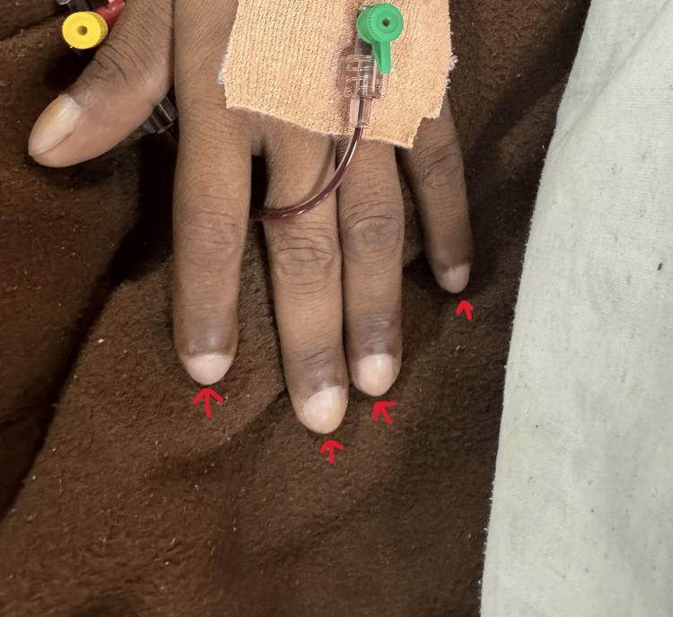
This clinical photograph displays a patient's hand demonstrating grade 3 digital clubbing, a common physical sign of chronic hypoxemia. The fingertips (distal phalanges) appear bulbous and enlarged, described as 'drumstick' morphology. The nails exhibit increased longitudinal curvature and a loss of the normal Lovibond angle at the nail base. Clinical features suggest cyanotic heart disease, supported by the visible medical equipment including a pulse oximetry sensor on the index finger and an intravenous (IV) catheter secured to the dorsal surface of the hand with adhesive dressing. There is evidence of peripheral cyanosis or hyperpigmentation of the distal digits. This visual is highly relevant for medical education regarding cardiovascular and respiratory physical examination findings and the assessment of chronic oxygen deprivation.
Physical Examination Findings in CA Lung
General Inspection (Bedside)
| Finding | Significance |
|---|---|
| Cachexia / muscle wasting | Advanced/metastatic disease |
| Pallor | Anaemia of chronic disease or bone marrow infiltration |
| Jaundice | Liver metastases |
| Plethora (flushing) | SVC syndrome or carcinoid |
| Tar staining of fingers | Smoking history (risk factor) |
Hands
- Tender, warm, swollen wrists and ankles
- Periosteal new bone formation on X-ray
- Classically associated with NSCLC (adenocarcinoma > squamous)
- Often accompanies clubbing
Eyes
- Unilateral ptosis (drooping upper lid)
- Miosis (constricted pupil)
- Enophthalmos (sunken eye)
- Anhidrosis (absent sweating on ipsilateral face)
Face and Head
- Puffy, congested face, particularly periorbital swelling
- "Coat-hanger" pattern of swelling (face, neck, upper arms)
Neck
| Finding | Significance |
|---|---|
| Distended neck veins (JVP↑) | SVC obstruction |
| Hard, non-tender lymph nodes in supraclavicular fossa (Virchow's node = left side) | Metastatic lymphadenopathy (N3 disease) |
| Cervical lymphadenopathy (>1 cm) | Regional or distant spread |
| Tracheal deviation | Collapse (towards), massive effusion or tension (away) |
| Hoarse voice | Recurrent laryngeal nerve palsy - left RLN loops under aortic arch; more common with left-sided tumours |
Chest Inspection
| Sign | Cause |
|---|---|
| Reduced chest expansion (unilateral) | Collapse, effusion, consolidation |
| Asymmetric chest movement | Phrenic nerve palsy (raised hemidiaphragm) |
| Prominent chest wall veins | SVC or subclavian vein obstruction |
| Visible chest wall mass | Direct chest wall invasion |
Chest Palpation
| Sign | Cause |
|---|---|
| Reduced expansion on affected side | Effusion, collapse, consolidation |
| Absent tactile vocal fremitus | Pleural effusion |
| Increased tactile vocal fremitus | Consolidation (post-obstructive pneumonia) |
| Mediastinal shift (trachea + apex beat) | Large effusion (pushed away) or collapse (pulled towards) |
| Tender chest wall / rib tenderness | Rib metastases, direct invasion |
Chest Percussion
| Finding | Cause |
|---|---|
| Stony dullness | Pleural effusion (most common sign on percussion) |
| Dullness | Consolidation (post-obstructive pneumonia), collapse |
| Hyper-resonance | Compensatory emphysema (rare) |
Chest Auscultation
| Finding | Cause |
|---|---|
| Absent/reduced breath sounds | Pleural effusion, complete collapse |
| Bronchial breathing | Consolidation / post-obstructive pneumonia |
| Localised wheeze (monophonic) | Endobronchial tumour partially obstructing a bronchus |
| Coarse crackles | Post-obstructive pneumonia |
| Pleural rub | Pleural involvement/inflammation |
| Reduced vocal resonance | Effusion |
| Pericardial rub | Pericardial extension |
Abdomen
| Finding | Cause |
|---|---|
| Hepatomegaly (>13 cm span) | Liver metastases (often irregular, nodular edge) |
| Right upper quadrant tenderness | Liver metastases |
| Ascites | Peritoneal metastases (less common) |
Neurological
| Finding | Cause |
|---|---|
| Proximal muscle weakness | Lambert-Eaton syndrome (SCLC), ectopic ACTH |
| Depressed/absent reflexes | Lambert-Eaton (unlike MG where reflexes improve with repetition) |
| Cerebellar ataxia | Paraneoplastic cerebellar degeneration |
| Peripheral sensory loss / glove-stocking | Paraneoplastic sensory neuropathy |
| Focal neurological deficits | Brain metastases |
| Upper limb weakness + paresthesia | Brachial plexus (Pancoast T1) |
| Signs of spinal cord compression | Vertebral metastases with epidural extension |
| Confusional state / altered GCS | Brain mets, SIADH (hyponatraemia), hypercalcaemia |
Musculoskeletal / Extremities
| Finding | Cause |
|---|---|
| Bone tenderness (spine, ribs, long bones) | Bone metastases |
| Pathological fracture | Lytic metastases |
| Tender, swollen ankles/wrists (HPOA) | Paraneoplastic periostitis |
| Peripheral oedema | SVC, hypoalbuminaemia, DVT (Trousseau's) |
| DVT/thrombophlebitis (Trousseau's sign) | Hypercoagulable state, especially with adenocarcinoma |
Skin
| Finding | Cause |
|---|---|
| Acanthosis nigricans | Paraneoplastic (any histology) |
| Dermatomyositis (heliotrope rash, Gottron's papules) | Paraneoplastic |
| Erythema gyratum repens | Paraneoplastic (rare, highly specific) |
| Subcutaneous nodules | Skin metastases |
Quick Reference: Signs by Syndrome
| Syndrome | Key Examination Signs |
|---|---|
| Pancoast | Horner's + hand muscle wasting + upper limb sensory loss |
| SVC syndrome | Facial oedema + dilated neck veins + arm swelling + plethora |
| SIADH | Confusion, reduced GCS (hyponatraemia) |
| Hypercalcaemia (PTHrP) | Confusion, dehydration, weakness - squamous cell CA |
| LEMS | Proximal weakness, hyporeflexia, improves with repeated movement |
| Ectopic Cushing's (SCLC) | Proximal myopathy, hypertension, but often WITHOUT Cushingoid features (rapid onset) |
| HPOA | Clubbing + tender wrists/ankles + periosteal pain |