


diagnosis
Reading File
Looking at the images, I can see three key findings:
- Image 1 (axilla/armpit): Hyperpigmented, thickened, velvety plaque in the axilla
- Image 2 (back): An annular/oval erythematous patch with central clearing and fine scaling on the back
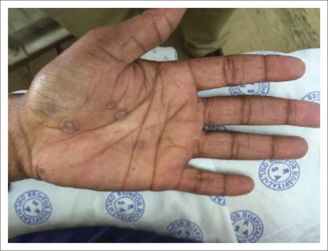
- Image 3 (palm): Desquamating, peeling skin along the palmar creases with erythema
Finding Sources
Finding Sources
Finding Sources
The combination of findings is very telling. Let me search further to confirm a unifying diagnosis — these three findings together (annular back lesion, axillary hyperpigmentation, palmar peeling) strongly suggest secondary syphilis or a dermatophyte + metabolic syndrome combination. The palmar peeling with the annular lesion is classic for syphilis.
Finding Sources
Finding Sources
The Fitzpatrick's textbook confirms it clearly: palmar lesions with annular scale crossing palmar creases in ~75% of cases is secondary syphilis. Let me read that section and get the relevant images.
Reading File
Searching Images
secondary syphilis palmar rash annular scaling
The diagnosis is clear. Here is a comprehensive analysis:
Diagnosis: Secondary Syphilis
The three images together form the classic triad of secondary syphilis:
Finding-by-Finding Analysis
| Image | Lesion | Significance |
|---|---|---|
| Axilla | Hyperpigmented, velvety plaque | Syphilitic papulosquamous lesion in intertriginous area; can also be condylomata lata in folds |
| Back | Oval/annular erythematous plaque with peripheral scale (Biett's collarette) | Papulosquamous syphilid — characteristic "collarette of scale" on trunk |
| Palm | Desquamating, peeling erythematous lesions crossing palmar creases | Pathognomonic palmar syphilid — present in ~75% of secondary syphilis cases |
Why This Is Secondary Syphilis
- Palmar involvement crossing the creases with copper-colored macules/desquamation is highly specific for secondary syphilis — very few other conditions cause this pattern
- Truncal oval plaques with collarette scaling (Biett's collarette) is characteristic of syphilid eruptions
- Intertriginous hyperpigmented lesion (axilla) fits the papulosquamous/lichenoid syphilid pattern
- The lesions are distributed on trunk + extremities + flexural folds — the classic disseminated pattern of Treponema pallidum bacteremia
"Erythematous to copper-colored round papules or macules, well demarcated and sometimes with an annular scale, are present on the palms and soles in nearly 75% of cases and classically cross the palmar creases." — Fitzpatrick's Dermatology, Chapter 170
Reference Images from Literature

Differential Diagnoses to Exclude
| Condition | Against it |
|---|---|
| Pityriasis rosea | No herald patch; palmar involvement unusual |
| Tinea corporis | Wouldn't explain palmar peeling |
| Psoriasis | Lacks silvery scale; palmar pattern different |
| Drug eruption | No palmar collarette crossing creases |
Recommended Workup
- RPR / VDRL (non-treponemal screening)
- TPHA or FTA-ABS (confirmatory treponemal test)
- HIV testing (co-infection is common)
- Partner notification and contact tracing
Treatment
- Benzathine penicillin G 2.4 million units IM — single dose (if no neurosyphilis)
- Penicillin-allergic: Doxycycline 100 mg BD × 14 days
- Monitor RPR titres at 3, 6, 12 months for treatment response
Note: This is a notifiable STI — sexual contacts need evaluation and treatment.
This is a shared conversation. Sign in to Orris to start your own chat.