
Rheumatic heart disease pathology explain with pictures
Reading File
Now let me search the medical library for rheumatic heart disease pathology, in parallel with a heading and paragraph search:
Finding Sources
Finding Sources
Excellent findings. Robbins Pathology has a dedicated section on Rheumatic Valvular Disease with images, and Sabiston has valve pathology images. Let me read the key sections in parallel:
Reading File
Reading File
Reading File
Now let me get the Robbins Pathology section on rheumatic valvular disease (the most image-rich pathology source) and the Sabiston images:
Reading File
Reading File
Now let me get more of the Robbins section which contains detailed pathology text and more images, and also fetch the key images:
Reading File
Loading Image
Loading Image
Excellent images retrieved. Now let me get the detailed pathology text from Robbins and also search for the Aschoff body image more specifically:
Reading File
Finding Sources
I have all the material I need. Let me fetch the Aschoff body histology image from Sherris Microbiology:
Loading Image
I now have comprehensive textbook content and high-quality pathology images. Let me compile the full answer.
Rheumatic Heart Disease (RHD) - Pathology with Images
1. Overview
Rheumatic heart disease is the cardiac sequela of acute rheumatic fever (ARF), itself triggered by group A beta-hemolytic streptococcal pharyngitis. It is the leading cause of acquired valvular heart disease in children and young adults worldwide, especially in low- and middle-income countries. It predominantly causes deforming mitral stenosis - RHD is essentially the only cause of acquired mitral stenosis.
2. Pathogenesis - Molecular Mimicry
The immune response to Group A Streptococcus M-protein cross-reacts with host cardiac proteins:
- Antibodies against streptococcal M proteins also recognize cardiac myosin, laminin, and tropomyosin in the myocardium and valves
- These cross-reactive antibodies bind valvular endothelium, upregulating adhesion molecules (VCAM-1, ICAM-1) that facilitate CD4+ T cell infiltration into the valve
- Cytokines from T cells drive macrophage activation, forming Aschoff bodies
- The characteristic 2-3 week delay in symptom onset after infection reflects the time needed to generate this immune response - by this point, no live streptococci remain in the lesions
- Only ~1-3% of infected patients develop ARF, suggesting genetic susceptibility
3. Acute Rheumatic Fever - Pancarditis
ARF causes inflammation of all three heart layers (pancarditis):
Pericarditis
- Fibrinous exudate on pericardium
- Usually resolves without sequelae ("bread-and-butter" pericarditis)
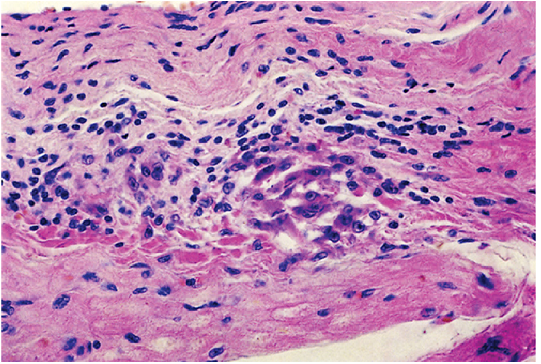
Myocarditis - Aschoff Bodies
The Aschoff body is the pathognomonic lesion of rheumatic fever:
- Collections of lymphocytes (mostly T cells), scattered plasma cells, and plump activated macrophages called Anitschkow cells (caterpillar cells)
- Anitschkow cells have abundant cytoplasm with nuclei showing centrally condensed chromatin in a wavy ribbon pattern - the "caterpillar nucleus"
- Zones of fibrinoid necrosis surround the cellular infiltrate
- Found in the interstitial connective tissue of the myocardium
Endocarditis / Valvulitis (Acute)
- Fibrinoid necrosis along the valve leaflet closure lines
- Formation of small 1-2 mm vegetations called verrucae along the lines of leaflet closure
- These vegetations cause little hemodynamic disturbance acutely
4. Gross and Microscopic Pathology - Key Images
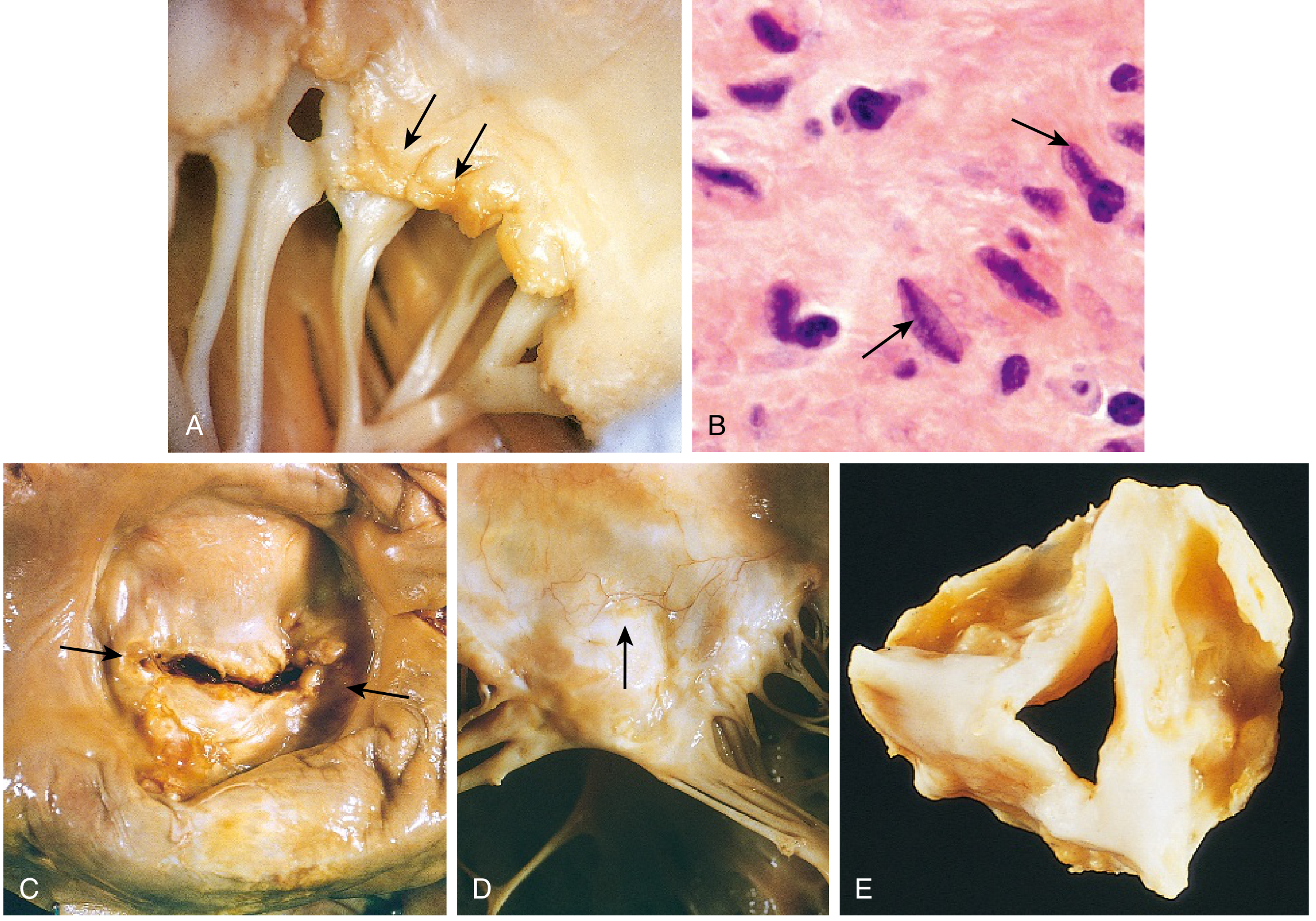
- (A) Acute rheumatic mitral valvulitis superimposed on chronic RHD - small verrucous vegetations visible along the line of leaflet closure (arrows); chordae tendineae already fibrous and fused from previous episodes
- (B) Microscopic Aschoff body - activated macrophages (Anitschkow cells) with prominent nucleoli and central wavy "caterpillar" chromatin (arrows)
- (C) Chronic mitral stenosis viewed from above - diffuse fibrous thickening, commissural fusion (arrows), and marked left atrial dilation
- (D) Anterior leaflet of rheumatic mitral valve showing neovascularization (arrow) - a hallmark of chronic inflammation/healing
- (E) Rheumatic aortic stenosis specimen - thickening and distortion of cusps
5. Chronic Rheumatic Heart Disease
As Aschoff bodies heal and scar, the valves undergo permanent destructive remodeling:
| Feature | Detail |
|---|---|
| Leaflet thickening | Fibrosis replaces normal leaflet architecture |
| Commissural fusion | Fibrous bridging between adjacent leaflets |
| Chordae tendineae | Thickening, shortening, and fusion |
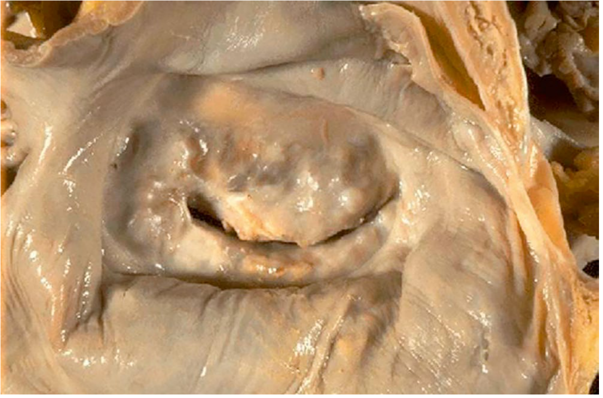
| Calcification | Progressive; can create "buttonhole" stenosis |
| Neovascularization | New vessels in leaflets (grossly and microscopically visible) |
The "Fish Mouth" / "Buttonhole" Valve
6. Valves Affected - Order of Frequency
| Valve Involvement | Frequency |
|---|---|
| Mitral alone | ~70% of cases |
| Mitral + Aortic (combined) | ~25% of cases |
| Tricuspid | Less frequent, less severe |
| Pulmonic | Almost never affected ("escapes injury") |
The mitral valve is predominately affected because it bears the highest hemodynamic stress.
7. Consequences of Chronic RHD
Mitral stenosis is the dominant functional consequence:
- Left atrial dilation due to pressure overload from obstructed inflow
- Atrial fibrillation from atrial dilation and fibrosis
- Left atrial mural thrombus - from AF + dilation (risk of systemic embolism and stroke)
- Pulmonary hypertension from passive venous congestion
- Right ventricular hypertrophy and eventual failure
- Left ventricle is typically normal in pure mitral stenosis
8. Summary Diagram of Pathologic Progression
Group A Strep pharyngitis
↓
Molecular mimicry (M-protein vs. cardiac myosin/laminin)
↓
ACUTE RHEUMATIC FEVER (2-3 weeks later)
↓
PANCARDITIS
├── Pericarditis (fibrinous, usually resolves)
├── Myocarditis (Aschoff bodies + Anitschkow cells)
└── Valvulitis (verrucae on line of closure)
↓ (repeated attacks → progressive scarring)
CHRONIC RHD
├── Leaflet fibrosis and thickening
├── Commissural fusion → stenosis
├── Chordal fusion and shortening
└── "Fish mouth" mitral valve
↓
COMPLICATIONS
├── LA dilation → AF → thrombus → embolism
├── Pulmonary hypertension
└── Right heart failure
Sources: Robbins & Kumar Basic Pathology, pp. 366-367 | Sabiston Textbook of Surgery, p. 2535 | Firestein & Kelley's Textbook of Rheumatology, pp. 300-315 | Sherris & Ryan's Medical Microbiology
This is a shared conversation. Sign in to Orris to start your own chat.