
Saricodisosi
What did you mean by 'Saricodisosi'? Here are some possibilities:
sarcoidosis diagnosis treatment
sarcoidosis non-caseating granuloma histology lung chest X-ray
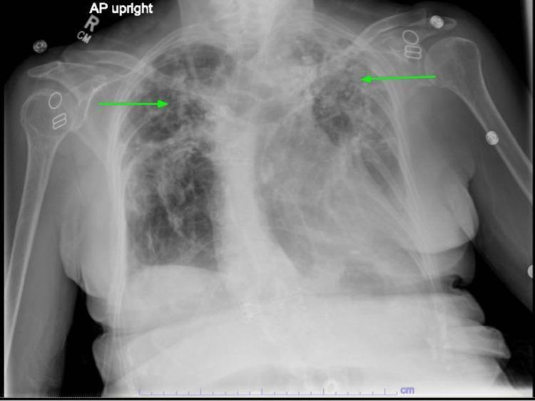
This diagnostic chest x-ray (AP upright projection) demonstrates bilateral upper lung predominant pathology, highlighted by green arrows. The imaging shows extensive areas of increased opacity and reticular markings consistent with chronic fibrotic changes, particularly pronounced in the left apical and upper lobe regions. These findings are characteristic of stage IV sarcoidosis with pulmonary fibrosis and associated architectural distortion, including apical scarring and traction bronchiectasis. The lower lung fields remain relatively lucent compared to the apices. Visible anatomical structures include the bony thorax, mediastinal silhouette, and bilateral humeri. Metallic densities are noted overlying the shoulder regions, likely representing external ECG leads or medical hardware. The radiograph serves as a clinical example of parenchymal lung disease progression and the typical upper-lobe distribution of sarcoidosis-related fibrosis.
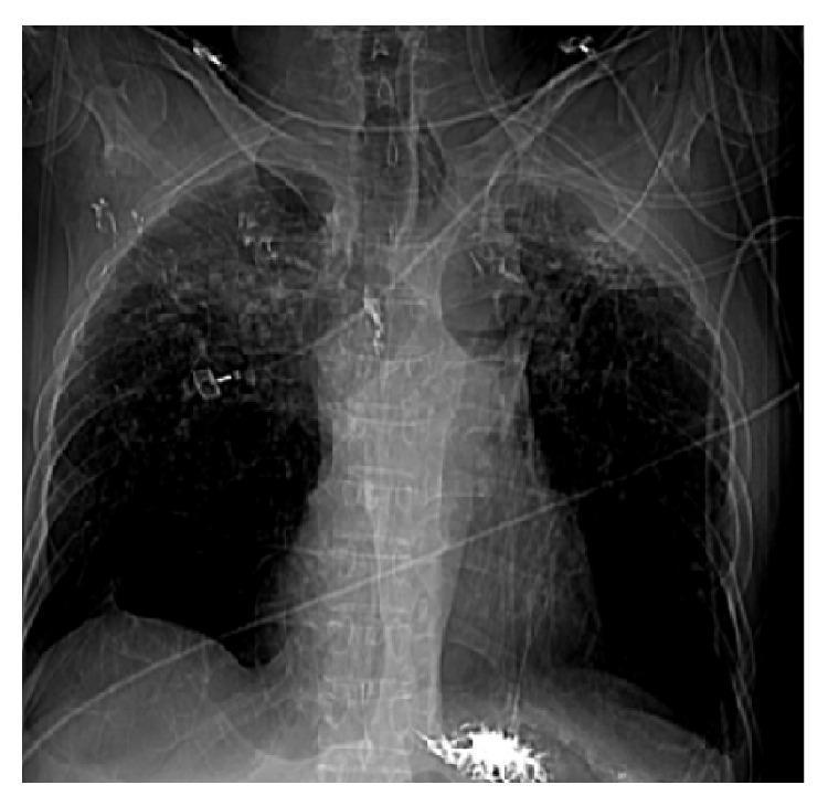
A frontal chest X-ray (CXR) demonstrating advanced pulmonary sarcoidosis manifestations. The upper lung fields bilaterally show extensive fibrocalcific scarring, characterized by dense, irregular, and mottled opacities consistent with stage IV sarcoidosis. There is significant volume loss in the upper lobes with superior retraction of the hila. In contrast, the lower lung zones exhibit increased lucency and hyperinflation, indicative of compensatory emphysema. Multiple medical devices are visible, including an endotracheal tube, enteric tube, and various monitoring leads and external cables, consistent with an intensive care clinical setting. The mediastinal contour appears distorted due to chronic fibrotic retraction. This image illustrates the end-stage progression of granulomatous lung disease, highlighting the transition from parenchymal inflammation to permanent structural fibrosis and secondary airflow obstruction.
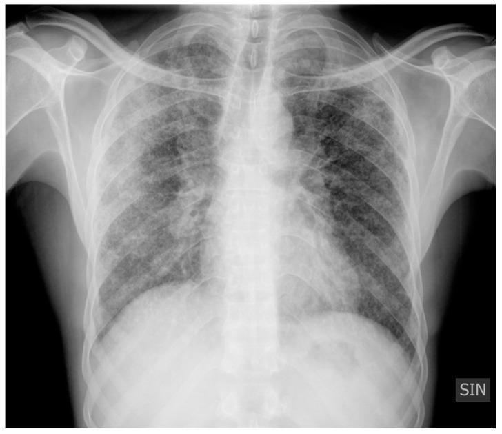
Anteroposterior (AP) chest X-ray demonstrating characteristic findings of pulmonary sarcoidosis, categorized as Scadding Stage II. The image reveals bilateral, symmetric hilar lymphadenopathy, characterized by prominent, well-defined densities at the lung roots. The lung parenchyma shows diffuse abnormalities, including multiple micronodules with a peribronchovascular distribution and extensive reticulonodular opacities scattered throughout both lung fields. Additionally, there is a localized area of pulmonary consolidation visible in the right lung. These findings represent a combination of thoracic adenopathy and active parenchymal infiltrates. The overall radiographic pattern is typical of a granulomatous lung disease, illustrating the transition from isolated nodal involvement to interstitial lung disease. This diagnostic image is intended for educational use in pulmonology and radiology to identify nodal and parenchymal patterns in sarcoidosis.
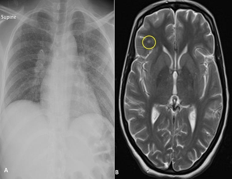
This composite diagnostic image consists of two panels labeled A and B, illustrating multi-organ manifestations of a systemic disease, likely tuberculosis or sarcoidosis. Panel A is a supine frontal chest X-ray (CXR) showing bilateral hilar lymphadenopathy and diffuse, fine, nodular opacities throughout both lung fields, characteristic of a miliary pattern. Panel B is a T2-weighted axial brain MRI (magnetic resonance imaging) showing a single, well-circumscribed hyperintense focal lesion in the left frontal lobe cortex (highlighted by a yellow circle), suggestive of a granuloma or tuberculoma. The MRI also demonstrates normal ventricular size and preserved sulcal patterns. This comparison is educationally significant for demonstrating the concurrent involvement of the respiratory and central nervous systems in disseminated granulomatous diseases. Target audience includes medical students and radiology residents focusing on infectious diseases and neuroradiology.
Sarcoidosis
Overview
- Fitzpatrick's Dermatology, p. 608
- Textbook of Family Medicine 9e, p. 481
Pathology
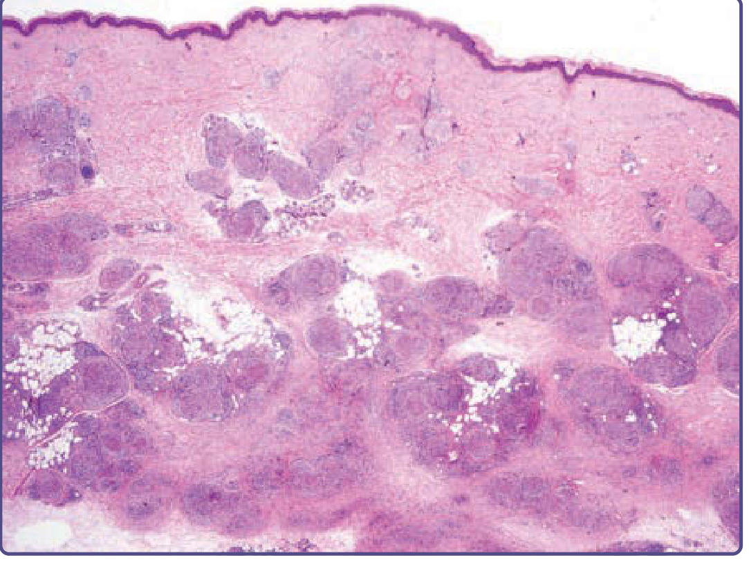
- Compact collection of mononuclear phagocytes (epithelioid cells - transformed monocytes)
- Multinucleated giant cells of the Langerhans type
- "Naked granulomas" - surrounded by very few lymphocytes (this is characteristic)
- Minimal or absent central necrosis (gross necrosis suggests tuberculosis or fungal infection instead)
- Progressive fibrosis can encapsulate granulomas, causing irreversible organ damage
Clinical Manifestations
Pulmonary (most common)
- Bilateral hilar lymphadenopathy in 50-85% of cases
- Symptoms: dyspnea, chronic non-productive cough, or in severe cases respiratory failure
- Can be asymptomatic
Skin (25-33%)
- Red-brown non-scaly papules and plaques, classically on the face around nose and mouth
- Lupus pernio: infiltration of the alar rim of the nose - highly associated with upper airway granulomatous infiltration and pulmonary fibrosis
- Lesions form within scars and tattoos
- Non-specific findings: erythema nodosum, ichthyosis, alopecia
Lofgren Syndrome
Other organ involvement
- Eyes: uveitis (can be severe)
- Cardiac: arrhythmias, heart block, cardiomyopathy
- Neurological (neurosarcoidosis): cranial nerve palsies, meningitis, mass lesions
- Renal/hepatic: hypercalcemia (from granuloma 1-alpha-hydroxylase activity), dysfunction
- Musculoskeletal: punched-out lytic lesions in hands/feet on X-ray, myopathy
Imaging - Scadding Chest X-Ray Stages
| Stage | Finding |
|---|---|
| 0 | Normal |
| I | Bilateral hilar +/- paratracheal adenopathy only |
| II | Adenopathy + pulmonary infiltrates |
| III | Pulmonary infiltrates only (no adenopathy) |
| IV | Pulmonary fibrosis |

Diagnosis
- Compatible clinical/radiographic picture
- Biopsy showing non-caseating granulomas (preferred biopsy site: skin lesions, lip mucosa, or transbronchial biopsy)
- Exclusion of other causes of granulomatous inflammation (especially TB and fungal infection - always send stains and cultures)
Useful tests
- Serum ACE (SACE): Reflects total granuloma burden. Elevated in all granulomatous diseases, so not specific enough for diagnosis alone. Useful for serial monitoring of disease course.
- Bronchoalveolar lavage: CD4/CD8 T-lymphocyte ratio >3.5 supports the diagnosis
- Gallium-67 scan: "Panda sign" (bilateral lacrimal + parotid uptake) + "Lambda sign" (bilateral hilar + right paratracheal uptake) are highly specific for sarcoidosis
- FDG-PET: More sensitive than gallium scan for detecting and monitoring active granulomatous inflammation
- MRI: Preferred for neurosarcoidosis and monitoring therapeutic response
- Fitzpatrick's Dermatology, pp. 608-609
- Cummings Otolaryngology, p. 1499
Treatment
When to treat
- Threatened organ failure (ocular, cardiac, neurological disease)
- Progressive, persistent, or symptomatic pulmonary disease
- Disfiguring skin disease (especially lupus pernio on the face)
- Persistent hypercalcemia, renal or hepatic dysfunction
- Severe fatigue/weight loss
Corticosteroids (first-line)
- Typical starting dose: prednisone 20-40 mg/day, then slow taper to a maintenance dose of 5-15 mg/day
- Duration: minimum 6-24 months (premature tapering causes relapse in >20% of cases)
- Inhaled corticosteroids alone are insufficient for chronic pulmonary sarcoidosis
Lofgren syndrome
Skin lesions
- Intralesional corticosteroid injections (every 2-4 weeks) - most effective
- Topical corticosteroids often insufficient (poor skin penetration)
- Widespread lesions: systemic corticosteroids
Second-line / steroid-sparing agents
| Drug | Use |
|---|---|
| Hydroxychloroquine | Mucocutaneous sarcoidosis, hypercalcemia, steroid-sparing |
| Methotrexate | Difficult cutaneous/pulmonary cases, steroid-sparing |
| Azathioprine | Steroid-sparing in chronic disease |
| Biologics (anti-TNF: infliximab, adalimumab) | Refractory disease - a 2025 meta-analysis (PMID 40393718) found evidence for efficacy of biologic and targeted synthetic therapies in sarcoidosis |
- Fishman's Pulmonary Diseases, pp. 604-627
- Textbook of Family Medicine 9e, p. 489
Prognosis
- Stage I: ~60-80% remit spontaneously within 2 years; systemic therapy usually not needed
- Stage II/III: About 50-60% show improvement; some develop chronic progressive disease
- Stage IV: Fibrosis is irreversible; about 5% of patients die from respiratory failure, cardiac sarcoidosis, or neurosarcoidosis
- Disease can be spontaneously self-limiting but can also relapse after apparent remission
Key Differentials
- Tuberculosis (caseating granulomas with necrosis; positive TB cultures/stains)
- Deep fungal infections (blastomycosis, histoplasmosis)
- Granuloma annulare
- Berylliosis (beryllium exposure history)
- Lymphoma (hilar adenopathy)
- Foreign body granuloma (tattoo pigment, paraffin)