
Explain cell injury completely with mote pictures
Reading File
Searching sources...
Finding Sources
Searching Images
cell injury mechanisms necrosis apoptosis pathology diagram
Reading File
Reading File
Searching Images
reversible irreversible cell injury swelling morphology
Reading File
Reading File
Reading File
Reading File
Reading File
Reading File
Now I have rich content from both Robbins textbooks. Let me fetch the key images and read the apoptosis/mechanisms sections.
Loading Image
Loading Image
Loading Image
Loading Image
Loading Image
Reading File
Loading Image
Loading Image
Reading File
Loading Image
Reading File
Reading File
Reading File
I now have comprehensive content from both Robbins textbooks. Let me fetch a couple more key images and then compile the full answer.
Loading Image
Loading Image
I now have comprehensive textbook content and all key diagrams. Here is the complete, image-rich explanation of cell injury.
Cell Injury - Complete Overview
Sources: Robbins & Kumar Basic Pathology (9780323790185); Robbins, Cotran & Kumar Pathologic Basis of Disease (9780443264528)
1. Overview: Cellular Responses to Stress
Cells constantly adjust their structure and function to maintain homeostasis. When faced with stress or injury, three outcomes are possible:
- Adaptation - new steady state achieved (hypertrophy, hyperplasia, atrophy, metaplasia)
- Reversible injury - cell injured but recovers when stress removed
- Irreversible injury - Cell death (necrosis or apoptosis)
Fig. 1.2 - The sequence of reversible and irreversible cell injury:
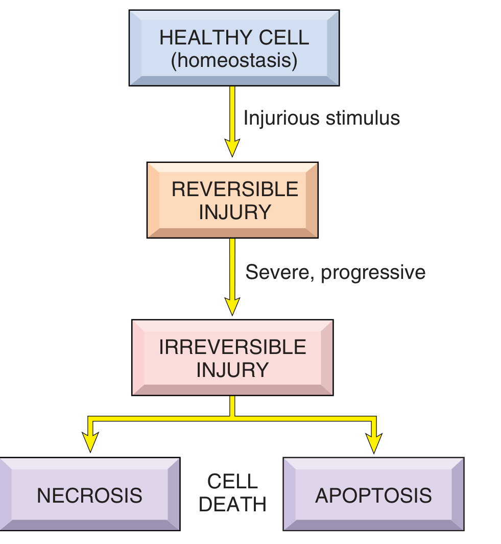
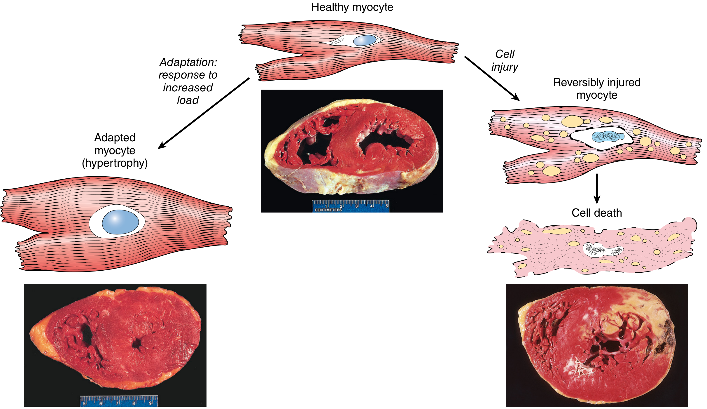
Fig. 2.2 - Relationship among healthy, adapted, reversibly injured, and dead myocardial cells (with gross cardiac specimen photos):
2. Causes of Cell Injury
Cell injury results from many different types of insult:
| Category | Examples |
|---|---|
| Hypoxia & Ischemia | Arterial obstruction, anemia, cardiorespiratory failure |
| Toxins | Alcohol, drugs, insecticides, CO, asbestos, cigarette smoke |
| Infectious agents | Bacteria, viruses, fungi, parasites |
| Immunologic reactions | Autoimmune diseases, allergic reactions, chronic inflammation |
| Genetic abnormalities | Down syndrome, sickle cell anemia, enzyme defects |
| Nutritional imbalances | Protein-calorie deficiency, vitamin deficiencies, obesity |
| Physical agents | Trauma, heat, cold, radiation, electric shock |
The cellular response depends on the type, duration, and severity of injury, and on the type and metabolic state of the target cell. For example, cardiac muscle dies after only 20-30 minutes of ischemia, whereas skeletal muscle survives 2-3 hours.
3. Sequence of Events in Cell Injury
Timing matters: Cell function is lost early, cell death follows later, and morphologic changes appear last.
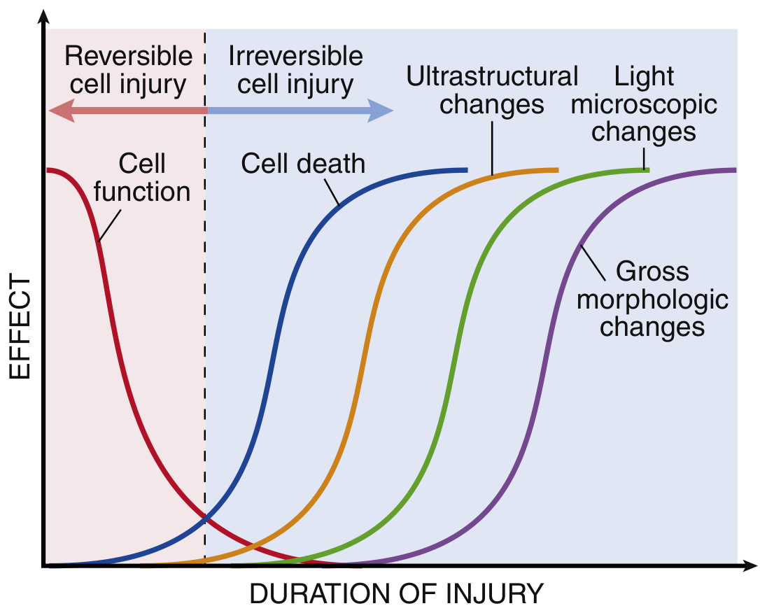
Key point: A cell can be functionally dead (e.g., an ischemic myocyte that stops contracting) before any morphologic changes are visible under the microscope.
4. Reversible Cell Injury
Reversible injury is a derangement of function and morphology from which the cell can recover if the damaging stimulus is removed.
Morphologic Changes
-
Cellular swelling (hydropic change / vacuolar degeneration)
- Most common change; seen with hypoxia and toxins
- Caused by failure of the ATP-dependent Na+/K+ pump → Na+ accumulates → osmotic water entry → cell and ER swell
- Small clear vacuoles appear in cytoplasm (distended ER segments)
-
Fatty change (steatosis)
- Lipid vacuoles appear in cytoplasm, especially in liver, heart, kidney
- Caused by toxins, malnutrition, diabetes, anoxia
Ultrastructural Changes (EM)
- Plasma membrane blebbing, blunting of microvilli
- Mitochondrial swelling with amorphous densities
- ER dilation with ribosome detachment
- Nuclear chromatin clumping
- Myelin figures (phospholipid collections from damaged membranes)
5. Cell Death
When injury exceeds the "point of no return," cells undergo irreversible injury and die by one of two main pathways:
| Feature | Necrosis | Apoptosis |
|---|---|---|
| Cell size | Increased (swelling) | Reduced (shrinkage) |
| Nucleus | Pyknosis → karyorrhexis → karyolysis | Fragmentation into nucleosome-sized pieces |
| Plasma membrane | Disrupted | Intact; altered structure |
| Cellular contents | Enzymatic digestion; may leak | Intact; released in apoptotic bodies |
| Inflammation | Frequent | No |
| Physiologic/pathologic | Always pathologic | Often physiologic; may be pathologic |
6. Necrosis
Necrosis occurs when injurious stimuli are severe or persistent and cause uncontrolled cell death with release of cellular contents, triggering inflammation.
Nuclear Changes in Necrosis
- Pyknosis - nuclear shrinkage, increased basophilia (DNA condenses into dark mass)
- Karyorrhexis - fragmentation of the pyknotic nucleus
- Karyolysis - fading of basophilia due to DNase digestion of DNA (complete dissolution in 1-2 days)
Cytoplasmic Changes
- Increased eosinophilia (denatured cytoplasmic proteins bind eosin)
- Glassy/homogeneous appearance
- Vacuolated "moth-eaten" cytoplasm
- Myelin figures more prominent than in reversible injury
Patterns of Tissue Necrosis
| Pattern | Morphology | Common Cause |
|---|---|---|
| Coagulative | Preserved architecture, firm texture, eosinophilic "ghost cells"; architecture maintained for days | Ischemia in most organs (infarcts); architecture preserved because enzymes are also denatured |
| Liquefactive | Digestion of dead cells → viscous liquid; creamy yellow pus | Bacterial/fungal infections; CNS hypoxic injury |
| Gangrenous | Coagulative necrosis of a limb; "wet" gangrene if superinfected | Loss of blood supply (usually lower limb) |
| Caseous | Soft, cheese-like, friable; structureless debris + granuloma | Tuberculosis (most common) |
| Fat necrosis | Focal fat destruction; chalky-white areas (saponification) | Acute pancreatitis |
| Fibrinoid | Bright pink amorphous deposits in vessel walls (immune complex deposition) | Immune vasculitis, malignant hypertension |
7. Apoptosis
Apoptosis is programmed cell death - an active process in which cells activate their own degradative machinery.
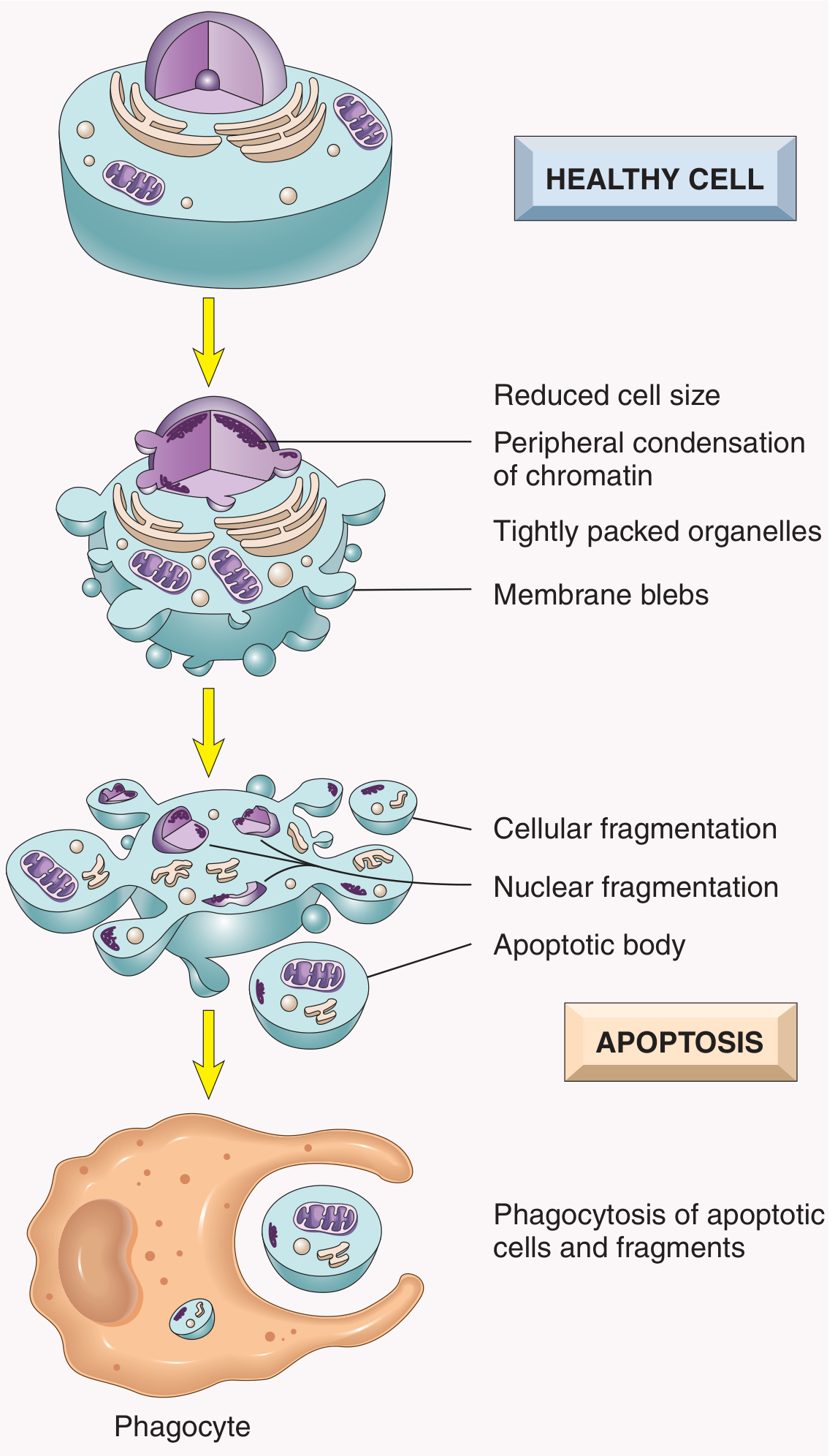
Key Features
- Cell shrinks and chromatin condenses peripherally
- Membrane blebs form; cell fragments into apoptotic bodies (membrane-bound fragments containing organelles and nuclear pieces)
- Plasma membrane remains intact throughout
- Apoptotic bodies are rapidly phagocytosed by macrophages - no leakage of contents, therefore no inflammation
Physiologic Causes
- Embryogenesis (organ sculpting, digit formation)
- Turnover of proliferative tissues (intestinal epithelium, lymphocytes)
- Involution of hormone-dependent tissues (endometrium)
- Elimination of autoreactive lymphocytes
- Decline of leukocytes at end of immune responses
Pathologic Causes
- DNA damage (radiation, chemotherapy)
- Accumulation of misfolded proteins (ER stress)
- Infections (viral cytopathic effects)
- Pathologic atrophy (duct obstruction)
Mechanisms of Apoptosis - Two Pathways
A. Mitochondrial (intrinsic) pathway - activated by DNA damage, misfolded proteins, oxidative stress, loss of survival signals:
- Pro-apoptotic proteins (BAX, BAK) increase; anti-apoptotic proteins (BCL-2) decrease
- Mitochondrial outer membrane becomes permeable
- Cytochrome c leaks into cytoplasm
- Cytochrome c + APAF-1 form the apoptosome → activates caspase-9 → activates executioner caspases (3, 6, 7)
B. Death receptor (extrinsic) pathway - activated by ligand binding (FasL, TNF):
- FasL binds Fas (CD95) → DISC complex forms → activates caspase-8 → activates executioner caspases
8. Mechanisms of Cell Injury
The four principal cellular targets are: mitochondria, cellular membranes, nucleus (DNA), and endoplasmic reticulum.
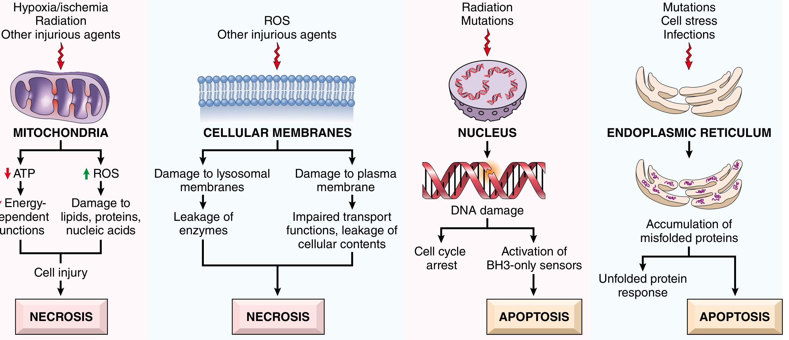
A. Mitochondrial Dysfunction
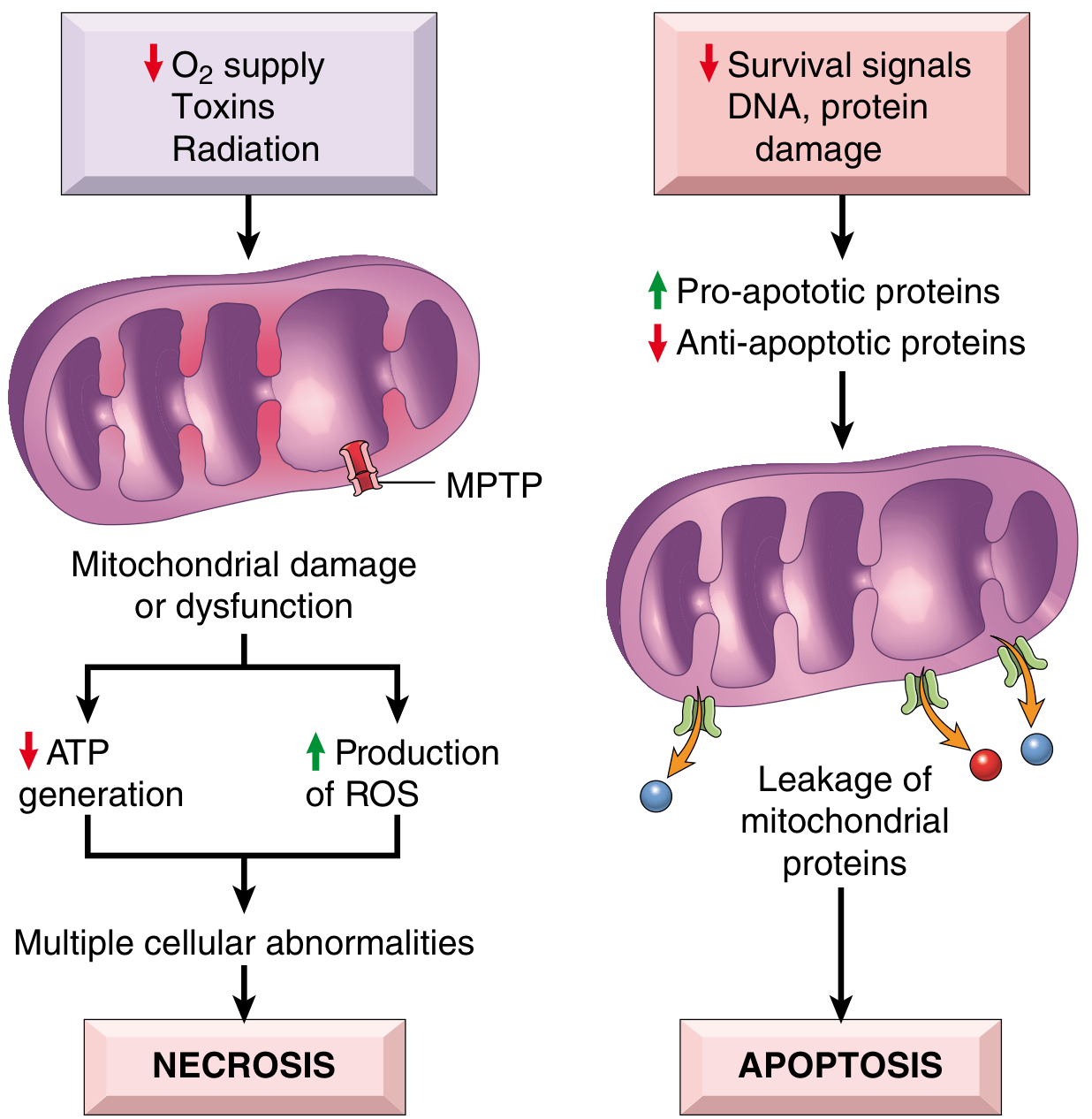
Two major pathways:
- Failure of oxidative phosphorylation → ATP depletion → necrosis
- Na+/K+-ATPase fails → Na+ accumulates → cell swelling
- Anaerobic glycolysis → lactic acid → decreased pH → enzyme inhibition
- Ribosomes detach from rough ER → protein synthesis fails
- Mitochondrial permeability transition pore (MPTP) opens → membrane potential lost → ATP depleted → necrosis
- Cytochrome c leakage → apoptosis (described above)
B. Oxidative Stress - Reactive Oxygen Species (ROS)
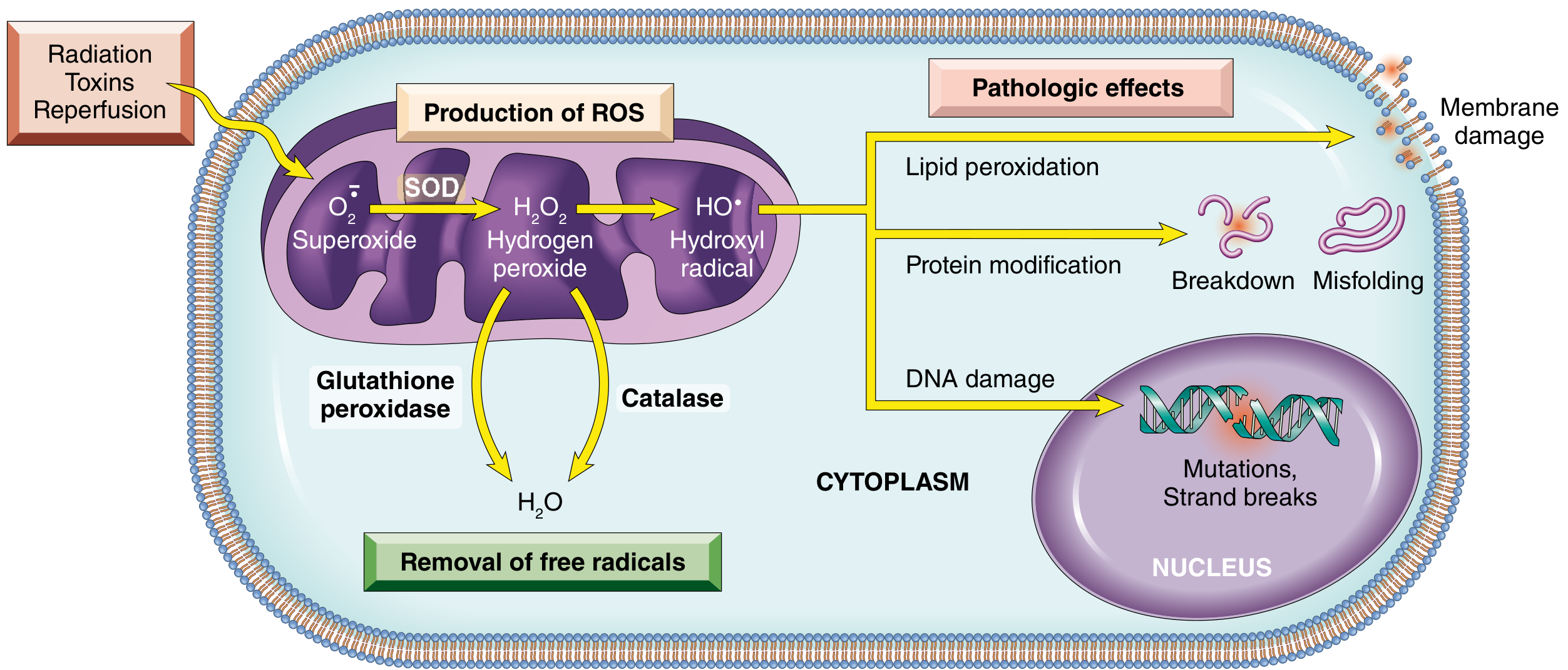
ROS are generated by:
- Mitochondrial electron transport (incomplete O2 reduction)
- Activated phagocytes (respiratory burst)
- Radiation, toxins, reperfusion
Principal ROS and their effects:
| Free Radical | Production | Removal | Effect |
|---|---|---|---|
| Superoxide (O2-) | Mitochondria, phagocytes | SOD converts to H2O2 | Direct damage to lipids, proteins, DNA |
| H2O2 | From superoxide via SOD | Catalase, glutathione peroxidase | Converted to •OH and ClO- which destroy cells |
| Hydroxyl radical (•OH) | From H2O2 | Glutathione peroxidase | Most reactive; direct damage to all macromolecules |
| Peroxynitrite (ONOO-) | O2- + NO | Mitochondrial enzymes | Damages lipids, proteins, DNA |
Pathologic effects of ROS:
- Lipid peroxidation of membranes (autocatalytic chain reaction)
- Protein modification - oxidation of side chains, cross-linking, protease cleavage
- DNA damage - strand breaks, mutations
Antioxidant defenses:
- Superoxide dismutase (SOD)
- Catalase (peroxisomes)
- Glutathione peroxidase
- Vitamins E, A, C, beta-carotene
- Transferrin, ferritin, ceruloplasmin (chelate free iron/copper)
C. Membrane Damage
Damage to membranes is a critical event in cell injury:
- Plasma membrane damage → loss of osmotic balance, impaired transport, leakage of cellular contents
- Lysosomal membrane damage → release of lysosomal enzymes (DNases, RNases, proteases, phosphatases) → enzymatic digestion of cell components
Mechanisms of membrane damage:
- ROS-induced lipid peroxidation
- Phospholipase activation by high cytosolic Ca2+
- Calpains (Ca2+-activated proteases) degrade membrane skeleton proteins
- Loss of phospholipid synthesis (ATP depletion)
D. Disturbance in Calcium Homeostasis
- Normal cytosolic Ca2+ is very low (~0.1 µmol/L); injury causes influx from extracellular space and release from ER/mitochondria
- Increased cytosolic Ca2+ activates:
- Phospholipases → membrane damage
- Proteases → cytoskeletal and membrane protein degradation
- ATPases → accelerates ATP depletion
- Endonucleases → nuclear chromatin fragmentation
- Mitochondrial permeability → leads to apoptosis or necrosis
E. ER Stress
- ER is responsible for protein folding; injury → accumulation of misfolded proteins → unfolded protein response (UPR)
- If excessive, UPR triggers apoptosis
- Example: diseases of misfolded proteins include alpha-1 antitrypsin deficiency, Alzheimer disease
F. DNA Damage
- Radiation, chemotherapy, ROS, toxins can damage DNA
- Mild damage → cell cycle arrest → DNA repair
- Severe DNA damage → activation of BH3-only pro-apoptotic sensors → apoptosis
- p53 is a critical mediator that senses DNA damage and drives either repair or apoptosis
9. Clinicopathologic Examples
Hypoxia and Ischemia
The classic and most common cause of cell injury. The sequence in ischemia:
- Reduced O2 → oxidative phosphorylation ceases → ATP falls
- Anaerobic glycolysis → glycogen depleted, lactic acid accumulates, pH falls
- Na+/K+-ATPase fails → cellular swelling
- Cytosolic Ca2+ rises → activates phospholipases, proteases
- Irreversible injury: MPTP opens, lysosomal rupture, membrane breakdown → necrosis
Ischemia-Reperfusion Injury
Paradoxically, restoring blood flow after ischemia can worsen injury:
- Burst of ROS from reperfused mitochondria and activated neutrophils
- MPTP opens with calcium influx on reperfusion
- Neutrophil-mediated inflammation amplifies damage
- Clinically relevant in MI, stroke, organ transplantation
Chemical (Toxic) Injury
- Direct toxicity: Mercury binds sulfhydryl groups; cyanide inhibits cytochrome oxidase
- Metabolic conversion to toxic products: CCl4 → CCl3 radical by smooth ER → lipid peroxidation → liver necrosis; acetaminophen overdose → toxic NAPQI metabolite
10. Newer Mechanisms of Cell Death (Beyond Necrosis and Apoptosis)
| Type | Key Feature | Trigger |
|---|---|---|
| Necroptosis | Programmed necrosis; caspase-independent; RIPK1/RIPK3 pathway; releases DAMPs | Viral infection, TNF |
| Pyroptosis | Caspase-1/11-mediated; releases IL-1β; highly inflammatory | Intracellular bacteria, danger signals |
| Ferroptosis | Iron-dependent; oxidative destruction of membrane lipids; glutathione/GPX4 axis | Oxidative stress, glutathione depletion |
Summary Table: Reversible vs. Irreversible Injury
| Feature | Reversible | Irreversible |
|---|---|---|
| Cellular swelling | + | ++ |
| Fatty change | + | + |
| Membrane blebs | + | ++ (rupture) |
| Mitochondrial changes | Swelling, small densities | Large amorphous densities, MPTP opening |
| Lysosomes | Intact | Ruptured |
| Nuclear changes | Chromatin clumping | Pyknosis → karyorrhexis → karyolysis |
| Membrane permeability | Mildly impaired | Severely disrupted |
| ATP | Decreased | Depleted |
| Outcome | Recovery if stimulus removed | Cell death (necrosis or apoptosis) |
References: Robbins & Kumar Basic Pathology, pp. 16-45 | Robbins, Cotran & Kumar Pathologic Basis of Disease, pp. 43-65
This is a shared conversation. Sign in to Orris to start your own chat.