
Thyroid swelling with anatomy and proper management in surgery
thyroid gland anatomy relations blood supply diagram
thyroid gland anterior view lobes isthmus trachea
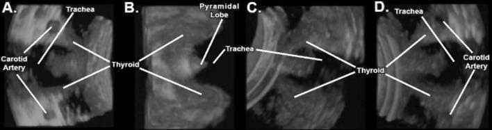
This diagnostic image set consists of four panels (A-D) showcasing 3D/4D ultrasound (US) reconstructions of the thyroid gland and adjacent cervical anatomy. The sequence demonstrates the multi-planar rotational capabilities of volume ultrasound for anatomical visualization.
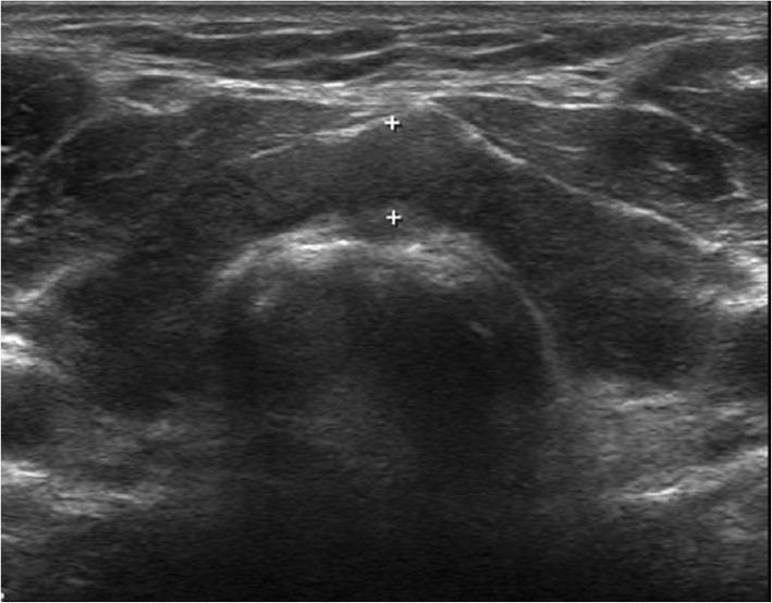
This diagnostic image is a B-mode ultrasound of the thyroid gland in a transverse (axial) view. The sonogram demonstrates diffuse enlargement of the thyroid parenchyma (goiter) with a notably heterogeneous echotexture across both the left and right lobes and the connecting isthmus. The thyroid tissue shows a mix of hypoechoic and hyperechoic regions, lacking the normal uniform fine-grained appearance. Measurement calipers (+) are positioned to assess the thickness of the isthmus overlying the trachea, which appears thickened. The irregular echo pattern and diffuse enlargement are hallmark sonographic features of chronic autoimmune thyroiditis, such as Hashimoto thyroiditis. Key anatomical landmarks visible include the anterior strap muscles and the underlying trachea, which presents with a characteristic curved, hyperechoic surface and posterior acoustic shadowing.
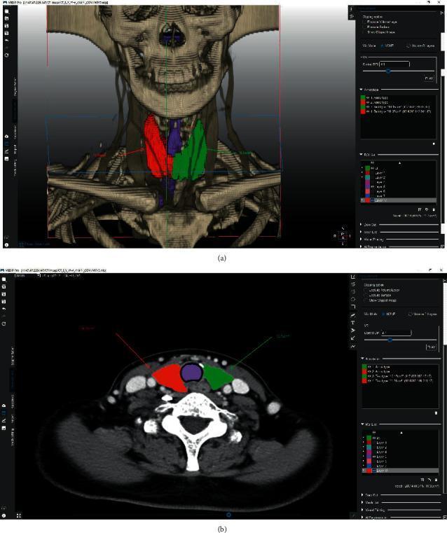
This medical visual demonstrates volumetric segmentation of the thyroid gland using specialized medical imaging software. Image (a) presents a 3D reconstruction overlay on a CT-derived skeletal model of the neck and upper thorax. The segmentation highlights the anatomical lobes: the right thyroid lobe is colored green, the left lobe is red, and the isthmus/central region is purple. Image (b) shows a corresponding axial CT slice at the level of the thyroid. In this cross-sectional view, the colored segmentation masks (red for left lobe, green for right lobe, and purple for the central isthmus) are superimposed on the soft tissue structures anterior to the trachea. The image illustrates the application of semi-automated graph-cut algorithms for preoperative planning in endocrine surgery, specifically for thyroid lobectomy. Key anatomical landmarks visible include the trachea, carotid arteries, jugular veins, and cervical vertebrae. This content is designed for intermediate to advanced medical training in radiology and surgical oncology, focusing on thyroid anatomy and computer-assisted volumetric measurement.
multinodular goitre neck swelling clinical photograph
thyroidectomy AND goiter management
Thyroid Swelling: Anatomy and Surgical Management
1. Anatomy of the Thyroid Gland
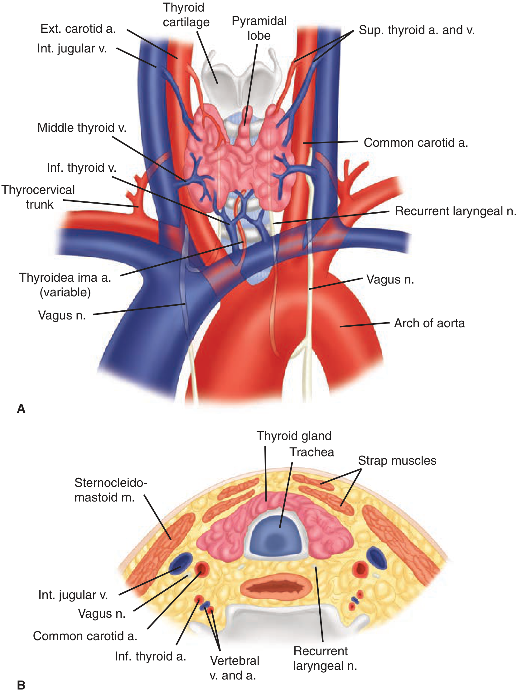
- Anterior: sternothyroid and sternohyoid (strap) muscles
- Lateral: carotid sheath (common carotid artery, internal jugular vein, vagus nerve)
- Posterior: trachea and esophagus (esophagus lies more to the left)
- Fascial attachments: the pretracheal fascia invests the gland and condenses superiorly as the suspensory (Berry's/median) ligament above the isthmus, and posteromedially as the ligament of Berry, which firmly binds the gland to the trachea/cricoid - this is where the recurrent laryngeal nerve (RLN) is most vulnerable during surgery.
- Arterial: superior thyroid artery (from external carotid), inferior thyroid artery (from thyrocervical trunk off the subclavian), and occasionally a thyroidea ima artery (direct from the aorta/innominate, in 1-4%).
- Venous: superior and middle thyroid veins drain to the internal jugular vein; inferior thyroid veins drain via a plexus into the brachiocephalic veins.
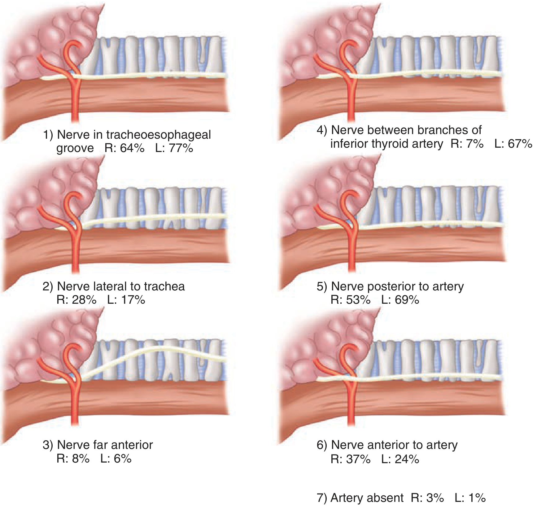
- Recurrent laryngeal nerve (RLN): the left RLN loops around the aortic arch, the right around the subclavian artery; both ascend in the tracheoesophageal groove, cross deep to the inferior thyroid artery, and enter the larynx below the inferior constrictor. A non-recurrent right RLN occurs in 0.5-1% (associated with aberrant subclavian artery) - identifying this variant preoperatively/intraoperatively is essential to avoid injury.
- External branch of the superior laryngeal nerve (EBSLN): runs close to the superior thyroid vessels; injury causes loss of voice pitch/projection.
2. Classification of Thyroid Swellings
| Category | Subtypes |
|---|---|
| Simple goitre (euthyroid) | Diffuse hyperplastic (physiological, pubertal, pregnancy); Multinodular goitre |
| Toxic | Diffuse (Graves' disease); Toxic multinodular goitre; Toxic adenoma |
| Neoplastic | Benign (adenoma); Malignant (papillary, follicular, medullary, anaplastic, lymphoma) |
| Inflammatory | Autoimmune (Hashimoto's), subacute (De Quervain's), acute suppurative, Riedel's thyroiditis |
3. Clinical Assessment
- History: duration, growth rate, pain, compressive symptoms (dysphagia, dyspnoea, stridor, voice change), thyroid status symptoms, family history, irradiation exposure
- Examination: swallowing test (moves with deglutition, confirms thyroid origin), tongue protrusion test (thyroglossal cyst), consistency, mobility, lymph nodes, tracheal deviation, retrosternal extension (Pemberton's sign)
- Red flags for malignancy: rapid growth, hardness, fixity, hoarseness, cervical lymphadenopathy, age extremes, male sex, history of neck irradiation
- Thyroid function tests (TSH, free T4) and thyroid antibodies
- Ultrasonography - gold standard first-line imaging; assesses nodule characteristics and guides biopsy
- FNAC (fine-needle aspiration cytology) under ultrasound guidance for nodules with suspicious sonographic features - reported using the Thy1-Thy5 system (Thy1 non-diagnostic, Thy2 benign, Thy3 follicular/indeterminate, Thy4 suspicious, Thy5 malignant)
- Cross-sectional imaging (CT/MRI) for retrosternal extension or large goitres, ideally in the surgical neck position
- Laryngoscopy for baseline vocal cord assessment, mandatory before and after thyroid surgery, especially with prior neck/chest surgery
4. Surgical Management
Indications for thyroidectomy
- Hyperthyroidism failing or unsuitable for medical/radioiodine therapy
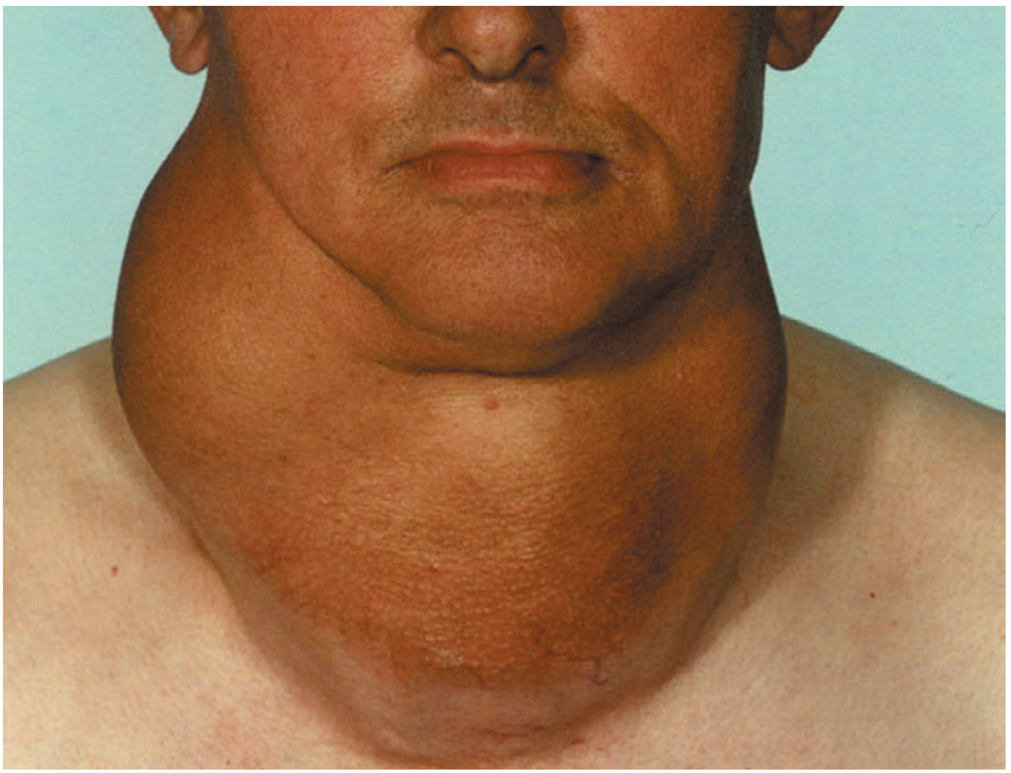
- Goitre with or without local compressive symptoms (dysphagia, dyspnoea, stridor)
- Thyroid nodules suspicious of, or diagnostic for, malignancy
- Retrosternal extension with airway compromise
- Cosmetic concern with large goitre
- Graves' disease in patients planning pregnancy within 6 months, or with a large goitre/nodule
Extent of resection
- Lobectomy/hemithyroidectomy: for a solitary indeterminate nodule or unilateral disease without proven malignancy
- Near-total/total thyroidectomy: for bilateral multinodular goitre, Graves' disease, or confirmed/high-risk thyroid cancer (per American Thyroid Association criteria: size >4 cm, extrathyroidal extension, nodal disease, or need for postoperative radioiodine)
- Subtotal thyroidectomy: largely historical, leaves 3-5 g of tissue - higher recurrence risk, now less used
- Isthmusectomy: isolated isthmus disease
Preoperative preparation
- Confirm euthyroid state before elective surgery (antithyroid drugs, beta-blockers, or iodine/Lugol's solution preoperatively in Graves' disease to reduce vascularity and risk of thyroid storm)
- Baseline vocal cord assessment
- Adequate cross-sectional imaging if retrosternal extension suspected
Operative technique (essentials)
- Transverse collar (Kocher's) incision
- Elevation of subplatysmal flaps, division of strap muscles if needed
- Ligation of superior and inferior thyroid vessels close to the gland capsule to preserve parathyroid blood supply
- Identification and preservation of the RLN throughout its course, particularly near the ligament of Berry
- Identification and preservation of parathyroid glands, with judicious autotransplantation if devascularized
- Intraoperative nerve monitoring is increasingly used to reduce RLN injury risk
Complications of thyroidectomy
- Recurrent laryngeal nerve injury (transient or permanent, 1-2% in experienced hands) - causes hoarseness or airway compromise if bilateral
- Hypoparathyroidism/hypocalcaemia - from devascularization or inadvertent removal of parathyroid glands during total thyroidectomy; managed with calcium and vitamin D supplementation, monitored via serum calcium/PTH
- Post-operative haemorrhage/haematoma - can cause airway compromise, requires clip removal at bedside if severe
- Thyroid storm - rare but life-threatening, can be precipitated by intraoperative gland manipulation in an inadequately prepared hyperthyroid patient; managed with beta-blockers, antithyroid drugs, steroids, and supportive care
- Wound infection, hypertrophic scarring
- Hungry bone syndrome - severe hypocalcaemia post-thyroidectomy in previously hyperthyroid patients due to high bone turnover
Special situations
- Graves' disease: total thyroidectomy gives immediate control and no recurrence risk but 100% postoperative hypothyroidism requiring lifelong replacement; subtotal thyroidectomy avoids this but carries lifelong recurrence risk (up to 5%) and progressive thyroid failure.
- Thyroid storm: managed medically first (beta-blockers, thionamides, iodine, steroids); surgery deferred until stable unless it is the precipitating/definitive treatment.
- Graves' disease in children: thyroidectomy reserved for those failing medical therapy, ideally performed by high-volume thyroid/paediatric surgeons given lifelong risk of hypoparathyroidism.
Pathophysiology of thyroid swelling
Pathophysiology of Thyroid Swelling
1. Normal Thyroid Physiology (Background)
2. Simple (Non-Toxic) Goitre - TSH-Driven Hyperplasia
- Iodine deficiency - the classical cause. Daily iodine requirement is ~0.1-0.15 mg; endemic goitre occurs in mountainous/inland regions with low soil/water iodide (Himalayas, Andes, Alps) or areas with high calcium content in water (also goitrogenic), where reduced hormone synthesis raises TSH chronically - Bailey and Love's Short Practice of Surgery, 28e.
- Dyshormonogenesis - inherited enzyme defects (e.g., in thyroid peroxidase, iodide transport) impair hormone synthesis despite adequate iodine intake, producing sporadic goitres, often with a family history. Environmental iodine excess can partly compensate (goitre is rare in iodine-rich Iceland despite genetic predisposition elsewhere).
- Puberty/pregnancy-associated goitre - physiological increased hormone demand causes transient TSH-driven hyperplasia.
3. Toxic Goitre - Autonomous Hormone Production
- An autoimmune disorder in which thyroid-stimulating immunoglobulins (TSI), present in ~90% of patients, bind and activate the TSH receptor, mimicking TSH action - stimulating adenylyl cyclase and driving both hormone hypersecretion and diffuse follicular hyperplasia.
- Strong genetic component (30-40% monozygotic twin concordance) with polymorphisms in immune-regulatory genes (CTLA4, PTPN22, IL2RA) and TSHR gene variants (GWAS-identified).
- Extrathyroidal manifestations: activated CD4+ T cells secrete cytokines that stimulate retro-orbital fibroblast proliferation and glycosaminoglycan deposition, causing exophthalmos (ophthalmopathy); similar glycosaminoglycan/lymphocytic infiltration in the pretibial dermis produces pretibial myxoedema.
- Histologically: diffuse hyperplasia, tall crowded follicular epithelium with papillary infoldings and scalloped colloid resorption at follicle margins.
4. Inflammatory (Thyroiditis)
- Breakdown of self-tolerance to thyroid autoantigens (thyroglobulin, thyroid peroxidase), with genetic predisposition (CTLA4, PTPN22, IL2RA polymorphisms - overlapping with Graves' susceptibility genes).
- Thyroid destruction occurs via CD8+ cytotoxic T-cell mediated killing, cytokine-mediated damage (Th1-driven interferon-γ recruiting macrophages), and possibly antibody-dependent mechanisms.
- Progressive lymphocytic infiltration (with germinal centres) and fibrosis replace parenchyma; atrophic follicles lined by oncocytes (Hürthle cells) are characteristic. Net result: goitre with progressive hypothyroidism (though an initial hyperthyroid "hashitoxicosis" phase can occur from follicular disruption).
5. Neoplastic Swellings
- Follicular adenoma - benign, encapsulated, monoclonal proliferation; most are non-functional, a minority are hormone-producing ("toxic adenoma").
- Thyroid carcinoma (papillary, follicular, medullary, anaplastic) - driven by acquired genetic alterations (e.g., RET/PTC rearrangements and BRAF mutations in papillary carcinoma; RAS mutations in follicular carcinoma; RET germline/somatic mutations in medullary carcinoma arising from parafollicular C-cells). Risk factors for malignancy in a nodule include solitary nodule, younger age, male sex, prior head/neck irradiation, and a "cold" (non-iodine-avid) nodule on scanning. Benign neoplasms outnumber carcinomas roughly 10:1, and most thyroid cancers carry an excellent prognosis (>90% survival at 20 years).
Summary Table
| Type | Core Mechanism | Result |
|---|---|---|
| Simple goitre | Chronic TSH stimulation (iodine deficiency, dyshormonogenesis) | Diffuse then nodular hyperplasia, euthyroid |
| Multinodular goitre | Clonal selection of autonomously growing follicular cells within a hyperplastic gland | Irregular asymmetric enlargement, may become toxic |
| Graves' disease | TSI autoantibody activates TSH receptor | Diffuse hyperplasia + thyrotoxicosis + ophthalmopathy/dermopathy |
| Toxic adenoma/MNG | Somatic TSHR/GNAS activating mutations | Autonomous "hot" nodule, thyrotoxicosis |
| Hashimoto thyroiditis | T-cell mediated autoimmune destruction | Goitre with progressive hypothyroidism |
| Neoplasia | Somatic driver mutations (BRAF, RET/PTC, RAS) | Discrete nodule, benign or malignant |