
Plantar warts treatment
"plantar wart" AND treatment
plantar wart verruca plantaris clinical photo
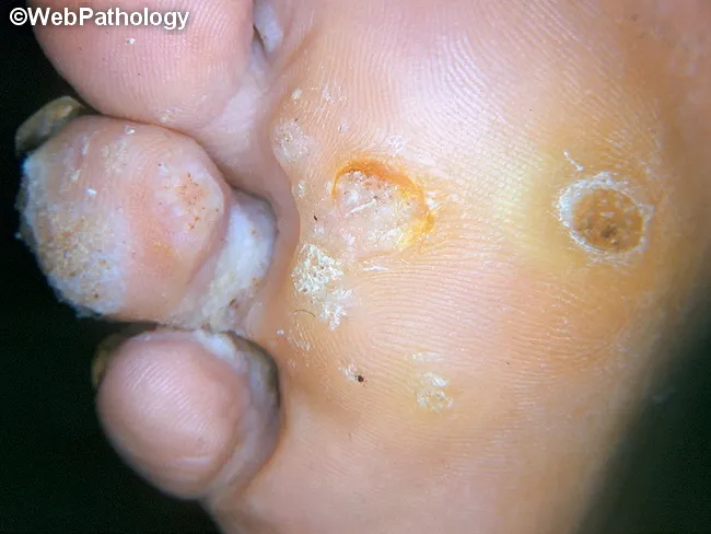
Clinical photography of a plantar foot displaying a verruca plantaris (plantar wart). Modality is a high‑quality gross image used in dermatology documentation. Precise location is the plantar surface of the foot (sole), with a focal, endophytic lesion that is rough and hyperkeratotic. Visual features include a circular to oval papule with a central crater or indent, irregular surface texture, and surrounding callus‑like thickening. Punctate black dots representing thrombosed capillaries may be seen within the lesion, while surrounding epidermis shows white scaling and fissuring. The lesion is typically mildly elevated above the surrounding skin and may be tender with pressure; it often coexists with other warts on the same sole. The clinical impression is verruca plantaris caused by human papillomavirus infection, with characteristic epidermal hyperplasia, papillomatosis, and hyperkeratosis evident on gross inspection. Differential diagnoses include callus/corn, plantar keratoderma, and foreign body reaction; histology would show koilocytosis and viral cytopathic effects if examined. This image supports clinical decision making, differential diagnosis, and treatment planning (topical salicylic acid, cryotherapy, or laser), and serves as an educational reference for students, residents, and clinicians evaluating plantar lesions. Clinical correlation includes assessing for additional lesions and patient history of HPV exposure; follow-up imaging is not required.
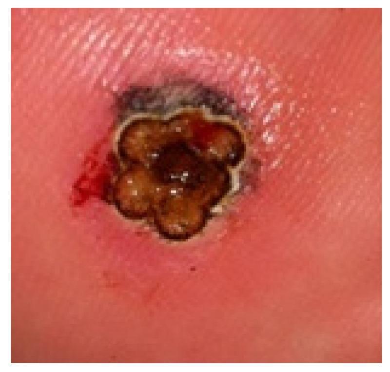
This clinical photograph displays a large plantar wart (verruca plantaris) measuring over 4 mm in diameter on the weight-bearing surface of the foot. The lesion is characterized by a raised, hyperkeratotic appearance with a clustered, multi-lobulated morphology resembling a 'mosaic' pattern. The central tissue exhibits dark brown to black discoloration, likely due to the application of India ink used in laser therapy protocols to enhance thermal absorption. A distinct, pale rim or halo surrounds the lobules, followed by a wider zone of erythematous, inflamed perilesional skin. Minor punctate bleeding is visible on the lateral margin, typical of exposed capillaries (thrombosed capillaries) following the removal of overlying hyperkeratosis. This image serves as a clinical example of a recalcitrant plantar wart prepared for laser treatment, illustrating the anatomical challenges of treating thick epidermal lesions on the plantar surface.
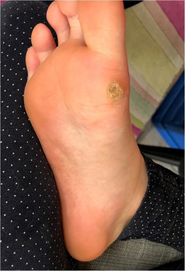
This clinical photograph displays a solitary, deep plantar wart (verruca plantaris) located on the weight-bearing plantar surface of the human foot, specifically over the metatarsal region. The lesion is well-circumscribed and approximately circular, characterized by a central area of significant hyperkeratosis. The surface texture is rough, verrucous, and uneven, exhibiting a beige to yellowish-tan discoloration that contrasts with the surrounding healthy pink skin. A slightly raised, indurated outer rim demarcates the lesion from the adjacent tissue. The visual findings are consistent with a myrmecia-type wart typically associated with Human Papillomavirus type 1 (HPV 1). Key educational features include the disruption of normal dermatoglyphics and the thick keratotic plug, which are hallmark diagnostic signs of plantar verrucae in dermatology.
"plantar wart" AND treatment
Plantar Warts (Verrucae Plantaris) - Treatment
Overview
Treatment Goals
- Wart resolution
- Avoidance of scarring or permanent sequelae
- Ideally, induction of lifelong immunity to that HPV type
Treatment Options
1. Salicylic Acid (First-Line, Patient-Applied)
- Concentrations of 10-26% OTC are effective and well-studied
- Soak the affected area in water for 5-10 min, apply topical, let dry, cover with a strip bandage for 24 hours - repeat daily
- Debride superficial keratinous debris with a pumice stone, emery board, or table knife (wash between uses)
- Results are conflicting - some studies show equal efficacy to cryotherapy, others show marked inferiority
- Good choice for patients who prefer self-treatment at home
2. Cryotherapy (First-Line, Office-Based)
- Cure rate: 20-50% with repeated applications over several months
- Liquid nitrogen is preferred (freezes to a lower temperature than other agents)
- Optimal technique: sustained 10-second freeze with a spray gun + a 30-45 second thaw time; produces a blister in 1-2 days
- Frequency: every 2-3 weeks (just as the old blister peels off)
- A single freeze-thaw cycle may be as effective as two cycles
- Complications: hypopigmentation, depigmentation, scarring, rarely digital nerve damage
- Contraindications/caution: Fanconi anemia, cryoglobulinemia, Raynaud phenomenon, poor peripheral circulation (risk of severe blistering)
3. Cantharidin (0.7%)
- Applied to the wart in office, allowed to dry, covered with occlusive tape for 24 hours (or until burning occurs)
- A blister forms in 24-72 hours; can be as painful as or more painful than cryotherapy blisters
- Effective and useful, though not FDA-approved in the US
- "Doughnut warts" (central clearing with annular recurrence) can complicate cantharone or cryotherapy
4. Immunotherapy
- Intralesional Candida antigen: efficacious, cost-effective, least traumatic of the alternatives, with virtually no residual scarring; can work on distant warts via systemic immune stimulation
- Imiquimod (5% cream): immune response modifier applied topically; useful especially for refractory cases
- Intralesional interferon: less commonly used due to systemic side effects
5. Intralesional Injections
- Bleomycin: highly effective, especially for recalcitrant plantar warts; painful on injection
- Candida antigen: as above, immunotherapy approach
- Recent RCT (2026, PMID: 40889709): intralesional acyclovir vs. cryotherapy - a new approach showing promise for plantar warts
6. Chemical Cautery / Topical Chemotherapy
- Trichloroacetic acid (TCA) and other caustic acids
- 5-Fluorouracil (5-FU): topical chemotherapy, useful in refractory cases
- Silver nitrate: cure rates higher than placebo; caution due to risk of silver impregnation into skin
- Nitric-zinc complex solution: 2024 case series of 72 patients showed promising results (PMID: 39205281)
7. Laser Therapy
- CO2 laser and pulsed-dye laser (PDL): used for refractory cases
- PDL selectively targets blood vessels supplying the wart
- Higher cost and more specialized equipment required
8. Occlusive Therapy (Duct Tape)
- Initial enthusiasm has not been substantiated by follow-up studies
- In adults, efficacy is very low - only 20% resolved in 2 months, with 75% recurrence
- If used, tape must be kept on at least 6.5-7 days/week
- Good option for young children (<12 years) where spontaneous resolution is high, or for patients unwilling to undergo other treatments
9. Surgical Excision
- Reserved for refractory cases
- Risk of scarring on the plantar surface which can cause post-excision pain
- A 2026 case report (PMID: 41988754) describes surgical excision technique
10. Electrosurgery / Infrared Coagulation
- Destructive modalities used when other options fail
- Generally not first-line due to pain and scarring risk
Special Considerations
| Population | Notes |
|---|---|
| Children <12 years (HPV-1) | High spontaneous resolution (>50%); favor conservative approaches (duct tape, watchful waiting) |
| Adults | More refractory; often require repeated destructive therapy or immunotherapy |
| Immunocompromised | Warts are more extensive and refractory; immunotherapy may be less effective; higher malignant transformation risk |
| Diabetics | Aggressive debridement should be avoided; ulceration risk |
| Mosaic warts | Larger clusters are notoriously difficult to treat; combination therapy often needed |
Treatment Algorithm (Summary)
Patient presents with plantar wart
        ↓
Consider watchful waiting (especially children <12)
        ↓
First-line: Salicylic acid (self-applied) OR cryotherapy (office)
        ↓
If refractory after 3 months:
  → Cantharidin, intralesional Candida antigen, bleomycin
        ↓
Further refractory:
  → 5-FU, imiquimod, pulsed-dye laser, CO2 laser
        ↓
Last resort: Surgical excision (with caution re: scarring)