
Endoscopic images while dissectomy
Reading File
Endoscopic discectomy involves removing herniated disc material through a small endoscope, visualizing the disc space, nerve roots, and surrounding structures. Let me search the library for relevant images and content.
Searching sources...
Finding Sources
Finding Sources
Good - found content in Campbell's Operative Orthopaedics. Let me read the key section on endoscopic discectomy techniques and look for images.
Reading File
Reading File
Reading File
Let me search specifically for endoscopic images of discectomy in the textbook.
Finding Sources
Let me search the block around the endoscopic discectomy section for images (markdown image syntax).
Reading File
I can see there's an image at line 7901 area. Let me also check the earlier section of block19 for endoscopic technique images, and read lines around 7893-7905:
Reading File
Reading File
I found one image. Let me also look at the block19 section which was referenced for TECHNIQUE 44.2 and check for more endoscopic images in both block19 and block20.
Reading File
Reading File
I can see several images. Let me read more of this section to find actual endoscopic view images:
Reading File
Reading File
I now have found multiple relevant images. Let me fetch the key endoscopic and intraoperative images:
Loading Image
Loading Image
Loading Image
Excellent! I have the key endoscopic intraoperative image showing the "Arch of Wagner" view. Let me also fetch the other images from that section:
Loading Image
Loading Image
I now have a comprehensive set of endoscopic discectomy images from Campbell's Operative Orthopaedics (15th Ed, 2026). Here is a full illustrated overview:
Endoscopic Images During Discectomy
Source: Campbell's Operative Orthopaedics, 15th Ed., 2026 - Chapters 44 & 46
1. Approach Planning - Surface Marking
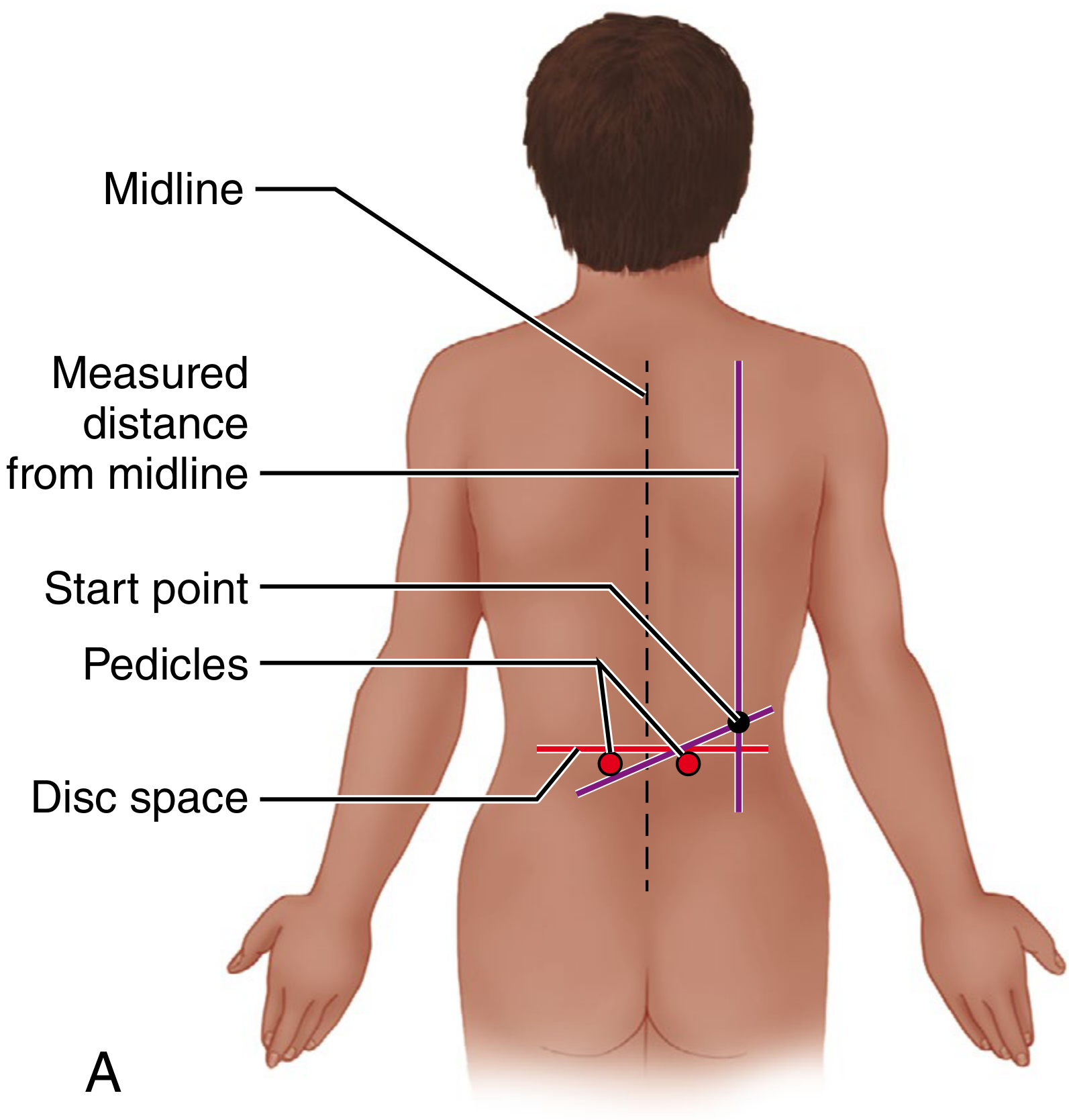
Figure 44.8A - Surface landmarks drawn using fluoroscopy before a transforaminal endoscopic discectomy. Shows midline (dashed line), the measured distance from midline to the start point, and the projected disc space and pedicle levels. Trajectory is drawn from the contralateral caudal pedicle edge to the ipsilateral cranial pedicle.
2. Intraoperative Setup - Right-Sided Approach Marking
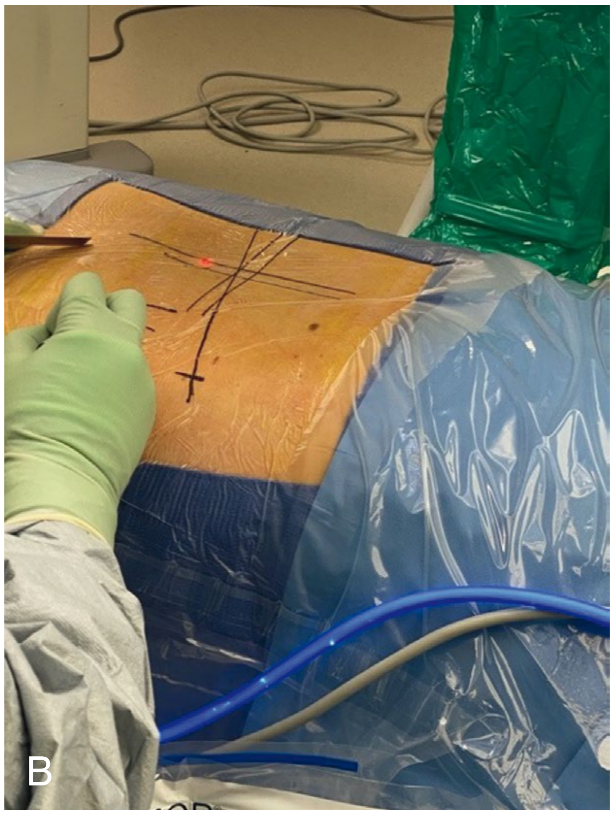
Figure 44.8B - Actual intraoperative photograph showing the trajectory lines drawn on the patient's back (prone position) for a right-sided transforaminal discectomy approach. A red laser dot marks the target start point.
3. Fluoroscopic Guidance - Needle/Probe Positioning
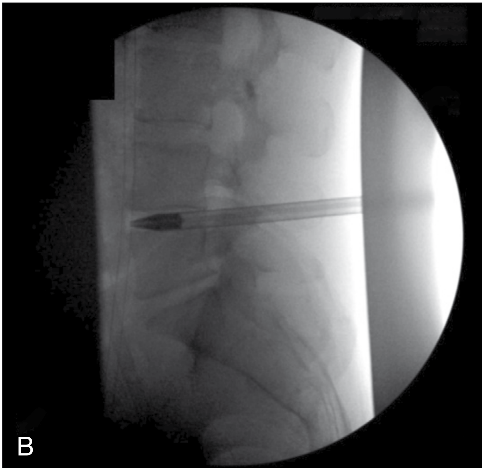
Figure 44.9/44.10B - Lateral fluoroscopic image confirming the probe is positioned within the foramen overlying the vertebral body of the caudal level. This confirms correct level and that the instrument is not inadvertently advancing into the disc space.
4. Key Endoscopic View - Arch of Wagner (Most Important Landmark)
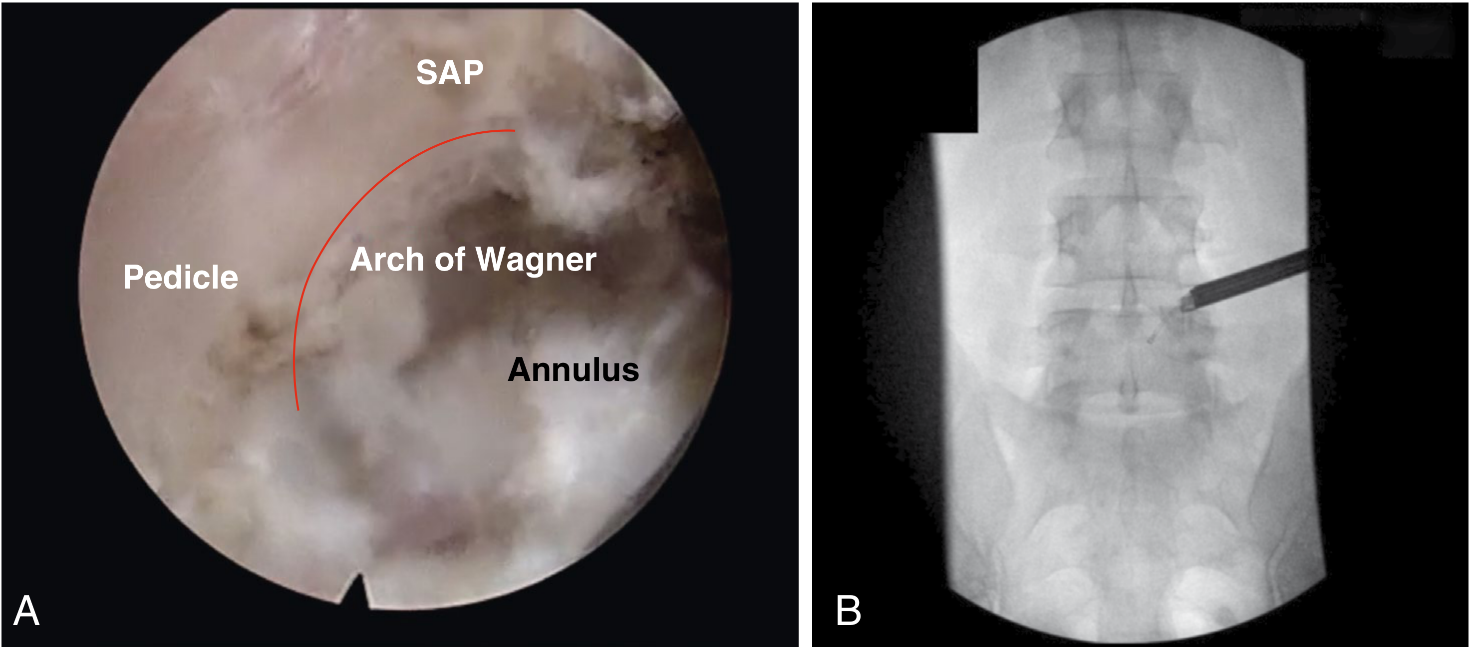
Figure 44.10A - This is the critical endoscopic view through the working cannula during transforaminal discectomy. The labeled structures are:
- SAP (Superior Articular Process) - at the top
- Pedicle - on the left
- Arch of Wagner (red arc) - the bony arch formed between the inferior pedicle and the superior articular process; this is the key orienting landmark in the foramen
- Annulus - visible centrally/inferiorly, representing the posterior disc
Once the Arch of Wagner is confirmed, instruments are directed to remove herniated disc material from under the nerve root.
Figure 44.10B - Corresponding AP fluoroscopic view showing the ball-tip probe lying within the foramen at the correct level.
5. Axial Diagram - Trajectory Angulation
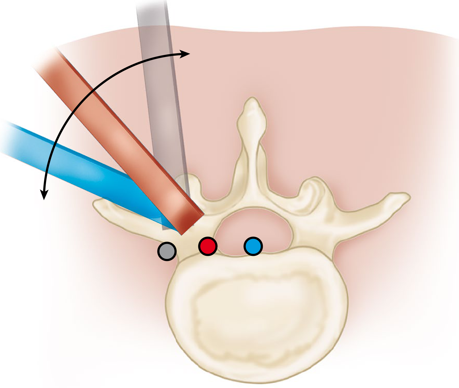
Figure 44.7 - Axial view illustrating how trajectory angulation affects target access:
- The blue tube (more lateral start point) is used for central herniations - flatter trajectory
- The red/brown tube (more medial start point, steeper trajectory) is used for far lateral herniations
- The colored dots in Kambin's triangle represent: gray = safe working zone, red = exiting nerve root, blue = traversing nerve root
- The gray instrument (top) represents an interlaminar approach for comparison
Key Endoscopic Anatomy Checklist During Discectomy
| Structure | What to Look For |
|---|---|
| Arch of Wagner | Bony arch (pedicle + SAP) - primary orienting landmark |
| Annulus fibrosus | Fibrous disc wall; incise to access nucleus |
| Epidural fat | Yellowish tissue around nerve structures |
| Traversing nerve root | Displaced dorsally by herniation; seen pulsating after decompression |
| Exiting nerve root | Identified for foraminal/far lateral herniations |
| Dura | Avoid injury; pulsation confirms decompression |
| Epidural veins | Control with fluid pressure (do not exceed diastolic BP) |
Endpoints for Adequate Decompression (Endoscopic)
The textbook describes decompression as complete when:
- The affected nerve root is seen pulsating to heartbeat without deformity
- The patient (awake under MAC) reports resolution of the typical radicular pain
- The nerve root is freely retractable medially and laterally, and is round
- Campbell's Operative Orthopaedics, 15th Ed., 2026 - Techniques 44.3, 46.5, 46.6
This is a shared conversation. Sign in to Orris to start your own chat.