
Dystonia
dystonia classification treatment
dystonia posturing movement disorder
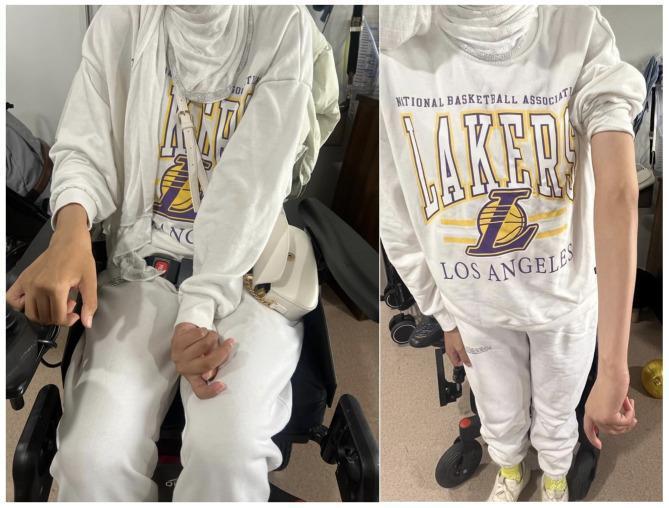
This clinical photograph consists of two side-by-side panels showing an individual in a seated position, illustrating movement disorder manifestations. The left panel shows the patient seated in a wheelchair, demonstrating a characteristic dystonic posture of the left upper extremity. This posturing is characterized by fixed internal rotation of the left shoulder and flexion at the elbow. In contrast, the right upper extremity appears more extended and relaxed. The right panel provides a closer view of the torso and limbs, highlighting the asymmetry between the flexed left arm and the extended right arm. The image serves as a visual representation of neurological signs associated with Woodhouse-Sakati Syndrome (WSS), specifically secondary dystonia and impaired mobility. The patient exhibits a female phenotype, though contextually noted for poor secondary sexual development. These images are medically significant for identifying phenotypic clinical signs of biallelic pathogenic variants in the DCAF17 gene, which manifest as progressive extrapyramidal symptoms and cognitive impairment.
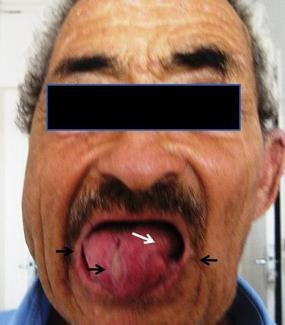
Clinical photograph of a 62-year-old male presenting with orofacial manifestations of Chorea-acanthocytosis (ChAc). The image highlights oromandibular dystonia and its chronic sequelae. A white arrow points to the tongue, which exhibits significant dystonic posturing and shape distortion. Black arrows indicate the sequelae of recurrent self-mutilation, including deep lacerations, irregular tissue loss, and chronic scarring along the lateral borders of the tongue and the commissures of the lips. The oral cavity shows evidence of repeated biting injuries, a characteristic clinical finding in neuroacanthocytosis syndromes. These findings are pedagogically significant for identifying movement-disorder-related self-mutilation, which distinguishes ChAc from other choreiform disorders. The photo is taken post-treatment for facial cellulitis, though the underlying neurological and mechanical tissue damage remains visible.
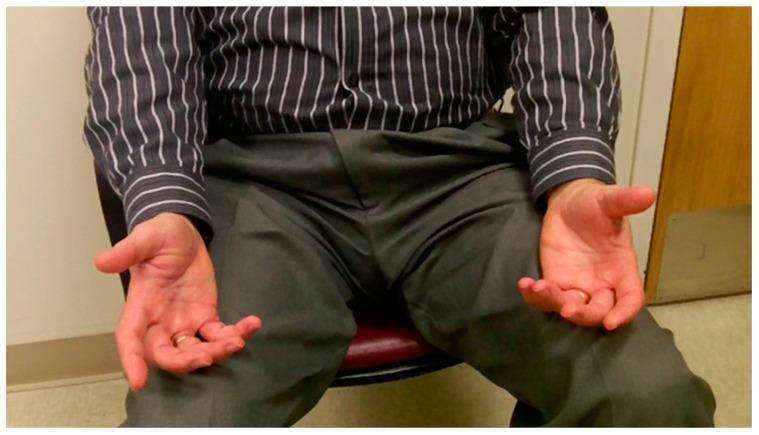
Clinical photograph of a patient exhibiting focal hand dystonia (FHD) at rest. The image shows a seated individual with both hands held in an abnormal, involuntary posture. The hands are positioned in front of the torso with palms facing medially. There is evidence of dystonic posturing characterized by partial flexion at the metacarpophalangeal (MCP) joints and more pronounced flexion at the proximal and distal interphalangeal (IP) joints of the fingers, particularly involving the third through fifth digits. The thumbs show subtle extension at the MCP joints with flexion at the IP joints. The wrists are maintained in a neutral to slightly extended position. This bilateral presentation illustrates the characteristic muscle co-contractions and tonic posturing associated with dystonia, used here to demonstrate a severe case where symptoms are present even without task-specific provocation. This clinical visual serves as an educational tool for neurology and physical therapy in identifying movement disorders and functional anatomy abnormalities during physical examination.
Dystonia
Definition
- Involuntary co-contraction of agonist and antagonist muscles
- Movements that are often patterned and repetitive (not random)
- Postures/movements that are initiated or worsened by voluntary action
Clinical Features
Phenomenology
- Movements may be slow and twisting or rapid (resembling myoclonic jerks)
- A characteristic feature is the "null point" - a position where dystonic tremor ceases when the patient relaxes and allows the limb to move freely
- Sensory tricks (gestes antagonistes): peculiar maneuvers patients discover to temporarily suppress dystonic movements (e.g., touching the chin in cervical dystonia)
- Task-specificity: dystonia may occur only during certain tasks - the classic example is writer's cramp (graphospasm), where hand dystonia occurs only during writing but not other manual tasks
Clinical image - focal hand dystonia at rest:
Affected body regions
| Region | Dystonia Type | Presentation |
|---|---|---|
| Eyelids | Blepharospasm | Forced involuntary closure |
| Jaw/tongue | Oromandibular dystonia | Jaw clenching, forced opening, tongue protrusion |
| Larynx | Spasmodic dysphonia | Harsh, strained, or breathy voice |
| Neck | Cervical dystonia (spasmodic torticollis) | Involuntary head deviation in any plane |
| Trunk | Axial dystonia | Spasms interfering with sitting, standing, walking |
| Hand | Writer's cramp, musician's dystonia | Task-specific occupational spasm |
| Foot | Foot dystonia | Inversion and plantar flexion, walking on toes |
Classification
By Distribution (Body Region)
- Focal: single body region (most common in adults)
- Segmental: two or more contiguous regions
- Multifocal: two or more non-contiguous regions
- Hemidystonia: one side of the body (usually secondary)
- Generalized: involves the trunk plus two other regions
By Etiology
1. Primary (Isolated) Dystonias
- Familial: multiple genetic causes (DYT gene family)
- DYT1 (TOR1A mutation): early-onset generalized dystonia (dystonia musculorum deformans) - autosomal dominant
- DYT5 (GCH1 mutation): Dopa-responsive dystonia (Segawa disease) - autosomal dominant, shows marked diurnal fluctuation (worse as day progresses, better after sleep), responds to tiny doses of L-dopa
- Sporadic (idiopathic): usually adult-onset, focal or segmental
2. Dystonia-Plus Syndromes
- Dystonia with parkinsonism
- Dopa-responsive dystonia
- Myoclonus-dystonia (DYT11, SGCE gene): myoclonic jerks + dystonia, alcohol-responsive
3. Heredodegenerative Dystonias
- Wilson disease (copper accumulation)
- Huntington disease
- Pantothenate kinase-associated neurodegeneration (formerly Hallervorden-Spatz)
- Lysosomal storage diseases
- Neuroacanthocytosis (chorea-acanthocytosis)
- Fahr disease (striatopallidodental calcification)
4. Secondary (Acquired) Dystonias
- Drug-induced (most common secondary cause):
- Neuroleptics (phenothiazines, butyrophenones, metoclopramide) - acute dystonic reactions
- L-dopa excess in Parkinson disease (tardive dyskinesia)
- Antiepileptic drugs, calcium channel blockers
- Acute reaction: retrocollis, arching of back, internal rotation of arms, extension of elbows/wrists (simulating opisthotonos) - responds to diphenhydramine or benztropine
- Structural: stroke, tumor, trauma, hypoxic injury (double athetosis/cerebral palsy), kernicterus
- Peripheral injury: dystonia following limb injury, associated with complex regional pain syndrome (CRPS)
- Thyroid disease
Genetic note: Monogenic variants are found in ~20% of all dystonia cases. The "DYT" classification prefix (DYT1-25) has known shortcomings and is under re-evaluation. - Goldman-Cecil Medicine
Pathophysiology
- Loss of inhibition: reduced surround inhibition in the motor cortex (decreased short-interval intracortical inhibition - SICI) leads to abnormal overflow contractions into adjacent muscles
- Abnormal plasticity: enhanced long-term potentiation (LTP)-like plasticity in motor cortex, with loss of topographic specificity (even non-dystonic body parts show abnormal plasticity in focal dystonias)
- Sensorimotor integration defects: sensory tricks work because altering sensory input temporarily normalizes the aberrant circuit
- SICI reduction is not site-specific in focal dystonia - it is observed in unaffected sides and in unaffected body regions, pointing to a diffuse cortical inhibitory deficit - Bradley and Daroff's Neurology
Important Clinical Syndromes
Dopa-Responsive Dystonia (Segawa Disease)
- Familial (usually autosomal dominant), onset in childhood
- Dystonia-athetosis combined with elements of parkinsonism
- Hallmark: marked diurnal fluctuation (worse in evening, better after sleep)
- Responds to very small doses of L-dopa
- Must always be tried in young patients with dystonia before assuming idiopathic disease
Rapid-Onset Dystonia-Parkinsonism
- Onset in adolescence/early adulthood
- Rapid evolution (hours to days) of severe dystonic spasms, dysarthria, dysphagia, postural instability with bradykinesia
- DYT12 (ATP1A3 mutation)
Occupational Dystonias
- Task-specific, associated with highly repetitive motor programs
- Musicians (pianists, reed players), dancers, writers
- Often career-threatening
Paroxysmal Dystonias
- Paroxysmal kinesigenic dyskinesia (PKD): attacks triggered by sudden movement/startle, autosomal dominant, PRRT2 gene, responds well to carbamazepine or phenytoin
- Paroxysmal non-kinesigenic dyskinesia: attacks triggered by caffeine/alcohol, longer duration
Differential Diagnosis
- Functional/psychogenic disorder: stress aggravates dystonia; sensory tricks and hypnosis can alleviate it, mimicking functional illness
- Hysteria (historically misinterpreted): e.g., cervical dystonia was once labeled as "turning away of responsibility"
- CRPS (complex regional pain syndrome): peripherally-induced fixed dystonia may occur with CRPS; neurophysiology (SICI, LICI) cannot reliably distinguish organic from functional dystonia
Treatment
1. Botulinum Toxin (First-line for Focal Dystonias)
- Periodic injection into overactive muscles is the most effective treatment for focal dystonias
- Indications: blepharospasm, cervical dystonia, spasmodic dysphonia, writer's cramp, limb dystonias
- Requires repeat injections every 3 months
- Adams and Victor's Principles of Neurology; Katzung's Basic and Clinical Pharmacology
2. Oral Medications (Especially for Generalized Dystonia)
| Drug | Notes |
|---|---|
| Trihexyphenidyl (anticholinergic) | High doses, more effective in children than adults; titrate slowly |
| Benztropine, Ethopropazine | Anticholinergic agents |
| L-dopa | Mandatory trial in children/young patients to rule out dopa-responsive dystonia |
| Tetrabenazine / Deutetrabenazine | Monoamine-depleting agents; effective for tardive dyskinesia |
| Baclofen | Muscle relaxant; intrathecal baclofen for severe cases |
| Clonazepam | Adjunctive |
- For acute drug-induced dystonic reactions: diphenhydramine (IV/IM) or benztropine rapidly effective
- For tardive dyskinesia: stop offending drug if possible (though this may worsen movements initially); deutetrabenazine, reserpine, or anticholinergics
3. Deep Brain Stimulation (DBS) - Surgery
- Target: posteroventral globus pallidus internus (GPi) bilaterally
- Controlled trial demonstrated ~50% improvement in dystonic movements over 1 year
- Preferred for severe generalized dystonia refractory to medical management
- Also being evaluated for severe cervical dystonia refractory to botulinum toxin and medications
- Adams and Victor's; Bradley and Daroff's Neurology
4. Stereotactic Surgery
- Pallidotomy and ventrolateral thalamotomy (older approach) - generally positive but unpredictable results; largely replaced by DBS
Treatment Algorithm (Summary)
Focal dystonia → Botulinum toxin (first-line)
                + Anticholinergics (adjunct)
                → DBS if refractory

Generalized dystonia (child/young adult) → L-dopa trial first (rule out DRD)
                                         → Trihexyphenidyl (high dose)
                                         → DBS if refractory

Acute drug-induced → Diphenhydramine or benztropine IV
Tardive dyskinesia → Stop offending drug + deutetrabenazine/reserpine
Key Points Summary
| Feature | Detail |
|---|---|
| Core mechanism | Agonist-antagonist co-contraction |
| Pathophysiology | Loss of cortical surround inhibition; abnormal plasticity |
| Most common adult onset | Focal, idiopathic |
| Most common secondary cause | Drug-induced (neuroleptics) |
| Most treatable hereditary form | Dopa-responsive dystonia (Segawa) |
| Best treatment for focal dystonia | Botulinum toxin injections |
| Surgical treatment | Bilateral GPi-DBS (~50% improvement) |
| Sensory trick | Geste antagoniste (hallmark of cervical dystonia) |
| Task-specific example | Writer's cramp, musician's dystonia |