
Meningitis - etiology pathogenesis
Reading File
Now let me search the medical library for meningitis etiology and pathogenesis content in parallel.
Finding Sources
Finding Sources
Good. Adams and Victor's Neurology and other neurology textbooks are the best sources here. Let me search for dedicated meningitis sections and read them in parallel.
Finding Sources
Finding Sources
Excellent. Let me now read the most comprehensive sources - Adams & Victor's Neurology, Robbins Pathology, and Rosen's Emergency Medicine meningitis sections simultaneously.
Reading File
Reading File
Reading File
Let me read more from Adams & Victor's for detailed pathogenesis, and also Robbins' pathology section:
Reading File
Reading File
I accidentally read a reference section from Adams & Victor's. Let me find the correct meningitis block in Adams & Victor's and also read more from Robbins:
Finding Sources
Reading File
Now let me read the key sections - "The Biology of Bacterial Meningitis" from Adams & Victor's (lines 1677-1740):
Reading File
Excellent! I now have very rich content. Let me also read the Robbins pathology Acute Pyogenic Meningitis morphology section and fetch the key images:
Finding Sources
Searching PubMed
bacterial meningitis pathogenesis blood brain barrier
Now let me fetch the key pathology image from Robbins' and also get the Robbins acute pyogenic meningitis morphology text:
Finding Sources
Loading Image
Now I have comprehensive information to compile a thorough answer. Let me also fetch the gross pathology image from Robbins:
Loading Image
I now have comprehensive information from the textbooks to compose a thorough, well-structured answer on meningitis etiology and pathogenesis. Let me compile this now.
Meningitis - Etiology and Pathogenesis
Definition
Meningitis is an inflammatory process involving the leptomeninges (pia mater and arachnoid) within the subarachnoid space. When infection extends into the underlying brain parenchyma, the condition is termed meningoencephalitis. The term also applies to non-infectious settings (chemical meningitis from ruptured epidermoid cyst debris, carcinomatous meningitis from metastatic cancer).
- Robbins & Kumar Basic Pathology, p. 833
Classification and Etiology
By Acuity and Cause
| Type | Usual Cause | Key Organisms |
|---|---|---|
| Acute pyogenic | Bacterial | See age-stratified table below |
| Aseptic (acute) | Viral | Enteroviruses, herpesviruses, HIV, LCM virus |
| Chronic | Tuberculosis, spirochetes, fungi | M. tuberculosis, Treponema pallidum, Cryptococcus |
| Non-infectious | Chemical, neoplastic | Epidermoid cysts, metastatic cancer |
Bacterial Etiology: Age-Stratified
| Age Group | Most Common Organisms |
|---|---|
| Neonates (< 6 weeks) | Escherichia coli, Group B streptococci |
| Infants and young children | Neisseria meningitidis, S. pneumoniae |
| Adolescents and young adults | Neisseria meningitidis (predominant) |
| Adults | Streptococcus pneumoniae (> 50% of adult cases) |
| Elderly and immunocompromised | S. pneumoniae, Listeria monocytogenes |
| Post-neurosurgery / CSF leak | Coagulase-negative staphylococci, S. aureus, Cutibacterium acnes, Pseudomonas aeruginosa |
- Robbins & Kumar Basic Pathology, p. 833; Rosen's Emergency Medicine, p. 2268
Important epidemiologic note: Haemophilus influenzae type b was previously a major cause in children but its incidence has sharply declined since the HiB vaccine. Meningococcal disease is most common in those living in close proximity (military barracks, college dormitories); serogroup B predominates in Europe, serogroup C in the United States. - Rosen's Emergency Medicine, p. 2268
Viral Etiology
- Enteroviruses are now the most common cause of meningitis overall (given bacterial decline from vaccination)
- Herpesviruses (HSV-1, HSV-2, CMV, EBV, VZV) - often in immunocompromised
- HIV, mumps, influenza, measles, LCM virus
- Prognosis for most viral meningitis is excellent
Pathogenesis
Step 1: Acquisition and Nasopharyngeal Colonization
Bacterial meningitis generally begins with nasopharyngeal colonization. Organisms like N. meningitidis and S. pneumoniae are carried asymptomatically in the nasopharynx of many individuals. Invasion occurs when bacteria penetrate the mucosal surface. Bacterial capsular polysaccharides are a key virulence factor that resist phagocytosis and allow the organism to survive in the bloodstream (bacteremia).
Step 2: Bacteremia and Blood-Brain Barrier (BBB) Crossing
Once in the bloodstream, bacteria must cross the BBB to enter the CSF. The subarachnoid space, once breached, supports rapid bacterial proliferation because it lacks adequate local immune defenses (low complement, low immunoglobulin levels in CSF). Bacteria multiply freely.
Step 3: Inflammatory Cascade in the Subarachnoid Space
This is the heart of pathogenesis. The immediate effect of bacteria (or their toxins, e.g., LPS, teichoic acid) in the subarachnoid space triggers a cascade:
- Hyperemia of meningeal venules and capillaries, with increased vascular permeability
- Exudation of protein and migration of neutrophils into the pia and subarachnoid space
- Cytokine release into CSF (IL-1, IL-6, TNF-alpha) - this inflammatory cascade promotes:
- Further increased permeability of the BBB
- Cerebral vasculitis
- Cerebral edema
- Increased intracranial pressure (ICP)
- Decreased cerebral blood flow → cerebral hypoxia
Because the subarachnoid space is continuous around the brain, spinal cord, and optic nerves, an organism gaining entry spreads rapidly throughout. Meningitis is therefore always cerebrospinal in extent. Infection also reaches the ventricles - either directly through the choroid plexuses or by reflux through the foramina of Magendie and Luschka.
- Adams and Victor's Principles of Neurology, 12th Ed., p. 708; Rosen's Emergency Medicine, p. 2268
Temporal Sequence of Inflammatory Events (Adams & Victor's)
This temporal understanding explains the clinical state and its sequelae:
| Timeframe | Pathological Event |
|---|---|
| Early (hours) | Hyperemia, increased vascular permeability, protein exudation; neutrophils migrate into pia and subarachnoid space |
| First few days | Subarachnoid exudate forms over brain base, into cranial and spinal nerve sheaths, and into cortical perivascular spaces. Neutrophils (mature and immature) predominate; many contain phagocytosed bacteria. Fibrinogen exudes and begins converting to fibrin |
| Days 3-5 | Lymphocytes and histiocytes increase in number |
| End of week 1 - week 2 | Plasma cells appear; exudate organizes into outer layer (neutrophils + fibrin, sub-arachnoid) and inner layer (lymphocytes, plasma cells, macrophages, next to pia). Fibroblasts proliferate. Thrombophlebitis of larger cortical veins may begin |
| Resolution | Inflammatory cells disappear in reverse order. Neutrophils disintegrate by days 4-5 with treatment. Lymphocytes/plasma cells/macrophages disappear slowly (may persist months) |
- Adams and Victor's Principles of Neurology, 12th Ed.
Vascular Changes
A striking feature of meningitis - appearing within 48-72 hours:
- Endothelial cells of subarachnoid arteries swell, multiply, and crowd the lumen
- Adventitial connective tissue sheath infiltrated by neutrophils
- Foci of arterial wall necrosis may occur
- Neutrophils and lymphocytes migrate from adventitia to sub-intimal region (subintimal fibrosis later)
- In veins: endothelial swelling, adventitial infiltration, diffuse wall infiltration; mural thrombi and focal wall necrosis in affected veins
- Thrombophlebitis of larger cortical veins may develop (usually not before end of second week)
This vascular reaction is most notably prominent in tuberculous and syphilitic meningitis (Heubner arteritis). It is anatomically explained by the fact that the adventitia of subarachnoid vessels is formed by an investment of the arachnoid membrane - which is invariably involved by the infectious process.
- Adams and Victor's Principles of Neurology, 12th Ed., p. 709
Gross and Histologic Pathology
Gross appearance:
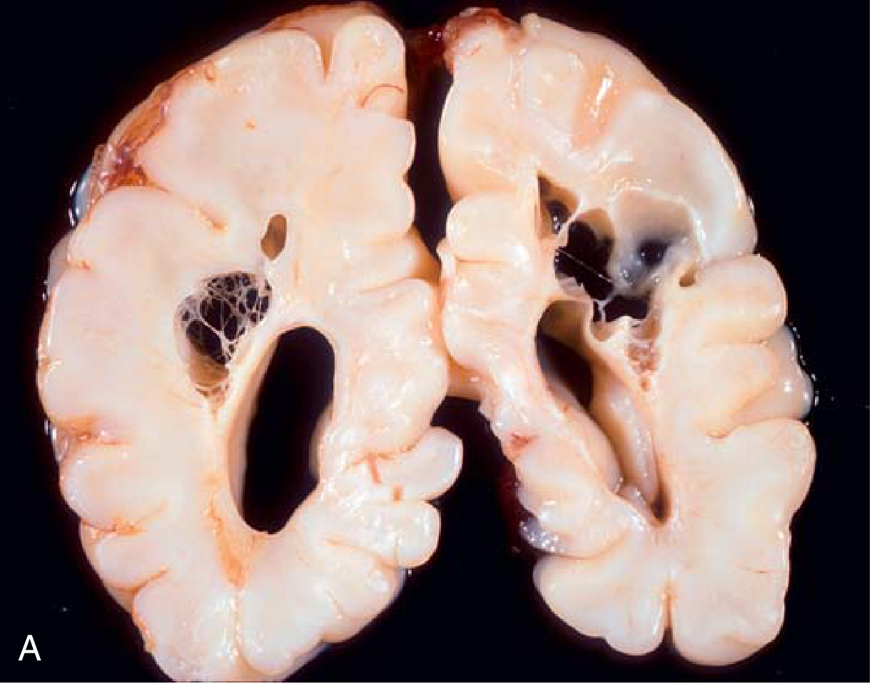
The brain surface shows cloudy, opaque arachnoid with purulent exudate filling the sulci; this exudate accumulates over the base of the brain particularly.
Histology:
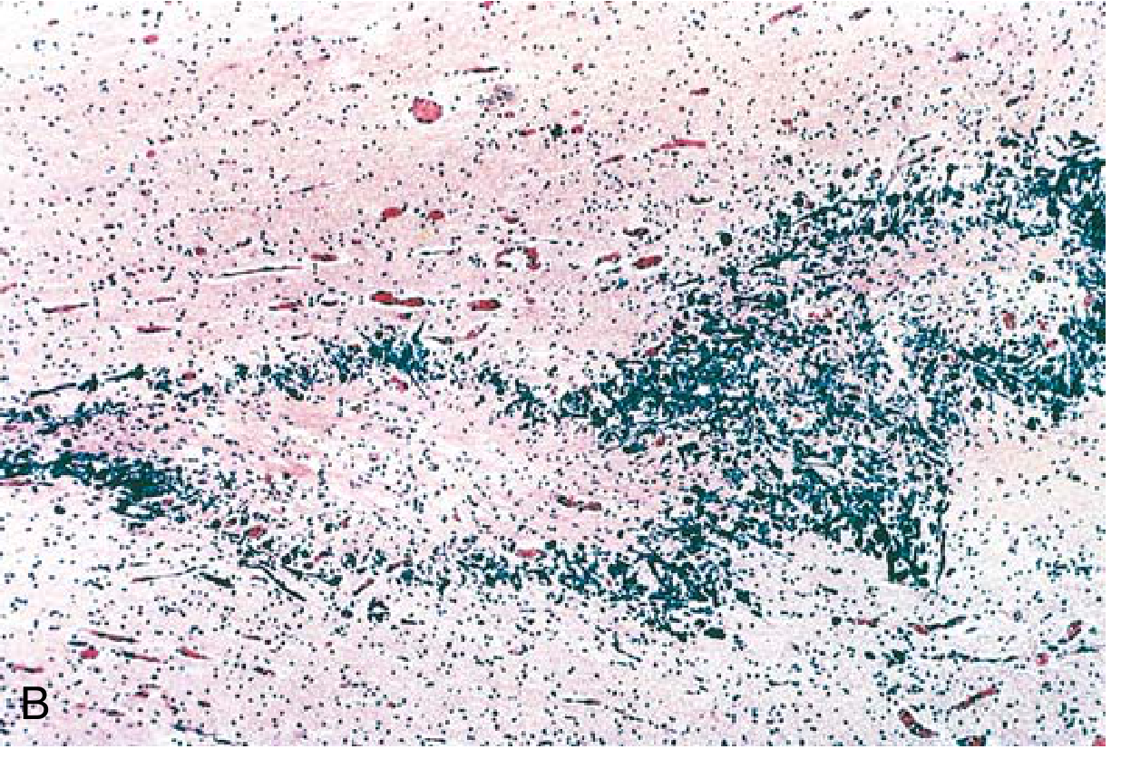
Dense neutrophilic infiltrate fills the subarachnoid space. The image shows neutrophils (blue-dark) concentrated in the meninges with relative sparing of the cortex (left).
Pathologic-Clinical Correlations
Acute Meningeal Inflammation
| Pathological Event | Clinical Manifestation |
|---|---|
| Pure pia-arachnoiditis | Headache, stiff neck, Kernig and Brudzinski signs |
| Subpial encephalopathy | Confusion, stupor, coma, convulsions |
| Cranial nerve root inflammation | Ocular palsies, facial weakness, deafness (also caused by middle ear infection, inner ear extension, or aminoglycoside toxicity) |
| Meningeal vein thrombosis | Focal seizures, hemiparesis, aphasia |
| Obstructed CSF outflow | Raised ICP: papilledema, CN VI palsy, altered consciousness |
| Cerebellar/cerebral herniation | Upper cervical cord compression, quadriplegia, midbrain-third nerve compression |
Subacute/Chronic Forms
| Complication | Mechanism |
|---|---|
| Hydrocephalus | Purulent exudate at base of brain blocking CSF flow; later meningeal fibrosis |
| Subdural effusion | Impaired meningeal drainage |
| Venous or arterial infarction | Infectious vasculitis at brain base; cortical vein thrombosis |
Late Sequelae
-
Meningeal fibrosis around optic nerves → blindness, optic atrophy
-
Spinal cord/root fibrosis → spastic paraparesis, sensory loss
-
Chronic meningoencephalitis with hydrocephalus → dementia, paralysis
-
Adams and Victor's Principles of Neurology, 12th Ed., pp. 708-710
Meningitis in Specific Contexts
Neonatal Meningitis
The most significant factor in the pathogenesis of neonatal meningitis is maternal infection (usually UTI or puerperal fever). In E. coli neonatal meningitis, pathogenesis involves colonization of the infant with maternal E. coli, with the K1 capsular antigen facilitating invasion.
- Adams and Victor's Principles of Neurology, 12th Ed.
Tuberculous Meningitis
A form of chronic meningitis with especially prominent vascular changes (Heubner arteritis). The granulomatous exudate is concentrated at the base of the brain. Results in a particularly high rate of cranial nerve palsies, hydrocephalus, and deep infarctions due to involvement of perforating arteries.
Viral (Aseptic) Meningitis
Enteroviruses and herpesviruses are most common. The inflammatory response is predominantly lymphocytic rather than neutrophilic. CSF shows lymphocytic pleocytosis, mildly elevated protein, and normal or only mildly reduced glucose. Prognosis is generally excellent.
CSF Summary
| Feature | Bacterial | Viral | TB/Fungal |
|---|---|---|---|
| Pressure | Elevated | Normal/mildly elevated | Elevated |
| Cells | Neutrophils (>1000/µL) | Lymphocytes (<500/µL) | Lymphocytes |
| Protein | Markedly elevated | Mildly elevated | Elevated |
| Glucose | Low (<45 mg/dL) | Normal | Very low |
| Appearance | Turbid/purulent | Clear | Clear/viscous |
Key Sources:
- Adams and Victor's Principles of Neurology, 12th Edition, Chapter 31 (Bacterial Meningitis section, pp. 708-710)
- Robbins & Kumar Basic Pathology, Chapter 21 (CNS Infections, p. 833)
- Rosen's Emergency Medicine, Chapter on Bacterial Meningitis (p. 2268)
This is a shared conversation. Sign in to Orris to start your own chat.