
Attic area boundaries
The query "Attic area boundaries" touches on anatomy (the attic of the middle ear), so I need to check the medical library. Let me look that up.
Quick answer: The attic (epitympanum) is the upper portion of the middle ear (tympanic cavity) located above the level of the tympanic membrane. Its boundaries are: roof - tegmen tympani; floor - a plane through the lateral wall at the level of the tympanic notch; medial wall - medial wall of the epitympanum containing the lateral semicircular canal prominence; lateral wall - scutum (bone) and Shrapnell's membrane; anterior - communicates with the Eustachian tube area; posterior - opens into the aditus ad antrum.
Searching sources...
Reading File
Finding Sources
Finding Sources
Reading File
Finding Sources
Reading File
Loading Image
Here is a complete breakdown of the Attic (Epitympanum) - Area and Boundaries, drawn directly from KJ Lee's Essential Otolaryngology:
Attic (Epitympanum) - Definition
The attic is the upper portion of the middle ear cavity located above the level of the tympanic membrane and above an imaginary plane drawn through the short process of the malleus. It communicates with the mastoid antrum posteriorly and plays a fundamental role in middle ear ventilation routes.
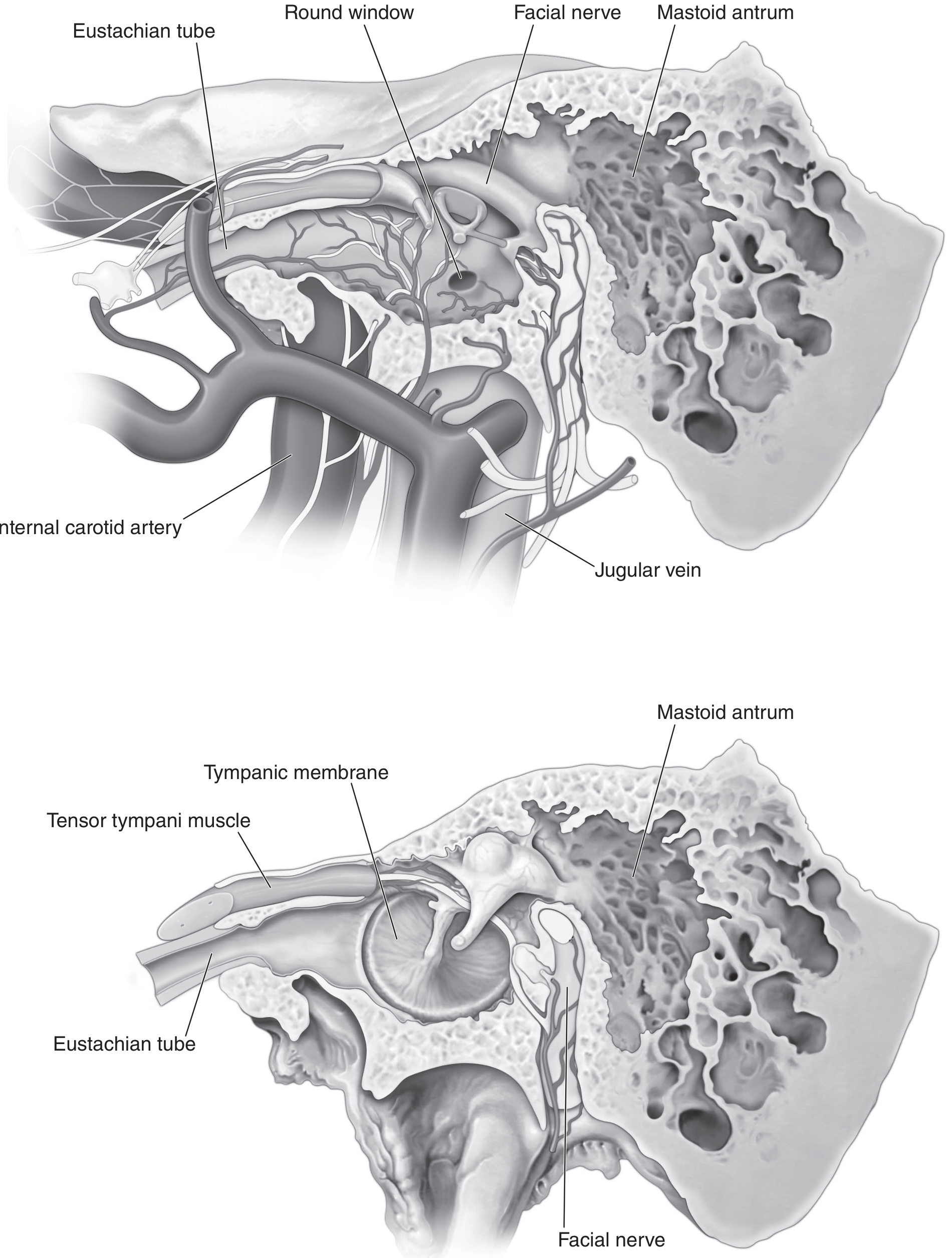
Boundaries (Limits) of the Attic
| Wall | Boundary |
|---|---|
| Superior | Tegmen tympani (thin bony plate separating it from the middle cranial fossa) |
| Inferior | Tympanic diaphragm and isthmus |
| Medial | Facial nerve canal and lateral semicircular canal |
| Lateral | Pars flaccida of the tympanic membrane + scutum (Chaussé spur) |
| Anterior | Zygomatic root |
| Posterior | Incudal fossa and aditus ad antrum |
Internal Subdivision: Anterior vs. Posterior Epitympanum
The attic is divided by the superior malleal fold into two compartments:
Posterior epitympanum (posterior to superior malleal fold):
- Posterior part of malleus head
- Incus body and short process
- Contains the posterior ventilation route - the most important pathway to the attic
Anterior epitympanum (anterior to superior malleal fold):
- Anterior portion of malleus head
- Contains the anterior ventilation route - an accessory pathway
Tympanic Diaphragm (inferior boundary)
The tympanic diaphragm (the floor of the attic) is composed of:
- Posterior incudal ligament
- Lateral incudomalleal fold
- Lateral malleal fold
- Lateral malleal ligament
- Anterior malleal fold
- Anterior malleal ligament
- Tensor tympani fold (posterior malleal fold)
- Tensor tympani muscle tendon
Tympanic Isthmus
A 2.5-mm opening in the tympanic diaphragm that ventilates the entire attic:
- Anterior tympanic isthmus: between the incudostapedial joint and tensor tympani muscle tendon - the largest and most important ventilation route. Blockage here causes attic selective dysventilation, chronic edema, retraction pockets, and cholesteatoma.
- Posterior tympanic isthmus: posterior to the incudostapedial joint, between the stapedial muscle tendon, pyramidal eminence, and incus short process.
Prussak Space (a key sub-space within the attic lateral wall)
| Wall | Boundary |
|---|---|
| Superior | Lateral malleal fold (tympanic diaphragm) |
| Inferior | Malleus neck |
| Medial | Malleus head |
Prussak space is clinically important as the most common site of origin for pars flaccida cholesteatoma.
KJ Lee's Essential Otolaryngology, p. 496-499
This is a shared conversation. Sign in to Orris to start your own chat.