I now have comprehensive data from all major surgical textbooks plus the latest PubMed evidence. Let me compile the complete, fully-detailed PG-level exam guide.---
🩺 SURGICAL AND BENIGN THYROID DISORDERS
Complete PG Surgery Examination Guide
Sources: Schwartz's Principles of Surgery 11e · Bailey & Love's Short Practice of Surgery 28e · Harrison's Principles of Internal Medicine 22e · Cummings Otolaryngology · PubMed Meta-Analyses 2023-2025
MODULE 1: SURGICAL ANATOMY OF THE THYROID
1.1 Gross Anatomy
┌─────────────────────────────────────────────────────────────┐
│              THYROID GLAND - GROSS ANATOMY                  │
│                                                             │
│  Hyoid bone                                                 │
│     │                                                       │
│  Thyroid cartilage ─── Pyramidal lobe (50% of patients)    │
│     │                         │                            │
│  Isthmus ────── Right lobe ───┤─── Left lobe               │
│     │                         │                            │
│  Tracheal rings 2-4           │                            │
│                               ▼                            │
│         Weight: 20-30 g  │  Dimensions: 4×2×2 cm per lobe  │
│         Enclosed in: True capsule (thyroid) +               │
│                       False capsule (pretracheal fascia)    │
└─────────────────────────────────────────────────────────────┘
- Lobes: Two lateral lobes joined by an isthmus overlying tracheal rings 2-4
- Pyramidal lobe: Present in ~50% of patients, extends superiorly from isthmus (remnant of thyroglossal duct)
- True capsule: Derived from thyroid parenchyma itself; sends septa into the gland
- False/surgical capsule: Derived from pretracheal fascia; forms the surgical plane of dissection
- Berry's ligament (Posterior suspensory ligament): Thick condensation of fascia anchoring the posteromedial aspect of each thyroid lobe to the cricoid cartilage and trachea - the most dangerous area during thyroidectomy
1.2 Blood Supply
┌──────────────────────────────────────────────────────────────────┐
│                    THYROID BLOOD SUPPLY                          │
│                                                                  │
│  ARTERIES:                                                       │
│  ┌─────────────────────────────────────────────────────────┐    │
│  │ Superior thyroid artery ──→ 1st branch External Carotid │    │
│  │   ↓ Supplies: Upper pole + isthmus                      │    │
│  │   ⚠ EBSLN runs along this artery → individual ligation  │    │
│  ├─────────────────────────────────────────────────────────┤    │
│  │ Inferior thyroid artery ──→ Thyrocervical trunk         │    │
│  │   ↓ Supplies: Lower pole + BOTH PARATHYROIDS            │    │
│  │   ⚠ RLN crosses near/behind it → most dangerous area    │    │
│  ├─────────────────────────────────────────────────────────┤    │
│  │ Thyroidea ima ──→ Aorta / Innominate (3% population)    │    │
│  │   ↓ Supplies: Isthmus                                    │    │
│  │   ⚠ Risk during emergency tracheostomy / median sternotomy│  │
│  └─────────────────────────────────────────────────────────┘    │
│                                                                  │
│  VEINS:                                                          │
│  Superior thyroid vein ──→ IJV (Internal Jugular Vein)          │
│  Middle thyroid vein ──→ IJV (no accompanying artery)           │
│  Inferior thyroid veins ──→ Innominate / Brachiocephalic vein   │
└──────────────────────────────────────────────────────────────────┘
Exam Pearl: The middle thyroid vein has NO accompanying artery. It is the first vessel divided when mobilising the thyroid lobe (lateral retraction reveals it).
1.3 Nerves - Critical Surgical Anatomy
Recurrent Laryngeal Nerve (RLN)
RIGHT RLN                              LEFT RLN
Loops around right subclavian artery   Loops around aortic arch
More oblique course                    More vertical course
Shorter intraneck course               Longer intraneck course
Non-recurrent in 0.5-1%               Non-recurrent is EXTREMELY RARE
(assoc. with aberrant subclavian)
Course in the neck:
- Lies in the tracheo-oesophageal groove (most consistent location)
- Crosses the inferior thyroid artery (usually posterior, sometimes anterior, sometimes between branches)
- Enters the larynx at the inferior margin of the cricothyroid joint (inferior cornu of thyroid cartilage)
- At Berry's Ligament: Nerve passes within or immediately adjacent to the ligament - the zone of maximum danger
Functions lost if injured:
- ALL intrinsic laryngeal muscles EXCEPT cricothyroid
- Adductors + Abductors of vocal cord
- Unilateral injury: hoarse/breathy voice, aspiration
- Bilateral injury: bilateral cord paralysis → stridor, respiratory distress → emergency tracheostomy
External Branch of Superior Laryngeal Nerve (EBSLN)
- Motor supply to cricothyroid muscle only (sole tensor/pitch-regulator of vocal cord)
- Runs in close proximity to superior thyroid artery and vein
- Cernea Classification (distance from EBSLN to superior thyroid artery):
- Type 1: EBSLN crosses >1 cm above superior thyroid artery - safe
- Type 2a: EBSLN crosses within 1 cm of superior thyroid artery - caution
- Type 2b: EBSLN crosses below the upper thyroid pole - highest risk of injury
- Injury: loss of high pitch, breathy voice, vocal fatigue (devastating for singers)
Ansa Cervicalis
- Loops from C1-C3 ventral rami; innervates strap muscles
- Dividing strap muscles superiorly preserves innervation (nerve enters inferiorly)
- Clinically relevant as a graft donor nerve for RLN reanastomosis
1.4 Parathyroid Glands - Surgical Anatomy
┌────────────────────────────────────────────────────────────┐
│              PARATHYROID GLANDS                            │
│                                                            │
│  SUPERIOR PARATHYROIDS (from 4th pharyngeal pouch)         │
│  • More CONSTANT position                                  │
│  • Posterior to upper thyroid lobe                         │
│  • Level of cricothyroid joint                             │
│  • Behind inferior thyroid artery, above RLN crossing      │
│                                                            │
│  INFERIOR PARATHYROIDS (from 3rd pharyngeal pouch)         │
│  • More VARIABLE position (migrate with thymus)            │
│  • Near lower thyroid pole (60%)                           │
│  • Within thymic tissue in mediastinum (10-20%)            │
│  • Intrathyroidal (rare, <2%)                              │
│                                                            │
│  BLOOD SUPPLY:                                             │
│  Both → Inferior thyroid artery (terminal branches)        │
│  ⚠ PRESERVE during thyroidectomy by ligating ITA           │
│    close to thyroid capsule (not at the main trunk)        │
│                                                            │
│  VIABILITY TEST:                                           │
│  Cut a tiny piece → if fresh blood bleeds = viable         │
│  If devascularised → AUTOTRANSPLANT into sternomastoid     │
└────────────────────────────────────────────────────────────┘
Autotransplantation technique: Mince devascularised parathyroid into 1mm³ fragments → implant into separate pockets in the sternocleidomastoid muscle → mark with non-absorbable suture or titanium clip for future identification
MODULE 2: DEFINITION AND CLASSIFICATION
2.1 Definition of Goiter
"Any enlargement of the thyroid gland is a goiter."
- Harrison's Principles of Internal Medicine 22E
Quantitative definition:
- Clinical: Lateral lobe volume greater than the thumb of the individual being examined
- Ultrasound: Total thyroid volume >30 mL is abnormal
2.2 Master Classification Table
| Axis | Types |
|---|
| By Function | Toxic (hyperthyroid) · Euthyroid · Hypothyroid |
| By Morphology | Diffuse · Uninodular · Multinodular |
| By Location | Cervical · Retrosternal/Substernal · Intrathoracic · Ectopic (lingual, sublingual) |
| By Epidemiology | Endemic (>5% population) · Sporadic |
| By Aetiology | See Table below |
2.3 WHO Goiter Grade Classification
| Grade | Description |
|---|
| 0 | No palpable or visible goiter |
| 1A | Palpable only (not visible even with neck extended) |
| 1B | Palpable AND visible only when neck is in extension |
| 2 | Visible with neck in normal position |
| 3 | Very large goiter visible at a distance ("Buffon goiter") |
2.4 Complete Classification of Benign Thyroid Disorders
BENIGN THYROID DISORDERS
├── 1. GOITERS
│   ├── Diffuse nontoxic (simple/colloid)
│   ├── Endemic goiter (iodine deficiency)
│   └── Multinodular goiter (MNG)
│       ├── Non-toxic MNG
│       └── Toxic MNG (TMNG)
├── 2. AUTOIMMUNE / INFLAMMATORY (THYROIDITIS)
│   ├── Hashimoto's thyroiditis (chronic lymphocytic)
│   ├── Graves' disease (diffuse toxic goiter)
│   ├── De Quervain's thyroiditis (subacute granulomatous)
│   ├── Riedel's thyroiditis (invasive fibrous - IgG4 disease)
│   ├── Postpartum thyroiditis
│   └── Silent/painless thyroiditis
├── 3. HYPERFUNCTIONING CONDITIONS
│   ├── Graves' disease
│   ├── Toxic multinodular goiter (Plummer's disease)
│   └── Solitary toxic (autonomous) adenoma
├── 4. BENIGN NODULES
│   ├── Follicular adenoma
│   ├── Colloid/hyperplastic nodule
│   ├── Thyroid cyst (simple/haemorrhagic)
│   └── Hurthle cell adenoma
└── 5. ECTOPIC THYROID
    ├── Lingual thyroid (at foramen caecum)
    ├── Sublingual thyroid
    ├── Retrosternal/intrathoracic goiter
    └── Struma ovarii
MODULE 3: CAUSES AND PATHOGENESIS
3.1 Causes of Nontoxic Goiter (Schwartz's Surgery Table 38-3)
| Classification | Specific Etiology | Mechanism |
|---|
| Endemic | Iodine deficiency | ↓ T4 synthesis → ↑ TSH → growth |
| Dietary goitrogens | Cassava (thiocyanate), Cruciferae vegetables (cabbage, cauliflower, Brussels sprouts) | Block iodide organification |
| Medications | Iodide excess, amiodarone, lithium, propylthiouracil, carbimazole | Block hormone synthesis |
| Thyroiditis | Hashimoto's, subacute thyroiditis | Acquired synthesis defects → ↑ TSH |
| Dyshormonogenesis (Familial) | NIS defect (iodide transport), TPO defect (organification), Tg synthesis defect, Dehalogenase deficiency | Enzyme mutations → ↑ TSH |
| Neoplasm | Follicular adenoma, carcinoma | Autonomous growth |
| Resistance to thyroid hormone | THRB gene mutation | TH cannot suppress TSH |
3.2 Pathogenesis Flowchart - Simple Goiter
TRIGGER (iodine deficiency / goitrogen / enzyme defect)
         │
         ▼
  ↓ Thyroid hormone synthesis
         │
         ▼
  ↑ TSH (compensatory) from pituitary
  [Note: in iodine deficiency, TSH may be normal/slightly ↑
   → other growth factors activated: IGF-1, EGF, FGF]
         │
         ▼
  Thyroid follicular cell HYPERPLASIA
         │
         ├──→ DIFFUSE GOITER (early, uniform)
         │
         ▼ (over years, with multiple cycles of stimulation/involution)
  Some follicles become AUTONOMOUS
         │
         ▼
  MULTINODULAR GOITER (MNG)
         │
         ▼ (if autonomous nodules overproduce hormone)
  TOXIC MNG (TMNG)
3.3 Graves' Disease - Detailed Pathogenesis
GENETIC PREDISPOSITION
(HLA-B8, DR3, DQA1*0501; CTLA-4, CD40, PTPN22, CD25 polymorphisms)
         │
         ▼
TRIGGER (postpartum, iodine excess, stress, viral/bacterial infection, lithium)
         │
         ▼
T-helper lymphocyte sensitisation → stimulate B-lymphocytes
         │
         ▼
B-cells produce anti-TSH-receptor antibodies:
  ┌──────────────────────────────────────────┐
  │ TSI (Thyroid Stimulating Immunoglobulins) │ → Stimulate TSH-R
  │ TBII (TSH Binding Inhibiting Ig)         │ → Block TSH-R
  └──────────────────────────────────────────┘
         │
         ▼
TSI binding to TSH-R → constitutive activation
    ├──→ ↑ Thyroid hormone synthesis (T3/T4)
    ├──→ Thyroid gland ENLARGEMENT (diffuse)
    └──→ Extrathyroidal manifestations (orbital fibroblasts have TSH-R)
              → Ophthalmopathy (most characteristic)
              → Pretibial myxedema
              → Thyroid acropachy
Graves' Disease - HLA Associations:
| Population | Susceptibility HLA | Protective HLA |
|---|
| Caucasian | HLA-B8, HLA-DR3, HLA-DQA1*0501 | HLA-DRB1*0701 |
| Japanese | HLA-Bw35 | - |
| Chinese | HLA-Bw46 | - |
MODULE 4: CLINICAL FEATURES
4.1 Symptoms of Goiter (Pressure Effects - "ABCDE" Mnemonic)
| Letter | Symptom | Anatomical Cause |
|---|
| A | Airway compression (dyspnoea, stridor) | Tracheal compression |
| B | Breathing difficulties (orthopnoea) | Intrathoracic extension |
| C | Choking / Dysphagia | Oesophageal compression |
| D | Dilated neck veins / SVC syndrome | Thoracic inlet obstruction |
| E | Emergency (superior vena cava syndrome, stridor) | Retrosternal extension |
Pemberton's Sign: Patient raises both arms above the head for 1-2 minutes → facial flushing, cyanosis, distension of neck veins = thoracic inlet obstruction by a retrosternal goiter. Indicative of SVC compression.
4.2 Features of Thyrotoxicosis
SYMPTOMS                    SIGNS
─────────────────────────   ──────────────────────────────
• Palpitations / AF         • Tachycardia / AF
• Weight loss with ↑ appetite • Fine tremor (outstretched hands)
• Heat intolerance          • Warm, moist, fine skin
• Excessive sweating        • Palmar erythema
• Diarrhoea                 • Onycholysis (Plummer's nails)
• Irritability / anxiety    • Proximal myopathy (cannot rise from squat)
• Oligomenorrhoea           • Pretibial myxedema (Graves' only)
• Decreased libido          • Exophthalmos / proptosis (Graves' only)
• Fatigue / weakness        • Thyroid bruit (Graves' - high vascularity)
                            • Thyroid acropachy (Graves' - clubbing + periostitis)
                            • Gynecomastia (males)
                            • Lid lag (von Graefe's sign)
                            • Lid retraction (Dalrymple's sign)
4.3 Graves' Ophthalmopathy (NOSPECS Classification)
| Class | Features |
|---|
| N - No signs | |
| O - Only signs | Lid lag, lid retraction |
| S - Soft tissue involvement | Chemosis, periorbital oedema |
| P - Proptosis | >20 mm (Hertel exophthalmometer) |
| E - Extraocular muscle involvement | Diplopia, restricted movements |
| C - Corneal involvement | Corneal exposure, ulceration |
| S - Sight loss | Optic nerve compression → visual field defect |
4.4 Features of Individual Thyroiditis
| Type | Pain | Gland | Function | Antibodies | Key Feature |
|---|
| Hashimoto's | No | Firm, lobulated | Hypo (eventual) | Anti-TPO ++, Anti-Tg + | Most common autoimmune thyroid disease |
| De Quervain's (Subacute) | YES (severe) | Tender, firm | Triphasic (hyper→eu→hypo→normal) | None specific | Raised ESR, preceded by viral URTI |
| Riedel's | No | Rock hard, fixed | Hypo | None | "Woody" thyroid; IgG4 disease |
| Postpartum | No | Enlarged | Triphasic | Anti-TPO | Within 1 year of delivery |
| Silent/Painless | No | Enlarged | Hyperthyroid initially | Anti-TPO | Resembles De Quervain's but painless |
MODULE 5: INVESTIGATIONS
5.1 Investigation Algorithm Flowchart
PATIENT WITH THYROID ENLARGEMENT / NODULE
              │
              ▼
    ┌─────────────────┐
    │  CLINICAL EXAM  │
    │ Assess: size,   │
    │ consistency,    │
    │ mobility, lymph │
    │ nodes, function │
    └────────┬────────┘
             │
             ▼
    ┌─────────────────────────────────────────────────┐
    │          STEP 1: THYROID FUNCTION TESTS          │
    │  TSH (most sensitive) + free T4 + free T3       │
    └──────┬──────────────┬──────────────┬────────────┘
           │              │              │
        TSH ↓           TSH ↑         TSH normal
      (Hyper-         (Hypo-         (Euthyroid)
       thyroid)        thyroid)
           │              │              │
           ▼              ▼              ▼
     Add: TRAb/     Add: Anti-     Proceed to
     TSI for        TPO Ab         Step 2
     Graves'        for Hashim.
           │
           ▼
    ┌─────────────────────────────────────────────────┐
    │         STEP 2: ULTRASOUND NECK                 │
    │  Size, echogenicity, nodule characterisation    │
    │  Lymph node assessment                          │
    │  ACR TI-RADS scoring of nodules                 │
    └──────────────────────┬──────────────────────────┘
                           │
             ┌─────────────┴─────────────┐
             │                           │
          SOLITARY              MULTINODULAR
          NODULE                  GOITER
             │                           │
             ▼                           ▼
    TI-RADS score +           Assess dominant nodule +
    Bethesda FNAC             assess for toxicity (RAIU)
5.2 Thyroid Function Tests - Interpretation Table
| TSH | fT4 | fT3 | Interpretation |
|---|
| ↓↓ | ↑ | ↑ | Primary hyperthyroidism (overt) |
| ↓ | Normal | Normal | Subclinical hyperthyroidism |
| ↑↑ | ↓ | ↓ | Primary hypothyroidism (overt) |
| ↑ | Normal | Normal | Subclinical hypothyroidism |
| ↓ | ↓ or normal | ↓ | Secondary (pituitary) hypothyroidism |
| Normal | Normal | ↑ | T3 toxicosis (early Graves') |
5.3 Antibody Panel for Thyroid Disorders
| Antibody | Condition | Clinical Use |
|---|
| Anti-TPO (anti-microsomal) | Hashimoto's (>95%), Graves' | Confirm autoimmune; present in 10% normals |
| Anti-Tg (anti-thyroglobulin) | Hashimoto's, Graves' | Less specific than anti-TPO |
| TRAb (TSH receptor Ab) | Graves' disease | Confirm Graves'; monitor treatment; predict relapse; crosses placenta (neonatal Graves') |
| TSI (thyroid stimulating Ig) | Graves' disease | More specific bioassay for stimulating Ab |
| Anti-TPO in pregnancy | Risk of neonatal hypothyroidism | Predicts foetal thyroid disease |
5.4 Ultrasound - ACR TI-RADS Scoring System
| Feature | Score |
|---|
| Composition | Cystic (0), Spongiform (0), Mixed (1), Solid (2) |
| Echogenicity | Anechoic (0), Hyperechoic/Isoechoic (1), Hypoechoic (2), Very hypoechoic (3) |
| Shape | Wider than tall (0), Taller than wide (3) |
| Margin | Smooth/ill-defined (0), Lobulated/irregular (2), Extra-thyroidal extension (3) |
| Echogenic foci | None (0), Comet-tail (0), Macrocalcification (1), Peripheral calcification (2), Punctate echogenic foci (3) |
Total score → TI-RADS Category:
| Category | Points | Risk | FNA Threshold |
|---|
| TR1 | 0 | Benign | No biopsy |
| TR2 | 0 | Not suspicious | No biopsy |
| TR3 | 3 | Mildly suspicious | FNA if ≥2.5 cm; follow if ≥1.5 cm |
| TR4 | 4-6 | Moderately suspicious | FNA if ≥1.5 cm; follow if ≥1 cm |
| TR5 | ≥7 | Highly suspicious | FNA if ≥1 cm; follow if ≥0.5 cm |
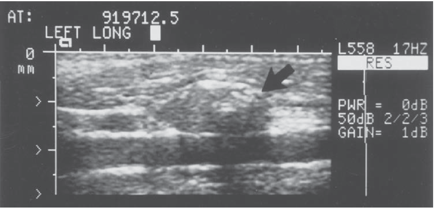
Thyroid ultrasound image (showing a lymph node along the carotid artery - from Schwartz's Surgery):
5.5 Radioactive Iodine (RAI) Scan
RAI UPTAKE AND SCAN INTERPRETATION

        ┌─────────────────────────────────────────┐
        │            RAIU PATTERN                 │
        ├──────────────┬──────────────────────────┤
        │  ↑ UPTAKE    │  Graves' disease (diffuse)│
        │  (hot scan)  │  TMNG (patchy)            │
        │              │  Solitary toxic adenoma   │
        ├──────────────┼──────────────────────────┤
        │  ↓ UPTAKE    │  Thyroiditis (all types)  │
        │  (cold scan) │  Exogenous thyroid hormone│
        │              │  Factitious thyrotoxicosis│
        │              │  Iodine excess            │
        └──────────────┴──────────────────────────┘

NODULE CHARACTERISATION:
  "Hot" nodule = hyperfunctioning = <1% malignancy risk
  "Cold" nodule = non-functioning = 5-15% malignancy risk
  "Warm" nodule = intermediate
Contraindications to RAI: Pregnancy, breastfeeding, active/severe ophthalmopathy (Graves'), poor compliance (radiation safety)
5.6 FNAC - The Bethesda System for Reporting Thyroid Cytopathology
BETHESDA SYSTEM (2017, 3rd Edition)
┌──────┬─────────────────────────────────┬────────────────┬─────────────────────────┐
│ Cat. │ Interpretation                  │ ROM (malignancy│ Recommended Action      │
│      │                                 │ risk)          │                         │
├──────┼─────────────────────────────────┼────────────────┼─────────────────────────┤
│  I   │ Non-diagnostic / Unsatisfactory │ 5-10%          │ Repeat USG-guided FNA   │
├──────┼─────────────────────────────────┼────────────────┼─────────────────────────┤
│  II  │ Benign                          │ 0-3%           │ Clinical follow-up      │
│      │ (colloid nodule, thyroiditis,   │                │ (USG in 12-24 months)   │
│      │  simple cyst)                   │                │                         │
├──────┼─────────────────────────────────┼────────────────┼─────────────────────────┤
│  III │ Atypia of Undetermined          │ 10-30%         │ Repeat FNA or           │
│      │ Significance (AUS/FLUS)         │                │ Molecular testing       │
├──────┼─────────────────────────────────┼────────────────┼─────────────────────────┤
│  IV  │ Follicular Neoplasm / SFN       │ 25-40%         │ Diagnostic lobectomy    │
│      │                                 │                │ or molecular testing    │
├──────┼─────────────────────────────────┼────────────────┼─────────────────────────┤
│  V   │ Suspicious for Malignancy       │ 50-75%         │ Near-total /            │
│      │                                 │                │ Total thyroidectomy     │
├──────┼─────────────────────────────────┼────────────────┼─────────────────────────┤
│  VI  │ Malignant                       │ 97-99%         │ Total thyroidectomy     │
└──────┴─────────────────────────────────┴────────────────┴─────────────────────────┘
Critical Exam Point: FNAC CANNOT distinguish follicular adenoma (benign) from follicular carcinoma (malignant). This distinction requires histopathological evidence of capsular or vascular invasion. This is why Bethesda IV → diagnostic lobectomy, not observation.
5.7 Additional Investigations
| Investigation | Indication | Information Obtained |
|---|
| Serum Calcium + PTH | Pre/post-thyroidectomy | Baseline parathyroid function |
| Calcitonin | Thyroid nodule workup | Screen for medullary thyroid ca (MTC) |
| Serum CEA | Suspected MTC | Marker for MTC |
| CT neck/chest | Retrosternal goiter, large MNG | Tracheal deviation/compression, mediastinal extension, tracheal calibre |
| MRI neck | Retrosternal goiter (preferred over CT) | Better soft tissue; no radiation; characterise extent |
| CXR | Routine pre-op; large goiter | Tracheal deviation, retrosternal shadow, calcification |
| Indirect/Fibreoptic Laryngoscopy | MANDATORY pre- and post-op | Vocal cord mobility assessment |
| Flow-volume loop | Suspected dynamic airway obstruction | Identifies extrathoracic vs. intrathoracic obstruction |
MODULE 6: DETAILED MANAGEMENT
6.1 Management of Hyperthyroidism - Overview
CONFIRMED HYPERTHYROIDISM (↓TSH + ↑fT4)
              │
              ▼
    ┌─────────────────────┐
    │ Identify Cause       │
    │  Graves' disease     │
    │  TMNG                │
    │  Toxic adenoma       │
    │  Thyroiditis         │
    └──────────┬──────────┘
               │
     ┌─────────┴──────────┐
     │                    │
  Graves'              TMNG /
  disease          Toxic adenoma
     │                    │
     ▼                    ▼
THREE OPTIONS:       TWO OPTIONS:
1. ATDs              1. Surgery (preferred
2. RAI                  if large, compressive)
3. Surgery           2. RAI (if small, no
                        compression)
6.2 Medical Management
Antithyroid Drugs (ATDs)
| Drug | Dose | Mechanism | Key Differences |
|---|
| Carbimazole | 20-40 mg/day (high dose); 5-15 mg/day (maintenance) | Blocks TPO - inhibits organification of iodide and coupling of iodotyrosines | Preferred in UK, India, Europe; prodrug of methimazole |
| Methimazole | 20-40 mg/day | Same as carbimazole | Used in USA/Japan |
| Propylthiouracil (PTU) | 100-200 mg 8-hourly | Blocks TPO + inhibits peripheral T4→T3 conversion (deiodinase) | Preferred in: 1st trimester pregnancy, thyroid storm, allergy to carbimazole; RISK: hepatotoxicity |
Titration vs. Block-and-Replace:
- Titration: Gradually reduce ATD dose as TSH normalises (preferred - lower side effects)
- Block-and-Replace: High dose ATD + levothyroxine (better compliance, fewer dose adjustments)
Side Effects:
- Agranulocytosis: 0.2-0.5% (warn patient - stop drug, check WBC if sore throat/fever)
- PTU-specific: Fulminant hepatic failure (rare but fatal; check LFTs)
- Rash, arthralgia, urticaria (switch drug if mild; stop if severe)
Radioactive Iodine (RAI, I-131)
- Mechanism: I-131 emits beta-particles destroying thyroid follicular cells
- Most effective for: Graves' disease, TMNG, solitary toxic adenoma
- Dose: 185-555 MBq (5-15 mCi) depending on gland volume
- Response: euthyroid in 6-12 weeks; hypothyroidism expected in majority long-term
- Absolute contraindications: Pregnancy, breastfeeding
- Relative contraindications: Active/moderate-severe Graves' ophthalmopathy (can worsen), large goiter with compression, suspected malignancy
Management of Hypothyroidism
- Levothyroxine (T4 replacement)
- Starting dose: 1.6 mcg/kg/day (in elderly or cardiac disease, start at 25-50 mcg/day)
- Target TSH: 0.5-2.5 mU/L for benign disease; 0.1-0.5 mU/L for cancer (suppression)
- Monitoring: TSH at 6-8 weeks after dose change, then annually
- Take 30 minutes before food (calcium, iron, PPIs reduce absorption)
Management of De Quervain's Thyroiditis
TRIPHASIC COURSE:
Phase 1 (hyperthyroid): 4-8 weeks → treat with beta-blockers only (NOT ATDs - hormone is pre-formed)
Phase 2 (euthyroid): weeks
Phase 3 (hypothyroid): weeks to months → levothyroxine if symptomatic
Phase 4 (recovery): 90% return to normal function

PAIN MANAGEMENT:
  Mild: NSAIDs (ibuprofen)
  Severe: Prednisolone 40 mg/day tapering over 4-6 weeks (dramatic response)
6.3 Indications for Surgery
Absolute Indications
- Suspected or confirmed malignancy (Bethesda V/VI; suspicious features on USG)
- Compressive symptoms (dyspnoea, stridor, dysphagia, Pemberton's sign)
- Retrosternal/substernal goiter
- Failed or contraindicated medical therapy for hyperthyroidism
- Graves' disease with:
- Very large goiter (>80 g)
- Desired rapid definitive control
- Active moderate-severe ophthalmopathy (RAI can worsen it)
- Pregnancy/planning pregnancy soon (RAI contraindicated)
- Riedel's thyroiditis with tracheal compression
Relative Indications
- Patient preference (avoidance of lifelong ATDs)
- TMNG - cosmetic concern, patient preference
- Large follicular neoplasm (Bethesda IV) where molecular testing unavailable
- Pregnancy with uncontrolled hyperthyroidism on ATDs (2nd trimester preferred)
- Unacceptable side effects to ATDs
MODULE 7: TYPES OF THYROID OPERATIONS
7.1 Classification of Thyroidectomy
THYROID OPERATIONS
│
├── HEMITHYROIDECTOMY (Lobectomy + Isthmusectomy)
│   Remove one complete lobe + isthmus
│   Indication: Unilateral benign nodule, Bethesda III/IV
│               Follicular adenoma, unilateral Hurthle cell
│
├── ISTHMECTOMY
│   Remove isthmus only
│   Indication: Riedel's (decompression), midline nodule,
│               tracheal biopsy access
│
├── NEAR-TOTAL THYROIDECTOMY
│   Remove all except 1-2g remnant posterolaterally on one side
│   (Protects parathyroid blood supply on that side)
│   Indication: Graves', bilateral MNG (historical; now less preferred)
│
├── TOTAL THYROIDECTOMY
│   Remove BOTH lobes completely (no thyroid tissue remains)
│   Indication: Graves', bilateral MNG, thyroid cancer
│               Retrosternal goiter, Hashimoto's + suspected cancer
│   Advantage: Eliminates recurrence risk; enables RAI post-op for cancer
│   Disadvantage: 100% need lifelong T4; highest risk of hypoparathyroidism
│
├── SUBTOTAL THYROIDECTOMY (Dunhill Operation)
│   Total on one side + subtotal (3-7g remnant) on contralateral side
│   Indication: Bilateral MNG (largely replaced by total thyroidectomy)
│   Disadvantage: 5-20% recurrence risk at 10 years
│
└── COMPLETION THYROIDECTOMY
    Remove the remaining lobe after initial lobectomy
    when malignancy found on histology
7.2 Choice of Operation by Condition
| Condition | Preferred Operation | Rationale |
|---|
| Unilateral benign nodule | Hemithyroidectomy | Preserve contralateral function |
| Bethesda IV (follicular neoplasm) | Hemithyroidectomy | Diagnostic; if benign - definitive |
| Graves' disease | Total thyroidectomy | No remnant → no recurrence |
| TMNG | Total thyroidectomy | Bilateral disease; no recurrence |
| Non-toxic MNG with compression | Total thyroidectomy | Bilateral; prevent recurrence |
| Retrosternal goiter | Total thyroidectomy (cervical) | +/- median sternotomy (<10%) |
| Riedel's thyroiditis | Isthmusectomy | Decompression only (not extensive) |
| Follicular carcinoma on histology after lobectomy | Completion thyroidectomy | Allows RAI, reduces recurrence |
MODULE 8: SURGICAL STEPS OF THYROIDECTOMY
(Based on Schwartz's Principles of Surgery 11e and Bailey & Love 28e)
8.1 Preoperative Preparation
Render Euthyroid (Critical for Graves'/TMNG)
- Antithyroid drugs until euthyroid (4-8 weeks typically): Carbimazole/methimazole + propranolol
- Lugol's iodine (Potassium iodide solution): 5-10 drops THREE times daily for 10 days BEFORE surgery
- Mechanism: Wolf-Chaikoff effect → iodine excess temporarily blocks thyroid hormone release AND reduces thyroid vascularity
- Result: Reduces intraoperative bleeding significantly
- IMPORTANT: Give ATDs BEFORE Lugol's to prevent iodine being used for hormone synthesis ("escaping" the Wolf-Chaikoff block)
Pre-op Checklist
| Item | Action |
|---|
| Vocal cord assessment | Indirect/fibreoptic laryngoscopy - document baseline |
| Serum calcium + PTH | Baseline parathyroid function |
| TFTs | Confirm euthyroid status |
| Blood group and save | Routine |
| Consent (specific risks) | Haemorrhage, RLN injury, hypoparathyroidism, hypothyroidism, scar, recurrence |
| Nerve monitor tube | NIM tube or electrode wrap organised |
| Mark incision | In skin crease before prepping |
8.2 Step-by-Step Operative Technique
STEP 1 - POSITIONING AND SETUP
┌─────────────────────────────────────────────────────────┐
│  PATIENT POSITIONING                                    │
│                                                         │
│  • Supine on operating table                           │
│  • Shoulder roll (pillow/inflatable bag) under scapulae │
│  • Head on ring/donut cushion                          │
│  • Neck EXTENDED (maximises exposure)                  │
│  • Head of table slightly elevated (10-15°)            │
│    → Reverse Trendelenburg → reduces venous bleeding   │
│  • Arms tucked at sides                                 │
│  • Prepare field: lower lip → upper chest              │
│  • NIM endotracheal tube positioned between vocal cords │
│    ⚠ CHECK tube position AFTER neck extension          │
│    (extension can withdraw tube, losing electrode contact)│
└─────────────────────────────────────────────────────────┘
STEP 2 - INCISION (Kocher's Collar Incision)
- Location: In a skin crease, at the level of the cricoid cartilage (superior edge of thyroid isthmus) - approximately 2 finger breadths above the sternal notch
- Length: 3-5 cm for routine cases; proportionate to goiter size; NOT beyond medial border of SCM
- Technique:
- Mark the incision before prepping
- Infiltrate with local anaesthetic + adrenaline (epinephrine) for haemostasis
- Scalpel through skin - commit to the full length of incision
- Incise through dermis in one stroke
STEP 3 - PLATYSMA AND SUBPLATYSMAL FLAPS
- Monopolar diathermy through platysma
- Raise subplatysmal flaps in the avascular plane:
- Superiorly: to thyroid notch of thyroid cartilage
- Inferiorly: to suprasternal notch
- Flaps held with self-retaining retractor (Joll's/Cloward)
STEP 4 - STRAP MUSCLES
- Identify midline (linea alba of neck) between the sternohyoid muscles
- Divide midline raphe with diathermy (avoid anterior jugular veins in midline)
- Use Langenbeck retractors to sweep superiorly and inferiorly
- Develop plane between muscle layers:
- Elevate sternohyoid laterally until ansa cervicalis nerve visible
- Mobilise sternothyroid from the gland with gentle dissection
- If more exposure needed: Divide strap muscles SUPERIORLY (preserves ansa cervicalis innervation which enters inferiorly)
- Identify IJV laterally → deep to find common carotid artery → vagus nerve between them → stimulate vagus to confirm NIM tube function
STEP 5 - MIDDLE THYROID VEIN
- Retract thyroid lobe medially and anteriorly
- Identify the middle thyroid vein (runs from lateral thyroid directly to IJV - NO accompanying artery)
- Ligate and divide - this allows full mobilisation of the lobe
STEP 6 - SUPERIOR POLE DISSECTION (Most Technically Demanding Part)
KEY PRINCIPLE: Ligate INDIVIDUALLY close to the thyroid capsule
               to protect the EBSLN
- Retract thyroid inferiorly and medially
- Identify the cricothyroid muscle (key landmark for EBSLN)
- EBSLN runs along the superior thyroid artery - identify it if possible (Cernea Type 2b = highest risk)
- Individually skeletonise, ligate and divide each superior thyroid vessel (artery and vein) as close to the thyroid capsule as possible
- NEVER mass-ligate the superior pedicle (risk of EBSLN injury)
- Once superior vessels divided, sweep tissues posteromedially from the upper pole (reduces risk to upper parathyroid blood supply)
STEP 7 - IDENTIFICATION OF THE RLN
RLN IDENTIFICATION PROTOCOL:
ATA 2015 Guidelines: Visual identification in ALL cases

LOCATE THE NERVE AT:
  1. Tracheo-oesophageal groove (most reliable primary location)
  2. Level of inferior thyroid artery crossing
  3. Cricoid cartilage level (most consistent anatomical landmark)

TRACE THE NERVE:
  → Superiorly towards its entry point at inferior cornu
    of thyroid cartilage / cricothyroid joint
  → At Berry's Ligament: MAXIMUM CAUTION

CONFIRM IDENTITY:
  → NIM tube: EMG spike on stimulation at 0.5-2 mA
  → Visual: pale white cord, no branching (vs. inferior thyroid artery branches)
  → "Cricket" sound of NIM monitor
Non-recurrent RLN:
- Right side only (0.5-1%)
- Associated with aberrant right subclavian artery (arteria lusoria)
- Suspect if: right inferior thyroid artery arises directly from the carotid
- Clinically important: RLN enters the field from a completely unexpected direction (lateral instead of inferior)
STEP 8 - INFERIOR POLE AND INFERIOR THYROID ARTERY
- Mobilise lower pole by gently sweeping all tissues dorsally
- Identify the inferior thyroid artery
- Ligate close to the thyroid capsule (NOT at the main trunk) - this preserves the terminal branches that supply the parathyroid glands
- Divide inferior thyroid veins (drain to innominate vein)
- Identify and preserve inferior parathyroids during this dissection
STEP 9 - PARATHYROID IDENTIFICATION AND PRESERVATION
PARATHYROID PRESERVATION STRATEGY:

  IDENTIFY:
    Superior: Posterior to upper thyroid, at cricothyroid level
    Inferior: Near lower thyroid pole, in surrounding fat

  PRESERVE:
    DO NOT skeletonise the inferior thyroid artery trunk
    Ligate ITA branches close to thyroid capsule
    Handle parathyroids with MINIMUM force (fragile)
    Preserve fat pad around each gland

  IF DEVASCULARISED:
    Test viability: tiny incision → fresh bleeding = viable
    If non-viable → AUTOTRANSPLANT into SCM
    Technique: Mince into 1mm³ fragments
    Create multiple pockets in SCM body
    Insert fragments; mark each pocket with non-absorbable suture

  INTRAOPERATIVE IDENTIFICATION AIDS:
    Near-infrared autofluorescence (NIRAF) - recent advance
    Parathyroids autofluoresce at 820nm wavelength
STEP 10 - BERRY'S LIGAMENT AND SEPARATION FROM TRACHEA
- With RLN clearly identified and visualised at all times:
- Carefully divide Berry's posterior suspensory ligament
- Bleeding in this area = controlled pressure FIRST, then careful haemostasis
- Do NOT use diathermy near the RLN (thermal spread injures nerve)
- Once ligament divided, separate thyroid from trachea by sharp dissection
- Pyramidal lobe: trace superiorly above thyroid cartilage notch and divide in continuity
STEP 11 - ISTHMUS AND CONTRALATERAL SIDE
- For hemithyroidectomy: Divide isthmus between ligatures at the trachea
- For total thyroidectomy: Repeat steps 5-10 on contralateral side
- Isthmus divided last (or early, to facilitate medial rotation)
STEP 12 - HAEMOSTASIS AND CLOSURE
HAEMOSTASIS:
  • Systematic check of all pedicles
  • Irrigation with warm saline
  • Bone wax to tracheal bleeding
  • Energy devices (harmonic/ligasure) for small bleeders
  • DO NOT rush this step

CLOSURE:
  • Strap muscles: LOOSELY approximated (NOT watertight)
    Reason: haematoma can escape anteriorly into subcutaneous space
             rather than compress trachea
  • Platysma: absorbable sutures
  • Skin: subcuticular absorbable suture = best cosmesis
         (or clips/staples in some centres)
  • DRAIN: NOT routinely recommended
    (No evidence of benefit; gives false reassurance)

SPECIMEN: Send immediately for formal histopathology
          Frozen section available if equivocal at operation
MODULE 9: POSTOPERATIVE COMPLICATIONS
9.1 Complications Summary Table
| Complication | Timing | Incidence | Mechanism | Management |
|---|
| Haematoma | 0-24h | 1-2% | Vessel slippage; coagulopathy | Open wound immediately + secure airway → theatre |
| Bilateral RLN palsy | Immediate | <0.5% | Bilateral nerve injury | Emergency intubation/tracheostomy |
| Transient hypocalcaemia | 24-72h | 20-30% (after total) | Parathyroid devascularisation | Oral Ca + Calcitriol |
| Permanent hypoparathyroidism | >6 months | <3% | Parathyroid removal/injury | Long-term Ca + Calcitriol |
| Transient RLN palsy | 0-4 weeks | 1.8% at 1 month | Traction, thermal injury | Observe; speech therapy |
| Permanent RLN palsy | >3 months | 0.5% | Nerve division/thermal injury | Medialisation procedures |
| EBSLN injury | Immediate | 0.4-14% | Damaged during superior pole dissection | Voice therapy; assess with laryngoscopy |
| Hypothyroidism | Weeks-months | 100% (total); ~30% (lobectomy) | No/inadequate remnant | Levothyroxine |
| Thyroid storm | Intraoperative/ immediate | Rare | Inadequate pre-op preparation | Emergency medical Rx (see below) |
| Wound infection | Days | <1% | Contamination | Antibiotics; wound care |
| Keloid/hypertrophic scar | Weeks-months | Variable | Skin type; tension | Silicone, steroid injection |
9.2 Haematoma - The Critical Emergency
POSTOPERATIVE HAEMATOMA - MANAGEMENT PROTOCOL

PRESENTATION:
  → Swelling at wound
  → Stridor/respiratory distress
  → Anxiety, increasing dyspnoea

⚠ FIRST ACTION (EVEN AT BEDSIDE):
  → OPEN SKIN CLIPS/SUTURES IMMEDIATELY
  → Release tension → haematoma decompresses anteriorly
  → This can be LIFE-SAVING while awaiting anaesthetist

THEN:
  → Call anaesthetist URGENTLY
  → Secure airway (orotracheal intubation preferred)
  → Rush to theatre for formal exploration and haemostasis
  → Evacuate haematoma, find bleeding point, re-ligate

WHY:
  Arterial bleed → rising tension → exceeds venous pressure
  → Venous congestion of larynx → glottic oedema
  → Airway obstruction → DEATH

Note: Drains do NOT prevent this - they clot and give false security
9.3 Hypocalcaemia - Diagnosis and Management
EARLY SYMPTOMS (within 24-48h):
  • Perioral tingling/numbness
  • Fingertip tingling
  • Muscle cramps
  • Anxiety

PROGRESSIVE:
  • Carpopedal spasm
  • Positive Chvostek's sign (tap facial nerve → facial twitch)
  • Positive Trousseau's sign (BP cuff inflated for 3 min → carpopedal spasm)
  • Tetany
  • Laryngospasm (emergency)
  • Seizures

MANAGEMENT:
  Asymptomatic / Mild: Oral calcium carbonate 1-2g 3x/day + Calcitriol 0.25mcg BD
  Symptomatic / Severe: IV Calcium gluconate 10ml 10% solution over 10min
                        → Followed by calcium infusion
  Prolonged: PTH assay + Magnesium level (hypomagnesaemia prevents PTH action)

MONITORING:
  Serum calcium 6 hours post-op, then daily × 3 days
9.4 RLN Injury - Management
| Situation | Management |
|---|
| Transient palsy (<3 months) | Observe, speech therapy, soft diet (aspiration prevention) |
| Permanent unilateral palsy | Speech therapy; if unacceptable voice → Medialisation laryngoplasty (Type I thyroplasty) or vocal cord injection (Teflon/fat/collagen) |
| Both ends identified at surgery | Immediate primary reanastomosis |
| Segment loss (e.g., cancer invasion) | Ansa cervicalis nerve graft - does not restore mobility but prevents atrophy, improves voice |
| Bilateral palsy | Emergency tracheostomy; later: arytenoidectomy, posterior cordotomy |
9.5 Thyroid Storm - Emergency Management
THYROID STORM (Thyrotoxic Crisis)
BURCH-WARTOFSKY SCORE: Score ≥45 = likely storm; 25-44 = impending

FEATURES:
  Hyperthermia (>38.5°C, often >40°C)
  Tachycardia/AF (HR >130-140)
  Agitation/delirium/coma
  Diarrhoea + vomiting
  Cardiac failure
  Precipitated by: surgery, infection, trauma, RAI, contrast agents

TREATMENT (sequential order MATTERS):
  Step 1: PTU 600mg loading → 200mg 4-hourly (blocks synthesis + T4→T3 conversion)
          OR Methimazole 60-80mg loading
  Step 2: Propranolol 80mg 6-hourly (or IV 0.5-1mg slowly) - rate control, blocks conversion
  Step 3: Hydrocortisone 300mg IV stat then 100mg 8-hourly
          (Prevents T4→T3 conversion; addresses relative adrenal insufficiency)
  Step 4: Lugol's iodine 8 drops TID orally
          ⚠ MUST give AFTER PTU (at least 1 hour) - otherwise iodine used for hormone synthesis!
  Step 5: Supportive care:
          • Cooling blanket, paracetamol (NOT aspirin - displaces T4 from TBG)
          • IV fluids, glucose
          • Treat precipitating cause
          • ICU monitoring
MODULE 10: SPECIFIC CONDITION MANAGEMENT
10.1 Graves' Disease - Complete Management Flowchart
GRAVES' DISEASE CONFIRMED (low TSH + high T4 + positive TRAb)
              │
              ▼
    ┌─────────────────────────────────────────────────────┐
    │           INITIAL CONTROL                           │
    │  Carbimazole + Propranolol (beta-blocker)           │
    │  (Render euthyroid in 4-8 weeks)                    │
    └──────────────────────────┬──────────────────────────┘
                               │
                ┌──────────────┴──────────────┐
                ▼                             ▼
        LONG-TERM ATD                 DEFINITIVE THERAPY
      (18-24 months course)           after euthyroid
        Remission rate: 30-50%              │
        Relapse: 50-60%                     │
                │                 ┌─────────┴──────────┐
                │                 ▼                    ▼
                │              RAI (I-131)         SURGERY
                │              Preferred if:      Preferred if:
                │              • Small goiter     • Large goiter (>80g)
                │              • No ophthalmopathy• Active ophthalmopathy
                │              • No pregnancy      • Pregnancy planned
                │              • Poor compliance   • Failed ATD/RAI
                │                  with surgery    • Patient preference
                │                                 • Suspicious nodule
                │                                 • Children
                │
                ▼
        Recurrence on stopping ATD → Definitive therapy

SURGERY SPECIFICS:
  Pre-op: ATDs + Propranolol → Euthyroid
          Lugol's iodine × 10 days (reduce vascularity)
  Operation: TOTAL THYROIDECTOMY
  Post-op: Levothyroxine started day 1 after surgery
           TRAb levels monitored (may persist years post-op)
           Ophthalmopathy: refer ophthalmology regardless
10.2 Toxic Multinodular Goiter (TMNG)
- Plummer's disease: Thomas Plummer first described autonomous thyroid nodules
- Mechanism: Long-standing MNG → some nodules acquire TSH receptor mutations causing constitutive (TSH-independent) activation
- Differs from Graves': No TRAb/TSI, no ophthalmopathy, no pretibial myxedema, older patients
- Treatment:
- Render euthyroid with ATDs (carbimazole/methimazole)
- Surgery preferred if: large goiter, compression, cosmesis, suspected malignancy
- RAI acceptable if: small TMNG, no compression, poor surgical candidate
- Operation: Total thyroidectomy
- Note: Subtotal thyroidectomy has high recurrence rate in TMNG
10.3 Retrosternal Goiter
DEFINITION: Goiter where >50% of the mass lies below the thoracic inlet

CLASSIFICATION (Pemberton):
  Grade I: Cervical goiter that descends into thorax on swallowing
  Grade II: Partial substernal goiter
  Grade III: Complete substernal goiter

PRESENTATION:
  Pemberton's sign (arms raised → facial flushing/cyanosis)
  Stridor, dyspnoea (especially lying flat)
  Dysphagia
  Superior vena cava syndrome (late)
  May be asymptomatic (discovered incidentally on CXR)

INVESTIGATIONS:
  CXR: Superior mediastinal widening, tracheal deviation/compression
  CT chest/neck: Defines extent, tracheal calibre, vessel involvement
  MRI: Preferred (no radiation; better soft tissue)
  TFTs + USG
  FNAC if suspicious nodule

MANAGEMENT - SURGICAL:
  INDICATION: All symptomatic substernal goiters; most asymptomatic ones

  APPROACH:
  ┌──────────────────────────────────────────────────────────┐
  │ >90% can be removed via CERVICAL APPROACH ALONE         │
  │ (blood supply = neck vessels → can deliver from below)  │
  │ Technique: sustained gentle traction + blunt dissection │
  │            + finger delivery of goiter from mediastinum │
  ├──────────────────────────────────────────────────────────┤
  │ <10% require MEDIAN STERNOTOMY:                         │
  │ Indications:                                             │
  │  • True intrathoracic blood supply from mediastinal vessels│
  │  • Unable to deliver via cervical route                  │
  │  • Recurrent/redo surgery                               │
  │  • Malignancy with mediastinal invasion                  │
  └──────────────────────────────────────────────────────────┘

  Special risks: High RLN injury rate, parathyroid injury,
                 great vessel injury, postoperative haematoma
                 (mediastinal expansion more dangerous)
10.4 Riedel's Thyroiditis - Management
- Now classified as IgG4-related systemic disease (elevated serum IgG4 >135 mg/dL; lymphoplasmacytic infiltrate with IgG4+ plasma cells)
- Associated with: Mediastinal fibrosis, retroperitoneal fibrosis, sclerosing cholangitis, orbital pseudotumour
- Diagnosis requires open biopsy (FNAC inadequate due to fibrous consistency)
- "Woody" thyroid, fixed, may encase RLN and parathyroids
Treatment:
- Surgery (isthmusectomy): Chief goal = tracheal decompression (NOT extensive resection - infiltrative process obscures landmarks)
- Corticosteroids (prednisolone 40-60 mg/day): First-line medical; dramatic response in most
- Tamoxifen: Anti-fibrotic; used in steroid-refractory cases
- Mycophenolate mofetil: Steroid-sparing agent
- Rituximab (anti-CD20): Emerging evidence in IgG4-RD; dramatic response in refractory cases
- Levothyroxine if hypothyroid
MODULE 11: POSTOPERATIVE FOLLOW-UP PROTOCOL
11.1 Follow-up Schedule
TIME POINT  │  ASSESSMENT
────────────┼──────────────────────────────────────────────────────
6 hours     │  Serum calcium (first check post-op)
post-op     │  Clinical assessment: voice, wound, breathing
────────────┼──────────────────────────────────────────────────────
24-48 hours │  Calcium again; laryngoscopy if hoarseness present
post-op     │  Wound check; discharge if calcium stable and euthyroid
────────────┼──────────────────────────────────────────────────────
2-4 weeks   │  FIRST CLINIC VISIT
            │  • Wound assessment
            │  • Voice assessment (indirect laryngoscopy)
            │  • TFTs (TSH + fT4)
            │  • Serum calcium + PTH
            │  • Histopathology result review
            │  • Start levothyroxine if total thyroidectomy
────────────┼──────────────────────────────────────────────────────
6-8 weeks   │  TFT recheck; adjust levothyroxine dose
────────────┼──────────────────────────────────────────────────────
3 months    │  TFTs; confirm euthyroid on replacement
            │  USG neck (baseline for future comparison)
            │  TRAb (Graves') - monitor
────────────┼──────────────────────────────────────────────────────
6-12 months │  Annual TFT; clinical review
            │  USG neck (for MNG - check for recurrence)
            │  Anti-TPO (Hashimoto's monitoring)
────────────┼──────────────────────────────────────────────────────
Lifelong    │  Annual TSH
            │  Bone density (if on suppressive T4)
            │  Ophthalmology (Graves' - annual)
11.2 Levothyroxine Target TSH by Indication
| Indication | Target TSH (mU/L) |
|---|
| Benign disease (post-total thyroidectomy) | 0.5 - 2.5 |
| Post-lobectomy (hypothyroid) | 0.5 - 2.5 |
| Low-risk differentiated thyroid cancer | 0.5 - 2.0 |
| High-risk differentiated thyroid cancer | 0.1 - 0.5 (suppression) |
| Graves' ophthalmopathy (active) | 0.5 - 1.0 (avoid suppression and excess) |
MODULE 12: RECENT ADVANCES AND CURRENT RESEARCH (2023-2026)
12.1 Non-Surgical Thermal Ablation Techniques
These techniques represent a paradigm shift - benign thyroid nodules can now be treated without surgery in selected patients.
Radiofrequency Ablation (RFA)
MECHANISM:
  High-frequency alternating current (350-500 kHz)
  → Ionic agitation → Frictional heat → Coagulation necrosis

TECHNIQUE:
  • USG-guided; local anaesthesia
  • "Moving shot" technique: probe moves through nodule
  • Hydrodissection: inject fluid between nodule and capsule
    to protect surrounding structures

INDICATIONS (Korean Society of Thyroid Radiology guidelines):
  • Benign solid/predominantly solid nodule (Bethesda II confirmed)
  • Symptomatic: compressive, cosmetic
  • Autonomously functioning thyroid nodule
  • Recurrent thyroid cyst

RESULTS (Meta-analysis 2024, PMID 37639985 - 5-year follow-up):
  • Volume reduction: 50-85% at 1 year; sustained at 5 years
  • Symptom relief: significant improvement
  • Complication rate: <4% (voice change, skin burn, haematoma)
  • Regrowth rate: 5-10% at 5 years (may need re-ablation)
Comparison: RFA vs. Lobectomy (Meta-analysis 2025, PMID 40849686)
| Outcome | RFA | Lobectomy |
|---|
| Volume reduction | Comparable | Complete removal |
| Hospital stay | Day procedure | 1-2 days |
| General anaesthesia | NOT required | Required |
| Complications | Lower overall | Higher (RLN, parathyroid) |
| Hypothyroidism | Rare | ~30% |
| Regrowth/recurrence | 5-10% | Nil (complete excision) |
| Scar | None | Neck scar |
| Cost | Lower | Higher |
| Malignancy exclusion | Limited | Definitive (histology) |
RFA vs. Microwave Ablation (MWA) (Meta-analysis 2024-2025, PMID 38413114, 39046500)
- Both are safe and effective for benign thyroid nodules
- MWA: Shorter operative time; higher peak temperature; may be superior for large nodules
- RFA: More data available; better validated long-term evidence
- No significant difference in complication rates
High-Intensity Focused Ultrasound (HIFU)
- Completely non-invasive (no needle insertion)
- Converges multiple ultrasound beams at focal point in nodule → thermal coagulation at focus
- Meta-analysis (PMID 37478539, 2023): Mean volume reduction 40-60% at 6 months; safe
- Limitation: Cannot treat nodules near critical structures (RLN, trachea) - limited safety margin
- Suitable for: Benign nodules in selected locations
Long-term Thermal Ablation Results (Meta-analysis 2025, PMID 40214733)
- Systematic review of all thermal ablation modalities (RFA, MWA, HIFU, LA)
- Long-term (>5 years): Volume reduction maintained; no increase in malignancy risk
- Conclusion: Safe alternative to surgery for correctly selected benign nodules
12.2 Remote Access / Scarless Thyroidectomy
| Approach | Access | Key Features | Evidence |
|---|
| TOETVA (Transoral Endoscopic Thyroidectomy Vestibular Approach) | 3 incisions inside lower lip (oral vestibule) | No neck scar; cosmetically superior; learning curve | Meta-analysis 2023 (PMID 37556081) |
| TORB (Transoral Robotic) | Oral vestibule + robotic system | Better articulation in tight space; more expensive | Compared with TOETVA: similar outcomes |
| BABA (Bilateral Axillo-Breast Approach) | Bilateral axillary + breast incisions | Popular in East Asia; longer dissection tunnel | Large Asian experience |
| Axilloscopic | Single axillary incision | Modified access | Less popular |
| Retroauricular (Facelift) | Behind ear | Combined with face-lift incision | Selected cases |
TOETVA vs Transoral Robotic (Meta-analysis 2023, PMID 37556081):
- Comparable safety and efficacy
- TOETVA: more widely practiced; lower cost
- Robotic: better ergonomics; more degrees of freedom
Track Recurrence (Meta-analysis 2024, PMID 39343618):
- Systematic review of cancer seeding along dissection tracks
- Overall incidence: <0.1% (rare but real complication)
- More common with malignant disease and violated capsule
- Implication: Remote access approaches should be used cautiously when malignancy suspected
Current Limitations of Remote Access:
- Longer operative time (1.5-2x conventional)
- Limited to small-volume, benign or low-risk disease
- Steep learning curve (>50 cases to competency)
- Increased risk of skin burn, subcutaneous emphysema, CO2 embolism (transoral)
12.3 Intraoperative Nerve Monitoring (IONM)
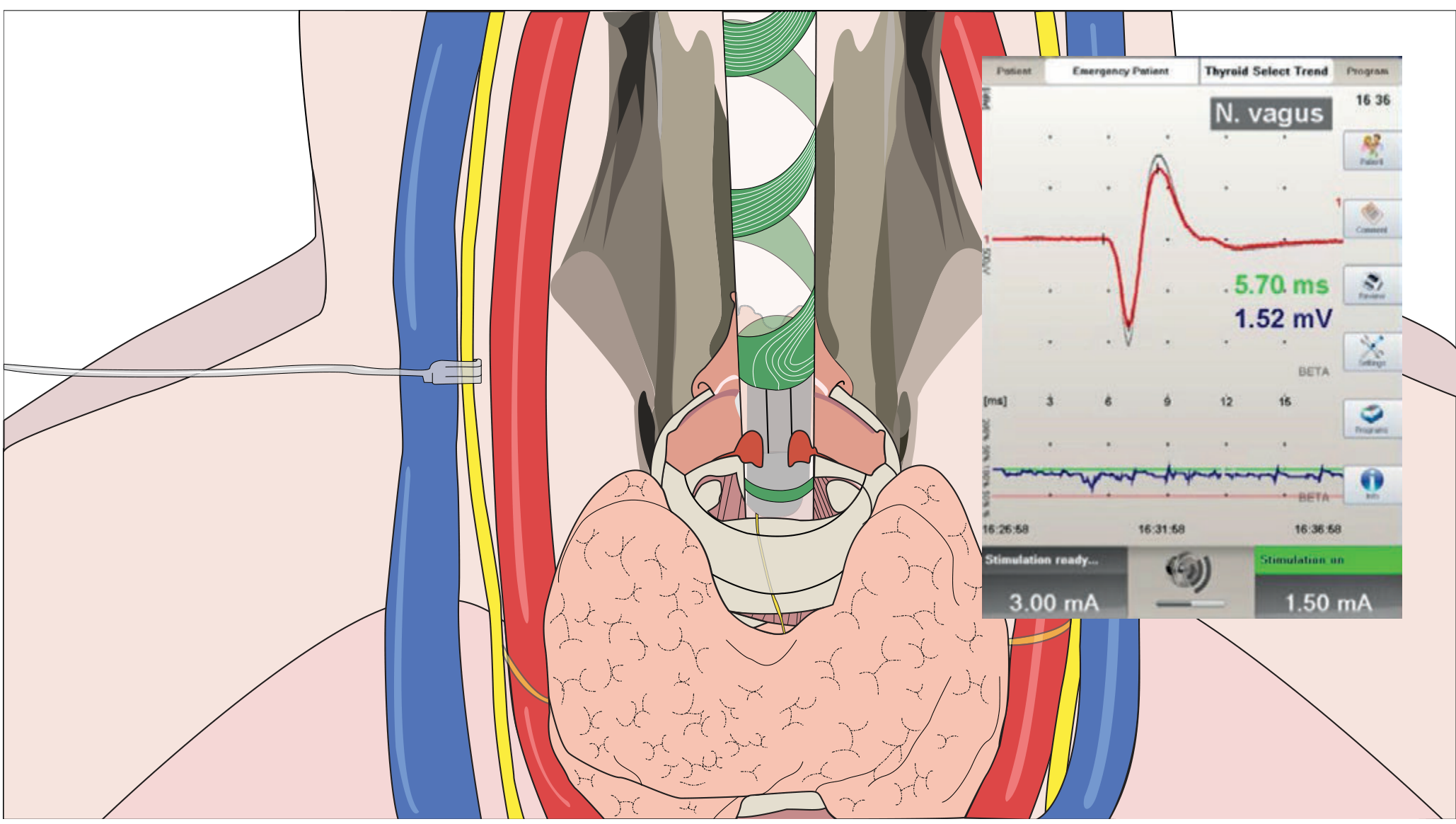
Continuous IONM (C-IONM) - The Latest Technology:
The diagram above shows continuous vagus nerve monitoring (C-IONM). An electrode is placed directly on the vagus nerve, providing real-time feedback of RLN function. The waveform panel shows the EMG signal recorded (5.70 ms latency, 1.52 mV amplitude) - a normal response confirming nerve integrity.
| Type | Technique | Advantage | Limitation |
|---|
| Intermittent IONM | NIM tube electrodes; surgeon periodically stimulates RLN/vagus | Identifies nerve; confirms identity; popular | Does not detect real-time injury |
| Continuous IONM (C-IONM) | Electrode on vagus nerve; continuous recording | Real-time alert for impending injury (traction, thermal) | Invasive electrode placement; not widely adopted |
Clinical significance of C-IONM:
- Detects "loss of signal" (LOS) before permanent injury
- Allows surgeon to pause, release traction, reassess
- Particularly valuable in bilateral thyroid surgery: if LOS on first side, consider staging the operation to prevent bilateral palsy
12.4 Near-Infrared Autofluorescence (NIRAF) for Parathyroid Identification
PRINCIPLE:
  Parathyroid glands contain calcium-sensing receptor and
  unique fluorophores that emit near-infrared light (800-900nm)
  when excited by a specific wavelength (785nm)
  → NO external contrast agent needed

CLINICAL USE:
  Intraoperative probe or camera system
  Real-time identification of parathyroids
  Distinguishes parathyroid from fat, lymph node, thyroid tissue
  Assesses relative perfusion/viability of preserved glands

EVIDENCE:
  Multiple prospective studies show:
  → 90-100% of parathyroids identified before removal
  → Significant reduction in inadvertent parathyroid excision
  → Trend towards reduced postoperative hypocalcaemia

STATUS: Commercially available (PTeye™, Fluobeam®);
        increasing adoption in high-volume centres
        Not yet standard of care but growing evidence base
12.5 Molecular Testing for Indeterminate Thyroid Nodules
PROBLEM: Bethesda III/IV nodules = "grey zone"
  Previously: ALL required diagnostic lobectomy
  Modern approach: Molecular testing on FNA specimen →
                   avoid surgery in ~50% of cases

AVAILABLE TESTS:
┌─────────────────┬──────────────────────────────────────────────┐
│ Afirma GSC      │ mRNA expression profiling (167 genes)       │
│ (Veracyte)      │ "Benign" result = avoids surgery (high NPV) │
│                 │ Sensitivity ~91%; Specificity ~68%          │
├─────────────────┼──────────────────────────────────────────────┤
│ ThyroSeq v3     │ DNA/RNA mutation analysis                    │
│ (CBLPath/Pitt)  │ Detects: BRAF, RAS, RET/PTC, PAX8/PPARγ,   │
│                 │ TERT, ALK fusions, etc.                     │
│                 │ Sensitivity ~94%; Specificity ~82%          │
├─────────────────┼──────────────────────────────────────────────┤
│ ThyGeNEXT+      │ Mutation/fusion + expression profiling      │
│ ThyraMIR        │ Combined approach (mutation + microRNA)      │
└─────────────────┴──────────────────────────────────────────────┘

CLINICAL IMPACT:
  "Negative/benign" result → avoid surgery; follow with USG
  "Suspicious/positive" result → proceed to surgery
  Reduces unnecessary lobectomies by 40-50%
  2023 ATA guidelines: Molecular testing recommended for AUS/FLUS (Bethesda III)
12.6 Active Surveillance for Small Thyroid Nodules
- Low-risk papillary microcarcinomas (<1 cm) may now be safely observed without surgery in carefully selected patients
- Major guideline groups (ATA 2023, European Thyroid Association) now endorse active surveillance as an option
- Not directly applicable to benign nodules - but represents a shift towards non-surgical management philosophies
12.7 Energy-Based Sealing Devices in Thyroid Surgery
| Device | Mechanism | Advantage vs. Tie-and-Cut |
|---|
| Harmonic Scalpel (ultrasonic) | Ultrasonic vibration → protein denaturation → haemostasis | Seals vessels up to 7mm; minimal thermal spread (< 1mm); no smoke |
| LigaSure (bipolar RF) | Bipolar RF energy + pressure → vessel wall fusion | Seals vessels up to 7mm; fast; proven efficacy |
| EnSeal | Bipolar RF + tissue sensing | Self-adjusting energy; minimal thermal spread |
- Meta-analyses confirm reduced operative time and comparable complication rates vs. conventional ligation
- Particularly useful in complex reoperative cases and when dealing with highly vascular Graves' glands
QUICK REVISION TABLES
Master Exam Pearls
| Topic | Exam Answer |
|---|
| Most common cause of goiter worldwide | Iodine deficiency |
| Most common cause of hyperthyroidism | Graves' disease (60-80%) |
| Most common cause of hypothyroidism (iodine-sufficient area) | Hashimoto's thyroiditis |
| Most sensitive thyroid function test | TSH |
| FNAC CANNOT distinguish | Follicular adenoma vs. follicular carcinoma (capsular/vascular invasion needed on histology) |
| Operation of choice for Graves' | Total thyroidectomy |
| Operation of choice for Riedel's | Isthmusectomy (decompression only) |
| Indication for Lugol's iodine | 10 days before thyroid surgery for Graves'/TMNG to reduce vascularity |
| First step for post-op haematoma | Open wound clips/sutures AT BEDSIDE immediately |
| Most common life-threatening complication of thyroidectomy | Haematoma (airway obstruction) |
| Most common complication of total thyroidectomy | Transient hypocalcaemia |
| RLN enters larynx at | Inferior cornu of thyroid cartilage / cricothyroid joint |
| EBSLN supplies | Cricothyroid muscle only (pitch/tension) |
| Parathyroids blood supply from | Inferior thyroid artery (both superior and inferior) |
| Pemberton's sign | Arms raised → facial flushing/cyanosis = retrosternal goiter |
| Cernea Type 2b | EBSLN crosses below upper thyroid pole = highest risk |
| Non-recurrent RLN | Right side (0.5-1%), associated with aberrant right subclavian artery |
| "Woody" fixed thyroid | Riedel's thyroiditis = IgG4-related systemic disease |
| Thyroid storm: Lugol's timing | AFTER PTU (minimum 1 hour) - PTU must block synthesis first |
| Hot nodule malignancy risk | <1% |
| Cold nodule malignancy risk | 5-15% |
| Kocher's incision | Transverse collar incision, cricoid level, 2 FB above sternal notch |
| Why NOT drain after thyroidectomy | No evidence of benefit; drains give false reassurance; haematoma can still develop |
| Remote access complication of concern | Track recurrence (seeding along dissection tract) |
| Novel parathyroid identification tool | Near-infrared autofluorescence (NIRAF) - 800-900 nm |
Differentiating Graves' vs. TMNG
| Feature | Graves' Disease | TMNG |
|---|
| Age | 40-60 years | >50 years (older) |
| Gender | Female:male = 5:1 | Female predominance, less marked |
| Onset | Acute/subacute | Insidious |
| Thyroid | Diffuse, smooth, soft | Nodular, irregular |
| Bruit | Yes (high vascularity) | Absent |
| Ophthalmopathy | Yes (characteristic) | No |
| Pretibial myxedema | Yes | No |
| Thyroid acropachy | Yes | No |
| TRAb/TSI | Positive | Negative |
| RAI scan | Diffuse uptake | Patchy "hot" nodules |
| Mechanism | Autoimmune (TSI) | TSH-R mutations (autonomous) |
| Relapse after ATD | Yes (50-60%) | Yes (high) |
| Treatment preference | ATD → RAI or Surgery | RAI or Surgery |
Sources: Schwartz's Principles of Surgery 11th Ed. (Chapter 38) · Bailey and Love's Short Practice of Surgery 28th Ed. (Chapter 55) · Harrison's Principles of Internal Medicine 22nd Ed. · Cummings Otolaryngology Head and Neck Surgery · Scott-Brown's Otorhinolaryngology · PubMed: PMID 40849686 (RFA vs. Lobectomy 2025) · PMID 37639985 (5-year thermal ablation 2024) · PMID 38413114 (RFA vs. MWA 2024) · PMID 39046500 (RFA vs. MWA 2025) · PMID 40214733 (Long-term thermal ablation 2025) · PMID 37556081 (TOETVA vs. Robotic 2023) · PMID 39343618 (Track recurrence 2024)Your credits are exhausted. You can purchase a top-up or subscribe to a plan to continue using Orris. If you're already subscribed, your daily credits will reset tomorrow.