
Branchial cyst acc to love and Bailey’s book of surgery for general surgery university exam, for 7 marks, for bds 3
Reading File
Now let me search the library for Branchial cyst in Bailey & Love's surgery textbook.
Finding Sources
Finding Sources
Found it in Bailey and Love's 28th Edition. Let me read the section and also search for more content on branchial cyst in that book.
Reading File
Finding Sources
Let me read more of the text and look for the image of the branchial cyst.
Reading File
Searching Images
branchial cyst neck anterior border sternomastoid
I have all the content I need from Bailey and Love's 28th Edition. Let me fetch the branchial cyst image from the textbook.
Loading Image
Loading Image
I now have everything needed to write a comprehensive 7-mark answer. Here is the complete exam answer:
Branchial Cyst
(Bailey and Love's Short Practice of Surgery, 28th Edition - Chapter 52)
Definition / Embryology
A branchial cyst develops from the vestigial remnants of the second branchial cleft. It is a congenital anomaly that results from failure of obliteration of the cervical sinus (formed by the second branchial arch growing over and burying the third and fourth clefts).
Pathology
- Lining: Squamous epithelium (stratified)
- Contents: Thick, turbid, cholesterol-rich fluid
- Lymphoid tissue is frequently found in the cyst wall, which has led to an alternative theory that the cyst arises from lymph node epithelial inclusions rather than the branchial cleft itself
Clinical Features
Age of presentation: Early to middle adulthood (20-40 years)
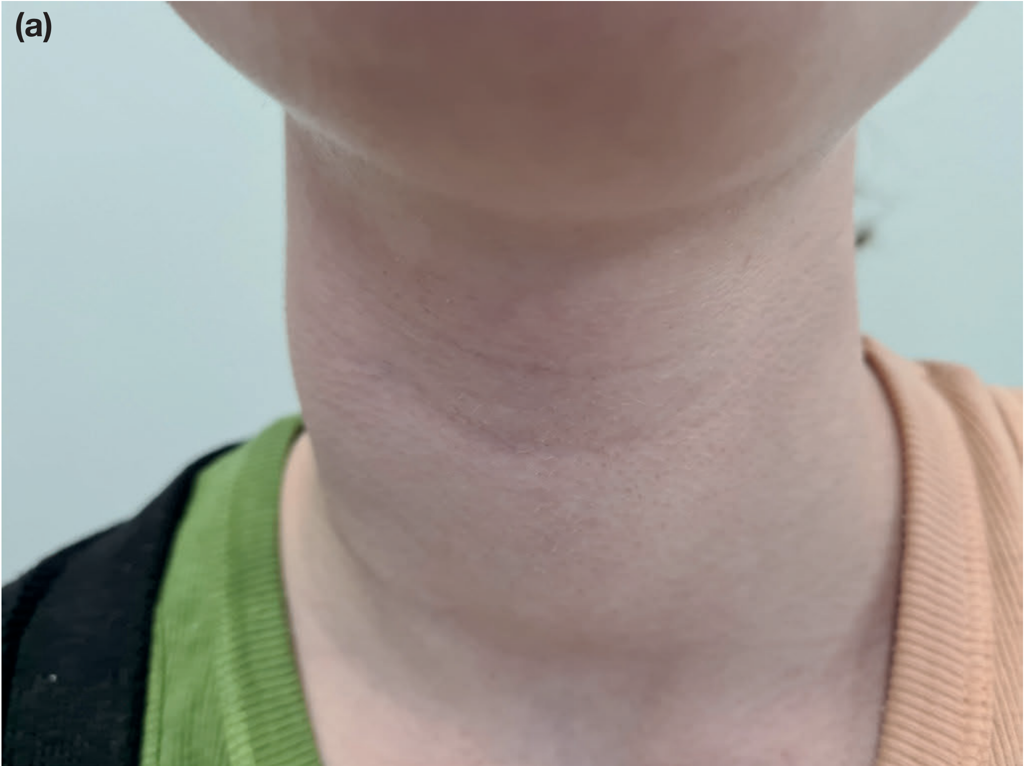
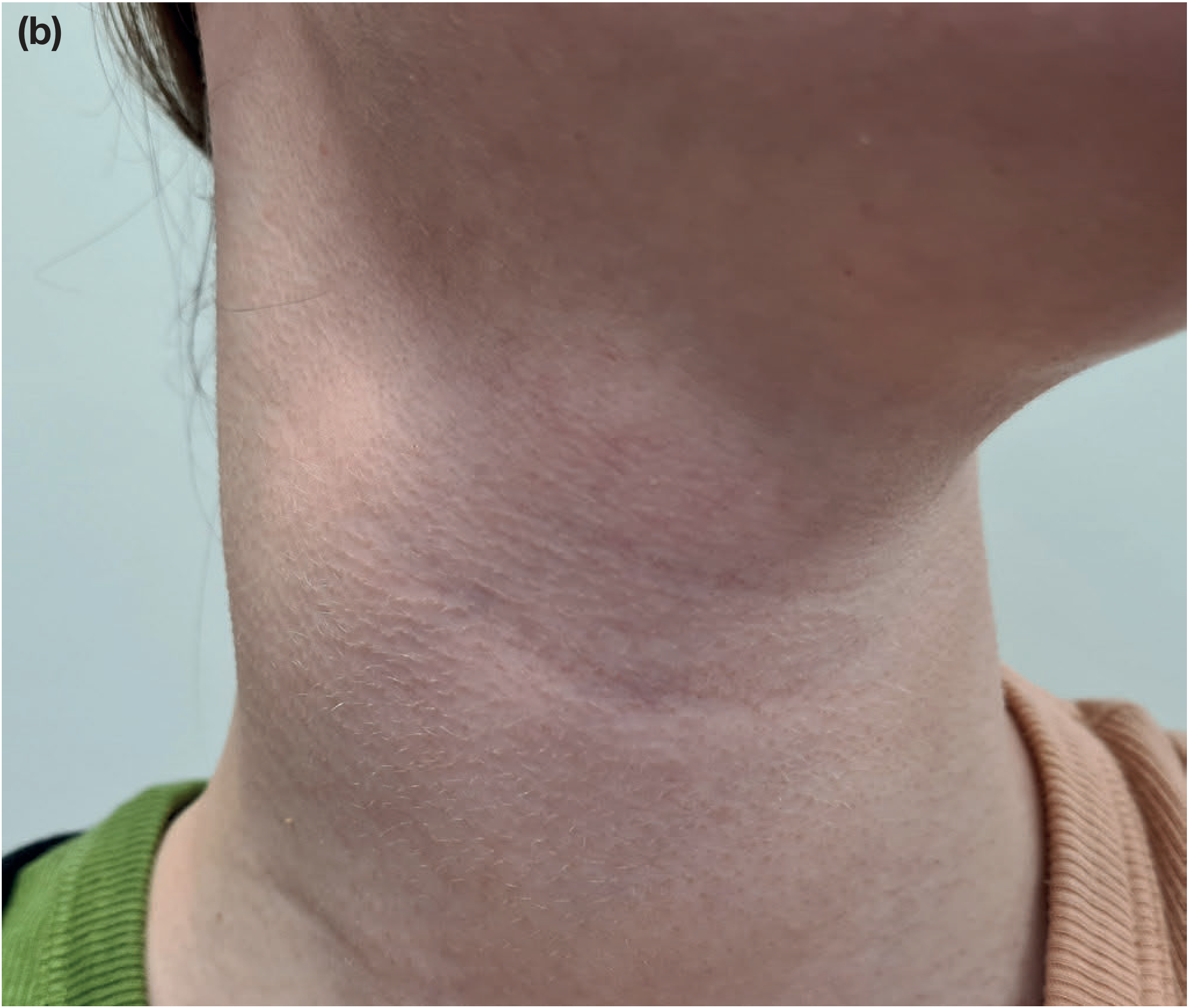
Site: Junction of the upper third and middle third of the sternomastoid muscle, at its anterior border - a classic and important exam point
Symptoms and Signs:
- Fluctuant swelling in the upper lateral neck
- May transilluminate (weakly)
- Often soft in early stages, making palpation difficult
- Usually non-tender and painless unless infected
Figure 52.63 from Bailey & Love - Right branchial cyst, anterior and oblique views:
Complications
- Infection: The cyst becomes erythematous, tender, and fluctuant. This broadens the differential diagnosis and makes excision more difficult.
- Malignant change: In patients over 35 years, a necrotic metastatic lymph node must be strongly considered and malignancy excluded before excision. A high index of suspicion is mandatory.
Differential Diagnosis (Lump in the Neck)
- Cervical lymphadenopathy (reactive, lymphoma, metastatic)
- Cystic hygroma (presents in neonates, brilliantly transilluminant)
- Thyroglossal cyst (midline, moves on tongue protrusion)
- Parotid tumour / salivary gland swelling
- Necrotic metastatic lymph node (especially if patient >35 years)
Investigations
- Ultrasound - confirms cystic nature, differentiates from solid lesions and necrotic lymph nodes
- Fine-needle aspiration (FNAC) - both aids diagnosis (cholesterol crystals in aspirate) and may be used therapeutically; important to exclude malignancy in older patients
Treatment
Complete surgical excision - the definitive treatment.
Timing: Best performed when the lesion is quiescent (not actively infected). If infected, antibiotics first, then excision once inflammation settles.
Surgical anatomy - structures at risk:
The cyst passes:
- Superficial to the hypoglossal nerve and glossopharyngeal nerve
- Deep to the posterior belly of the digastric muscle
The following structures must be positively identified to avoid damage:
- Hypoglossal nerve (CN XII)
- Glossopharyngeal nerve (CN IX)
- Spinal accessory nerve (CN XI)
Branchial Fistula (Associated Condition - BDS Exam Relevant)
A branchial fistula represents a persistent second branchial cleft.
- External orifice: lower third of neck, anterior border of sternocleidomastoid
- Internal orifice: anterior aspect of posterior faucial pillar (just behind the tonsil)
- The tract passes through the bifurcation of the common carotid artery toward the pharyngeal constrictors
- Lined by ciliated columnar epithelium with mucopurulent discharge
- Treatment: complete excision (same tract path as branchial cyst)
Summary for 7-Mark Exam Answer
| Feature | Detail |
|---|---|
| Origin | 2nd branchial cleft remnant |
| Lining | Squamous epithelium |
| Contents | Thick turbid, cholesterol-rich fluid |
| Age | Young to middle-aged adults |
| Site | Junction of upper 1/3 and middle 1/3 of sternomastoid (anterior border) |
| Signs | Fluctuant, transilluminant, soft swelling |
| Investigation | Ultrasound + FNAC |
| Treatment | Complete surgical excision (when quiescent) |
| Structures at risk | CN IX, CN XII, Spinal accessory nerve (CN XI) |
| Important caution | Exclude malignancy in patients >35 years |
Source: Bailey and Love's Short Practice of Surgery, 28th Edition, Chapter 52 - Lump in the Neck, p. 826
Marsupiliazition in detail with steps
Marsupialization is a surgical procedure where a cyst is opened, drained, and its walls are sutured to the surrounding tissue to create a permanent open pouch - preventing re-accumulation. It is commonly used for ranulas, Bartholin's cyst, dental/jaw cysts, and pancreatic pseudocysts.
Searching sources...
Finding Sources
Finding Sources
Good - I have several key sources. Since this is a BDS exam question, the most relevant applications are ranula/oral cysts and Bartholin's cyst. Let me read the most detailed sections in parallel.
Reading File
Reading File
Reading File
This is a shared conversation. Sign in to Orris to start your own chat.