
Fetal circulation
Reading File
Finding Sources
Finding Sources
Reading File
Reading File
Reading File
Reading File
Reading File
Reading File
Loading Image
Loading Image
Reading File
Loading Image
Loading Image
I now have comprehensive information from multiple authoritative textbooks and excellent diagrams. Let me compile the full answer.
Fetal Circulation
Overview
The fetal cardiovascular system is designed to serve prenatal needs with three bypass shunts that allow blood to avoid the non-functioning lungs and partially bypass the liver. Gas exchange occurs at the placenta - not the lungs. The three key structures are the ductus venosus, foramen ovale, and ductus arteriosus.
Diagram 1 - Fetal Circulation (color-coded by oxygen saturation)
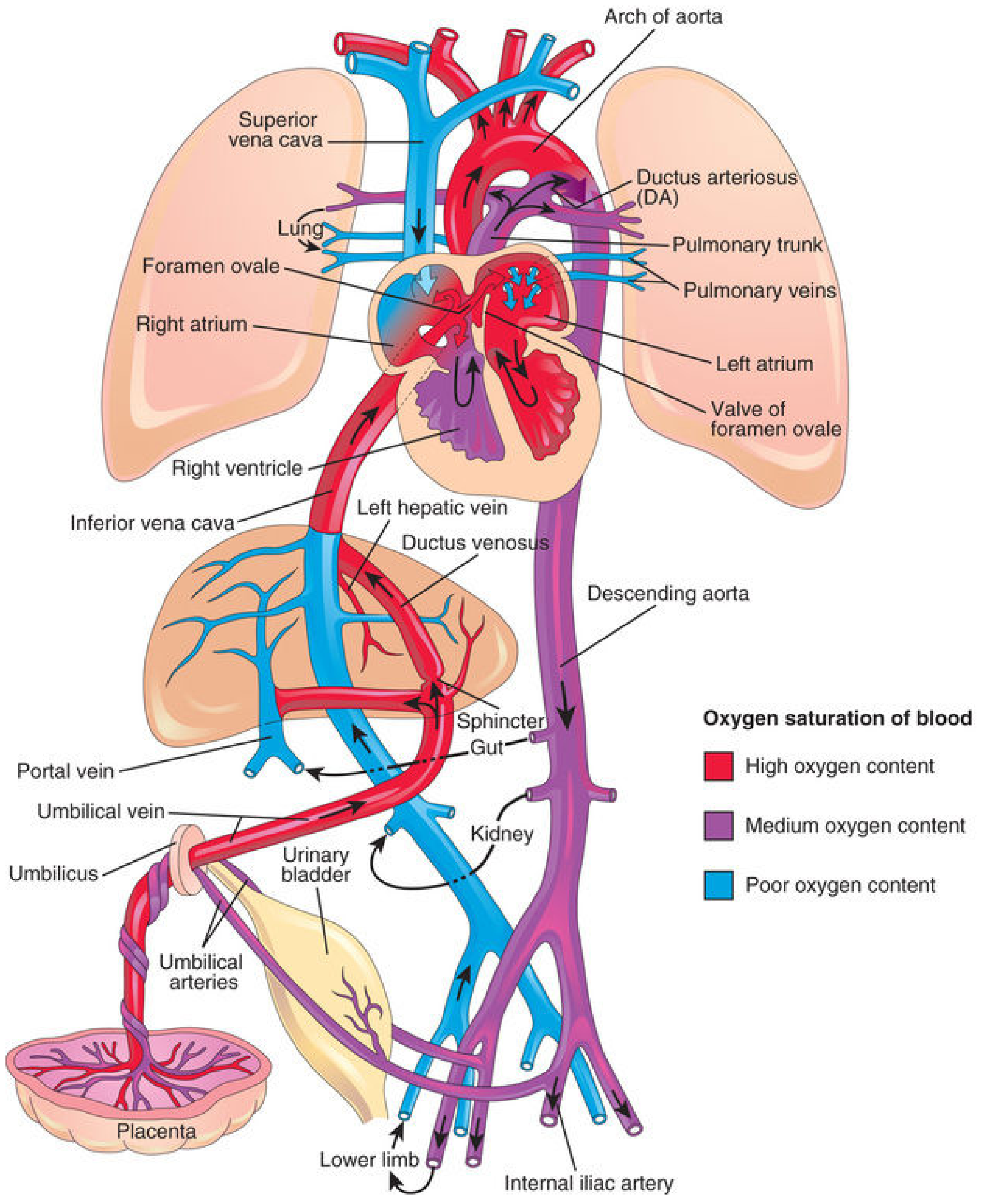
Diagram 2 - Blood Flow as % of Combined Cardiac Output (CCO)
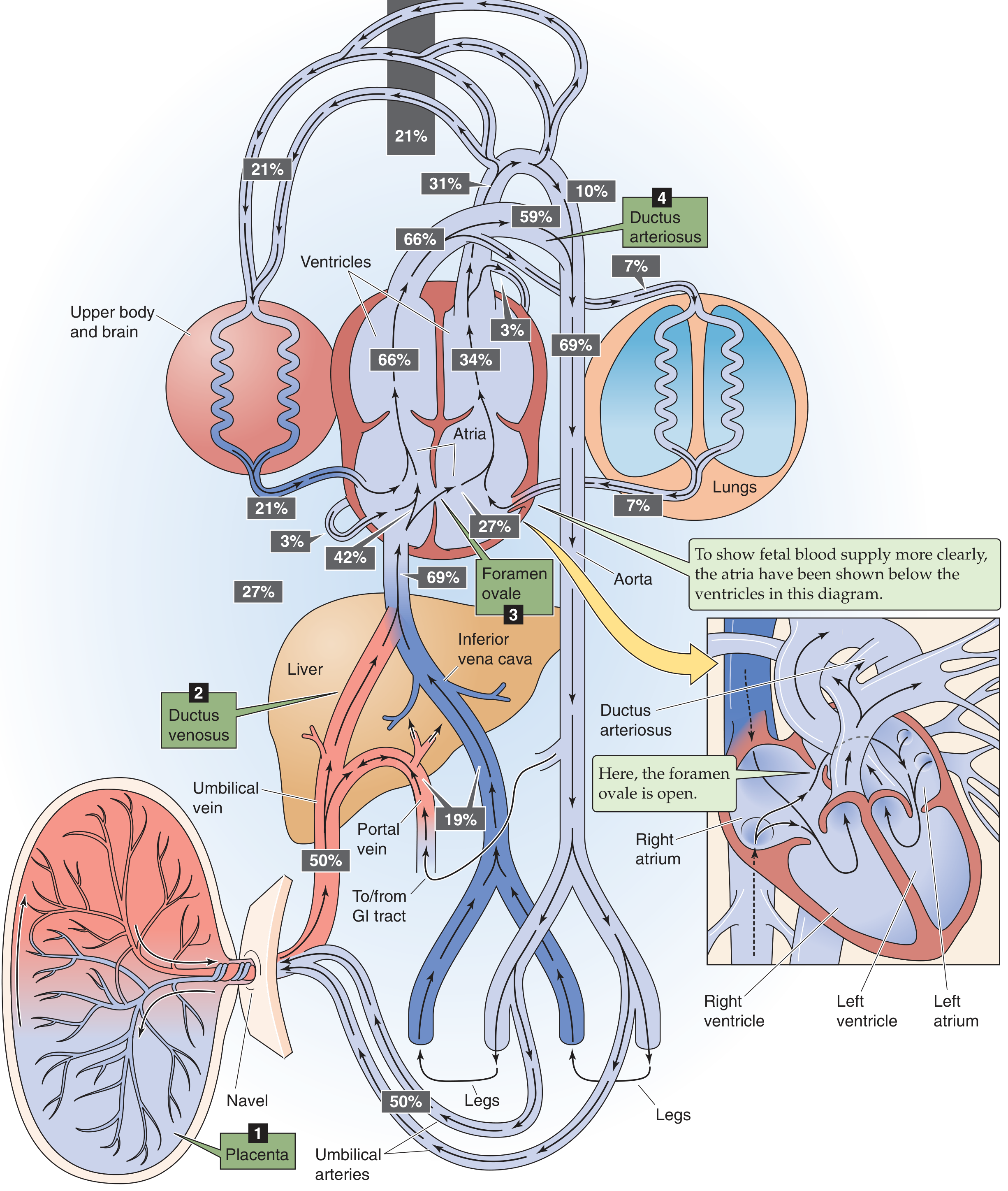
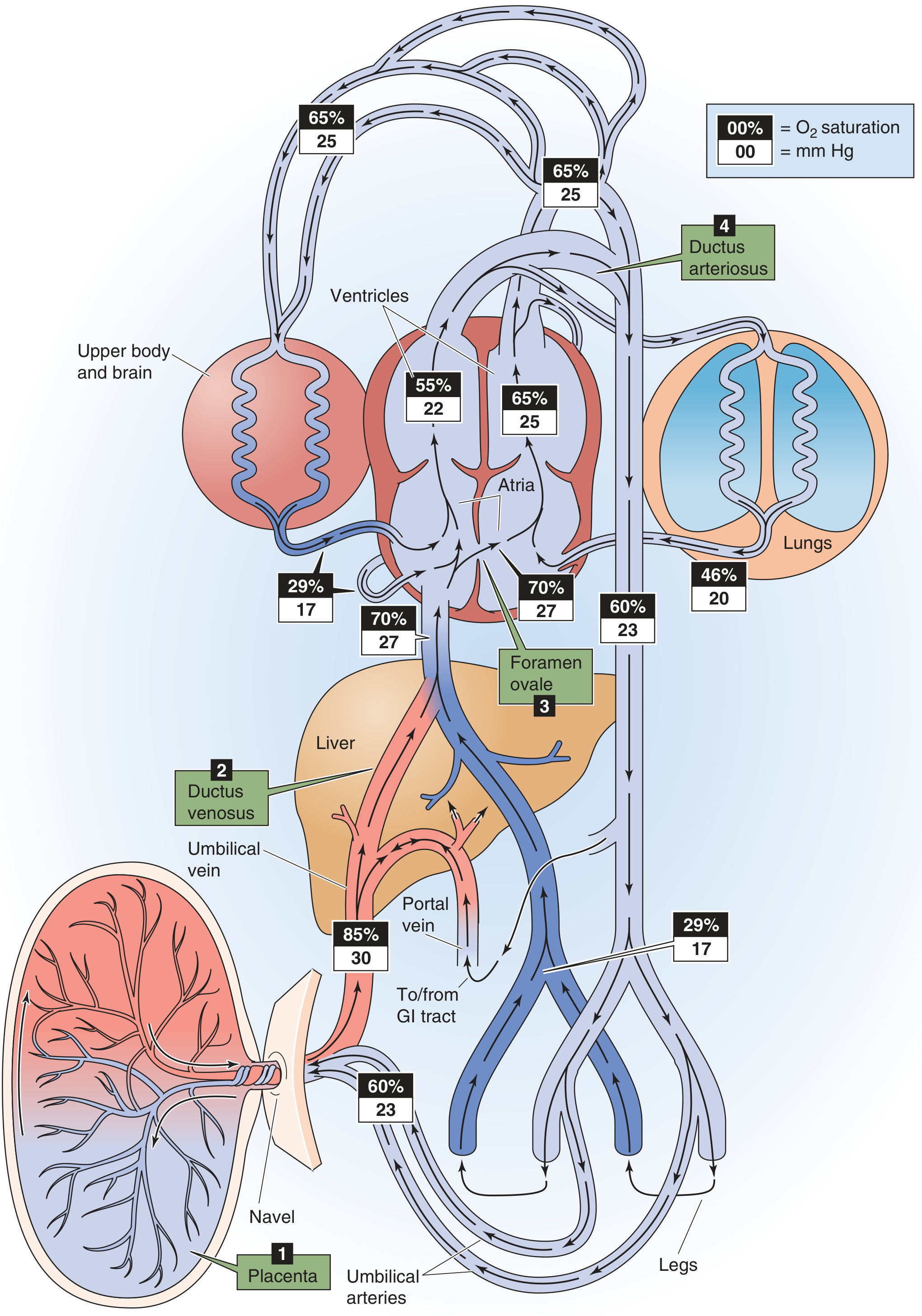
Diagram 3 - O₂ Saturation and PO₂ at Each Checkpoint
Step-by-Step Blood Flow
1. Placenta → Umbilical Vein
- Oxygenated, nutrient-rich blood leaves the placenta via the single umbilical vein
- O₂ saturation ~80-85%, PO₂ ~30 mmHg (highest in the fetal circuit)
- This is the only vessel in fetal life that carries oxygenated blood with a venous name
2. Umbilical Vein → Liver / Ductus Venosus (Shunt 1)
- On reaching the liver, blood splits:
- ~50% enters the ductus venosus - a direct channel from umbilical vein to inferior vena cava (IVC), bypassing the liver sinusoids
- ~50% flows into the portal system and liver sinusoids, then drains via hepatic veins into the IVC
- A physiologic sphincter near the umbilical vein regulates this split - it prevents cardiac overloading during uterine contractions by diverting more blood to the liver
3. IVC → Right Atrium
- Blood in the IVC is now a mixture of:
- Well-oxygenated blood from ductus venosus + hepatic veins
- Poorly oxygenated blood from lower limbs, pelvis, kidneys
- Net O₂ saturation in IVC ~70%, PO₂ ~27 mmHg
- Blood enters the right atrium
4. Right Atrium → Foramen Ovale → Left Atrium (Shunt 2)
- The crista dividens (lower edge of the septum secundum) acts as a flow divider:
- The larger stream (well-oxygenated IVC blood) is directed by the valve of the inferior vena cava through the foramen ovale directly into the left atrium
- The smaller stream (poorly oxygenated IVC blood) stays in the right atrium and mixes with SVC blood returning from the head and upper limbs
- In the left atrium, blood mixes with the small volume of pulmonary venous return (~7% CCO from the lungs) - O₂ saturation ~65%, PO₂ ~25 mmHg
5. Left Atrium → Left Ventricle → Ascending Aorta
- Blood passes through the left ventricle and is pumped into the ascending aorta
- The first branches off the ascending aorta are the coronary arteries and the carotid arteries
- This means the heart muscle and brain receive the best-oxygenated blood available in fetal life - a critical preferential arrangement
6. Right Atrium → Right Ventricle → Pulmonary Trunk
- The poorly oxygenated stream (from SVC + small IVC portion) passes from right atrium → right ventricle → pulmonary trunk
- The right ventricle handles ~66% of the combined cardiac output (CCO); the left ventricle handles ~34%
- Pulmonary vascular resistance is very high in fetal life (due to hypoxic vasoconstriction), so only ~7-10% of this blood goes to the lungs
7. Pulmonary Trunk → Ductus Arteriosus → Descending Aorta (Shunt 3)
- ~90% of pulmonary trunk blood bypasses the lungs via the ductus arteriosus - a wide channel connecting the pulmonary trunk directly to the descending aorta
- This blood (O₂ saturation ~55-60%) enters the descending aorta and mixes with the smaller amount of blood coming from the ascending aorta
8. Descending Aorta → Body + Umbilical Arteries
- Blood in the descending aorta (~60% O₂ saturation, PO₂ ~23 mmHg) distributes to:
- Viscera and lower body (~35%)
- Two umbilical arteries (~65%) - which return deoxygenated blood back to the placenta (O₂ saturation ~58% in umbilical arteries)
- The umbilical arteries are branches of the internal iliac arteries
Sites of Oxygen Dilution (Langman's Five Mixing Points)
Oxygenated blood from the umbilical vein is progressively diluted as it mixes with deoxygenated blood at five points:
| Site | Mixing event |
|---|---|
| I. Liver | Small amount of portal (GI) blood mixes in |
| II. IVC | Blood from lower limbs, pelvis, kidneys |
| III. Right atrium | Blood from head and upper limbs (SVC) |
| IV. Left atrium | Small amount of pulmonary venous return |
| V. Descending aorta | Ductus arteriosus flow mixes with aortic stream |
The Three Shunts - Summary Table
| Shunt | Connects | Purpose | Stimulus for closure at birth |
|---|---|---|---|
| Ductus venosus | Umbilical vein → IVC | Bypasses liver (~50% blood) | Umbilical flow stops; smooth muscle contraction (days) |
| Foramen ovale | Right atrium → Left atrium | Bypasses lungs | ↑ Left atrial pressure > right atrial pressure |
| Ductus arteriosus | Pulmonary trunk → Descending aorta | Bypasses lungs (~90% of pulmonary output) | ↑ O₂ tension + ↓ prostaglandins (hours-days) |
Why the Fetus Tolerates Low PO₂
Adult blood would be critically hypoxic at PO₂ 25-30 mmHg, yet the fetus does fine because of:
- Fetal hemoglobin (HbF) - has higher O₂ affinity than adult HbA (curve shifted left), allowing adequate saturation at lower PO₂
- Elevated hematocrit - increases total O₂-carrying capacity
- High cardiac output - maintains O₂ delivery despite lower saturation
- Preferential streaming - the best-oxygenated blood is selectively directed to the coronary arteries and brain
Circulatory Changes at Birth
At birth, two events trigger the transition:
- Clamping of the umbilical cord - removes the low-resistance placental circulation, doubling total peripheral resistance and raising aortic pressure
- First breath / lung expansion - pulmonary vascular resistance falls >5-fold due to: mechanical expansion, rising alveolar PO₂, falling PCO₂, and release of prostacyclin (PGI₂)
Consequences:
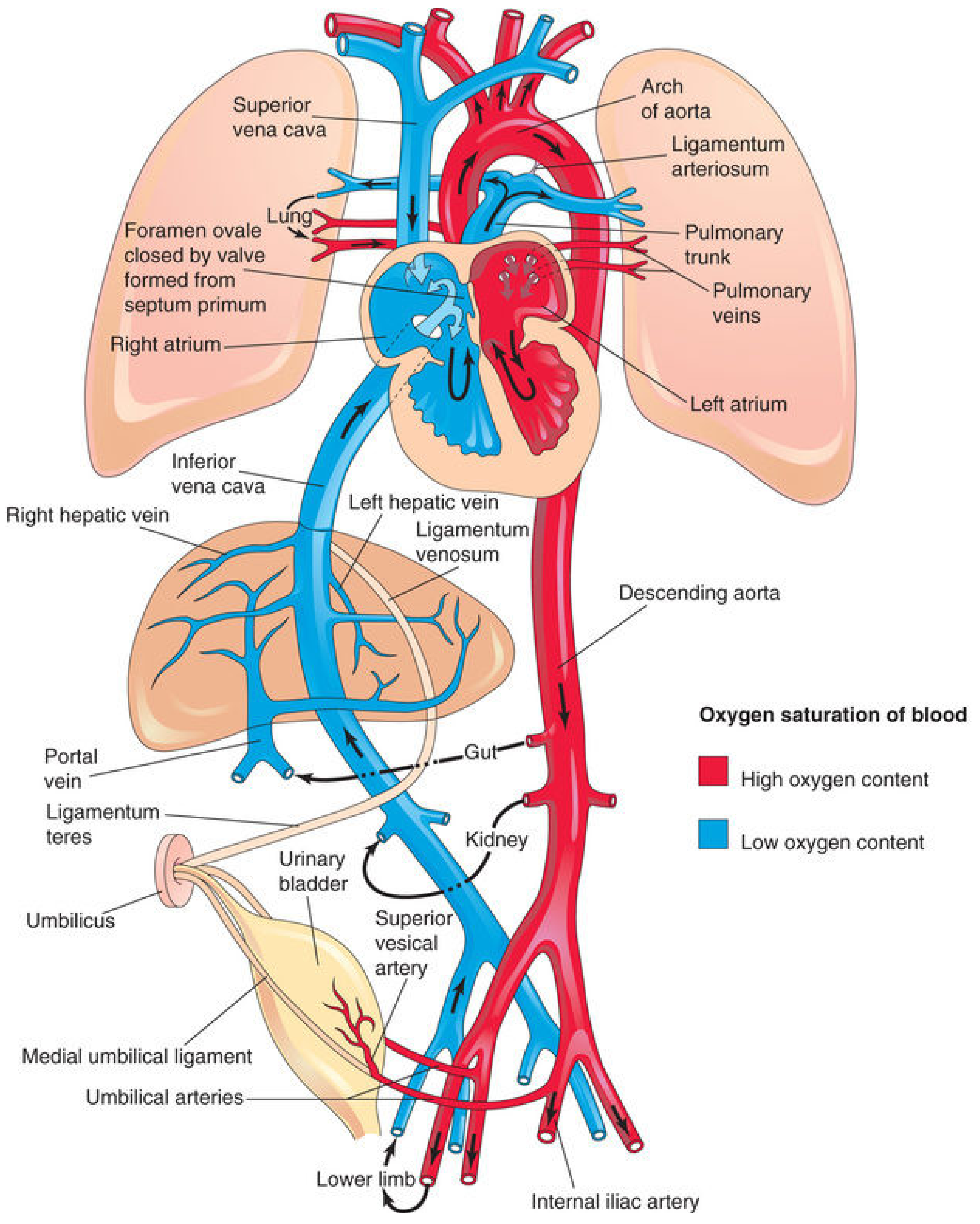
Foramen ovale closes:
- Pulmonary blood flow increases 4-fold → more blood returns via pulmonary veins → left atrial pressure rises above right atrial pressure → septum primum is pressed against septum secundum (functional closure)
- Anatomical closure (fusion) occurs over weeks to months
- Adult remnant: fossa ovalis
Ductus arteriosus closes:
- Rising PO₂ causes smooth muscle contraction (hours)
- Falling prostaglandin E₂ levels (prostaglandins kept it open in utero)
- Functional closure: 10-15 hours after birth; anatomical closure: 2-3 weeks
- Adult remnant: ligamentum arteriosum
Ductus venosus closes:
- Loss of umbilical venous flow, smooth muscle constriction over days
- Adult remnant: ligamentum venosum
Umbilical vessels:
- Umbilical vein → ligamentum teres hepatis (round ligament of liver)
- Umbilical arteries → medial umbilical ligaments (proximal portions persist as superior vesical arteries)
Neonatal Circulation (Post-Birth)
Key Clinical Points
- Persistent fetal circulation (Persistent Pulmonary Hypertension of the Newborn, PPHN): If pulmonary vascular resistance fails to fall after birth, right-to-left shunting continues through a patent ductus arteriosus and/or foramen ovale, causing cyanosis
- Patent ductus arteriosus (PDA): Failure of ductus arteriosus to close; common in premature infants; treated with indomethacin (inhibits prostaglandin synthesis) or surgical ligation
- Patent foramen ovale (PFO): Present in ~25% of adults as a probe-patent remnant; usually asymptomatic but may permit paradoxical embolism
- Indomethacin/prostaglandins in CHD: PGE₁ is used therapeutically to keep the ductus arteriosus open in duct-dependent congenital heart defects (e.g., pulmonary atresia, transposition)
Sources: The Developing Human (Moore), 9th ed. | Langman's Medical Embryology, 14th ed. | Ganong's Review of Medical Physiology, 26th ed. | Medical Physiology (Boron & Boulpaep)
This is a shared conversation. Sign in to Orris to start your own chat.