
from Guyton and Hall textbook of medical physiology, 4th South Asia edition write a 5 mark short note on erythroblastosis foetalis
erythroblastosis fetalis Rh incompatibility hemolytic disease newborn
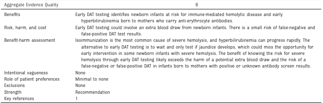
<table> <tr> <td>Aggregate Evidence Quality</td> <td>B</td> </tr> <tr> <td>Benefits</td> <td>Early DAT testing identifies newborn infants at risk for immune-mediated hemolytic disease and early hyperbilirubinemia born to mothers who carry anti-erythrocyte antibodies.</td> </tr> <tr> <td>Risk, harm, and cost</td> <td>Early DAT testing could involve an extra blood draw from newborn infants. There is a small risk of false-negative and false-positive DAT test results.</td> </tr> <tr> <td>Benefit-harm assessment</td> <td>Isoimmunization is the most common cause of severe hemolysis, and hyperbilirubinemia can progress rapidly. The alternative to early DAT testing is to wait and only test if jaundice develops, which could miss the opportunity for early intervention in some newborn infants with severe hemolysis. The benefit of knowing the risk for severe hemolysis through early DAT testing likely exceeds the harm of a potential extra blood draw and the risk of a false-negative or false-positive DAT in infants born to mothers with positive or unknown antibody screen results.</td> </tr> <tr> <td>Intentional vagueness</td> <td>None</td> </tr> <tr> <td>Role of patient preferences</td> <td>Minimal to none</td> </tr> <tr> <td>Exclusions</td> <td>None</td> </tr> <tr> <td>Strength</td> <td>Recommendation</td> </tr> <tr> <td>Key references</td> <td>1</td> </tr> </table>
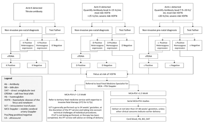
Summary : This flowchart outlines the diagnostic and management pathway for pregnancies affected by maternal alloimmunization to Anti-K, Anti-D, or Anti-c antibodies, focusing on the risk assessment and monitoring for hemolytic disease of the fetus and newborn (HDFN). flowchart: # Nodes : • Anti-K detected (rectangle) • Titrate antibody (rectangle) • Non-invasive pre-natal diagnosis (rectangle) • Test father (rectangle) • K Positive Heterozygous expression (rectangle) • K Positive Homozygous expression (rectangle) • K Negative (rectangle) • cffDNA (rectangle) • K Positive (rectangle) • K Negative (rectangle) • Anti-D detected (rectangle) • Quantify Antibody level 4–15 IU/mL mod risk HDFN >15 IU/mL severe risk HDFN (rectangle) • Non-invasive pre-natal diagnosis (rectangle) • Test father (rectangle) • D Positive Heterozygous expression (rectangle) • D Positive Homozygous expression (rectangle) • D Negative (rectangle) • cffDNA (rectangle) • D Positive (rectangle) • D Negative (rectangle) • Fetus at risk of HDFN (rectangle) • Referral to local Fetal Medicine Specialist for MCA – PSV Doppler (rectangle) • MCA-PSV > 1.5 MoM (rectangle) • Refer to tertiary fetal medicine service with expertise in invasive fetal therapy (IFT) for IUT(s) (rectangle) • MCA-PSV < 1.5 MoM (rectangle) • Serial MCA-PSV studies (rectangle) • Deliver at not later than 37–38 weeks’ gestation, unless other clinical concerns indicate earlier delivery (rectangle) • Cord blood, Hb, Bili, DAT (rectangle) • Anti-c detected (rectangle) • Quantify Antibody level 7.5–20 IU/mL mod risk HDFN >20 IU/mL severe risk HDFN (rectangle) • Non-invasive pre-natal diagnosis (rectangle) • Test father (rectangle) • c Positive Heterozygous expression (rectangle) • c Positive Homozygous expression (rectangle) • c-Negative (rectangle) • cffDNA (rectangle) • c Positive (rectangle) • c Negative (rectangle) • Legend (rectangle) – defines abbreviations used in the chart # Connectors : • Arrows indicate the flow from antibody detection to titration, then to non-invasive diagnosis or paternal testing. • Branches split based on paternal genotype (heterozygous, homozygous, negative). • If the father is negative, the pathway ends (no risk). • If the father is positive (heterozygous or homozygous), cffDNA is used to determine fetal antigen status. • If the fetus is antigen negative, the pathway ends (no risk). • If the fetus is antigen positive, the fetus is at risk of HDFN and referred for further monitoring. • For at-risk fetuses, referral to fetal medicine for MCA-PSV Doppler is indicated. • If MCA-PSV > 1.5 MoM, refer for invasive fetal therapy (IUT). • If MCA-PSV < 1.5 MoM, continue serial monitoring. • Delivery is recommended at 37–38 weeks unless earlier delivery is clinically indicated. # Layout : • The chart is organized into three parallel vertical pathways for Anti-K, Anti-D, and Anti-c, each with similar diagnostic and management steps. • Each pathway splits into non-invasive diagnosis and paternal testing, then converges on fetal risk assessment and management. • The bottom section merges all pathways for final fetal monitoring and delivery planning. • A legend box is present at the bottom left, defining abbreviations. # Analysis : • The flowchart provides a clear, stepwise approach for managing pregnancies at risk of HDFN due to Anti-K, Anti-D, or Anti-c alloimmunization. • The process emphasizes non-invasive testing first, with paternal testing to stratify risk. • Only fetuses at risk (antigen positive) are referred for intensive monitoring and possible intervention. • The use of MCA-PSV Doppler is central for ongoing fetal surveillance, with invasive therapy reserved for those with evidence of fetal anemia. • The chart standardizes care and minimizes unnecessary interventions for low-risk pregnancies.
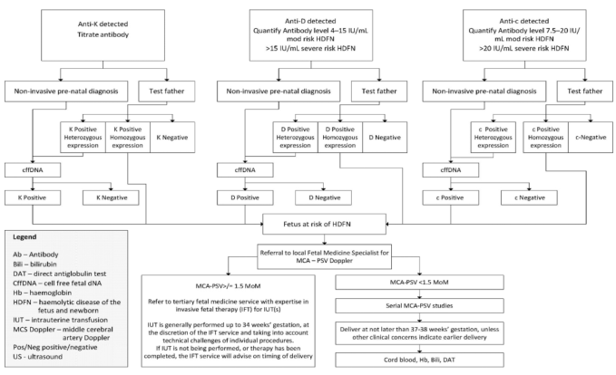
Summary : This flowchart outlines the diagnostic and management pathway for pregnancies affected by maternal alloimmunization to K, D, or c antigens, focusing on the risk assessment and monitoring for hemolytic disease of the fetus and newborn (HDFN). It details the steps for antibody titration, non-invasive prenatal diagnosis, paternal testing, and subsequent fetal monitoring and intervention. flowchart: # Main Pathways : • Three parallel pathways for Anti-K, Anti-D, and Anti-c detected in maternal blood. • Each pathway begins with antibody detection and titration/quantification. • Decision points for non-invasive prenatal diagnosis and paternal antigen testing. • Use of cell-free fetal DNA (cffDNA) for fetal antigen status determination. • Referral to fetal medicine specialist if fetus is at risk of HDFN. # Nodes : • Anti-K detected (rectangle) • Titrate antibody (rectangle) • Non-invasive pre-natal diagnosis (rectangle) • Test father (rectangle) • K Positive Heterozygous expression (rectangle) • K Positive Homozygous expression (rectangle) • K Negative (rectangle) • cffDNA (rectangle) • K Positive (rectangle) • K Negative (rectangle) • Anti-D detected (rectangle) • Quantify Antibody level 4–15 IU/mL mod risk HDFN, >15 IU/mL severe risk HDFN (rectangle) • D Positive Heterozygous expression (rectangle) • D Positive Homozygous expression (rectangle) • D Negative (rectangle) • D Positive (rectangle) • D Negative (rectangle) • Fetus at risk of HDFN (rectangle) • Referral to local Fetal Medicine Specialist for MCA – PSV Doppler (rectangle) • Anti-c detected (rectangle) • Quantify Antibody level 7.5–20 IU/mL mod risk HDFN, >20 IU/mL severe risk HDFN (rectangle) • c Positive Heterozygous expression (rectangle) • c Positive Homozygous expression (rectangle) • c-Negative (rectangle) • c Positive (rectangle) • c Negative (rectangle) # Connectors : • Arrows indicate progression from antibody detection to diagnosis, paternal testing, and fetal risk assessment. • Branching based on test results (e.g., positive/negative, heterozygous/homozygous). • Merging of pathways at "Fetus at risk of HDFN" leading to specialist referral. # Management Pathway (Bottom Section) : • MCA-PSV > 1.5 MoM: Refer to tertiary fetal medicine service for intrauterine transfusion (IUT) if indicated. • IUT generally performed up to 34 weeks’ gestation, with timing and delivery advice from the specialist service. • MCA-PSV < 1.5 MoM: Serial MCA-PSV studies. • Delivery recommended at 37–38 weeks’ gestation unless earlier delivery is clinically indicated. • At delivery: Cord blood, hemoglobin, bilirubin, direct antiglobulin test (DAT). # Legend : • Ab – Antibody • Bili – bilirubin • DAT – direct antiglobulin test • CffDNA – cell free fetal DNA • Hb – hemoglobin • HDFN – hemolytic disease of the fetus and newborn • IUT – intrauterine transfusion • MCS Doppler – middle cerebral artery Doppler • Pos/Neg – positive/negative • US – ultrasound # Layout : • Three main vertical columns for Anti-K, Anti-D, and Anti-c. • Each column splits into non-invasive prenatal diagnosis and paternal testing. • All columns converge at fetal risk assessment and management. • Bottom section provides management steps based on Doppler results. Analysis : • The flowchart provides a clear, stepwise approach for managing pregnancies at risk of HDFN due to maternal alloimmunization. • It emphasizes early risk stratification using both non-invasive and paternal testing, with escalation to specialist care if the fetus is at risk. • The use of MCA-PSV Doppler as a key decision point for intervention is highlighted. • The chart ensures that all possible antigen combinations and test results are accounted for, minimizing missed diagnoses and optimizing fetal outcomes.
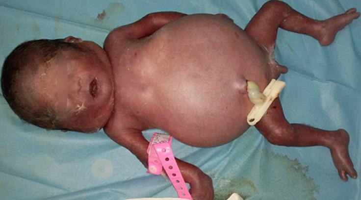
A clinical photograph of a neonate demonstrating Hydrops Fetalis (anasarque fœto-placentaire). The primary visual finding is severe, generalized edema, most notably presenting as marked abdominal distension (ascites). The newborn's skin appears erythematous and translucent, characteristic of significant subcutaneous fluid accumulation. The extremities appear swollen, and the facial features show mild puffiness. A standard white plastic umbilical clamp is visible on the umbilical cord stump. The infant is placed on a blue surgical drape, and a pink identification band is attached to the right wrist. This image serves as an educational illustration of fetal hydrops, a life-threatening condition defined by abnormal fluid accumulation in two or more fetal compartments, often associated with severe anemia, heart failure, or polyserositis. This specimen is relevant to neonatal intensive care, obstetrics, and pediatric pathology.
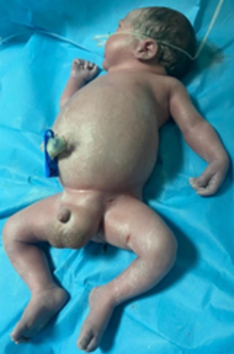
Clinical photograph of a newborn in the immediate postnatal period demonstrating hydrops fetalis with severe anasarca. The infant exhibits generalized, massive soft tissue edema. Key visual features include a markedly distended abdomen consistent with significant ascites, and severe swelling of the upper and lower extremities resulting in the loss of normal skin folds and obscured anatomical landmarks. The skin appears taut and shiny due to subcutaneous fluid accumulation. Pronounced scrotal edema is also visible. The infant is placed on a blue sterile field with a clamped umbilical cord and an orogastric tube in situ. This presentation is clinically significant for systemic fetal fluid overload, often secondary to cardiac failure (such as from a rhabdomyoma), severe anemia, or immune/non-immune hydrops. The image serves as a teaching example of the phenotypic manifestations of neonatal anasarca and its association with Mirror syndrome in the mother.
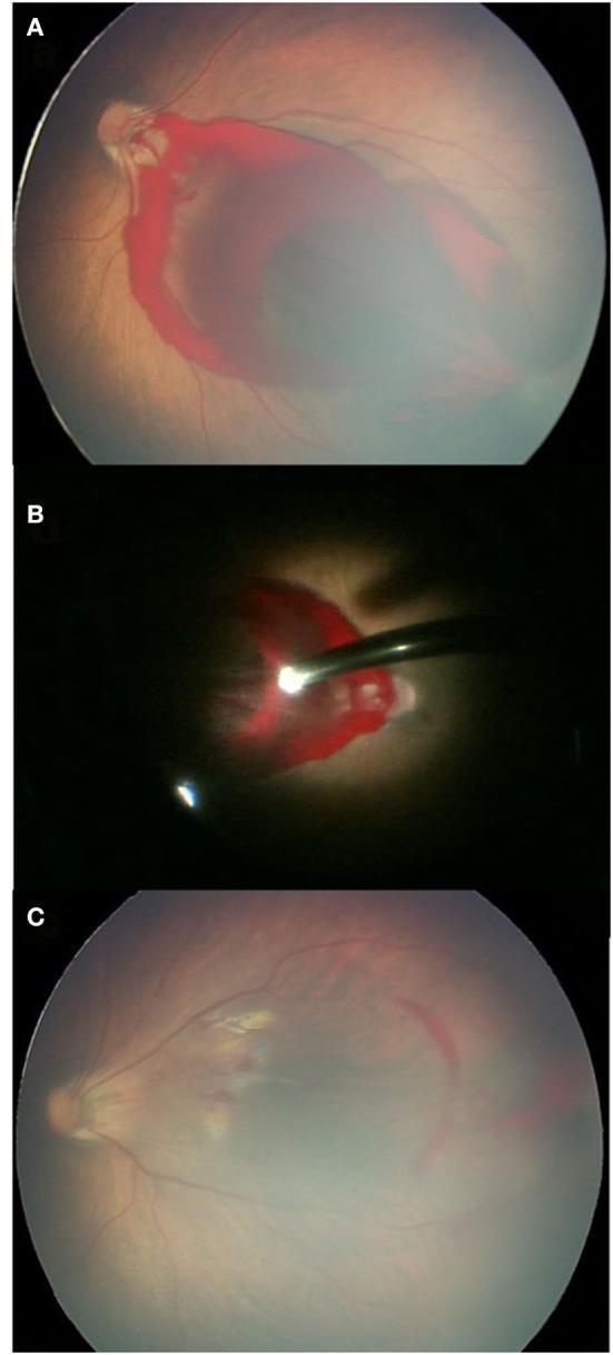
This composite of clinical ophthalmological images illustrates the management and surgical resolution of a dense pre-macular retinal hemorrhage (RH) in a newborn. Panel A shows a preoperative fundus photograph featuring a large, dense, boat-shaped subhyaloid or preretinal hemorrhage located at the posterior pole. The hemorrhage is opaque and completely obscures the underlying macula and optic disc details, presenting a significant threat to visual development. Panel B provides an intraoperative view during a lens-sparing vitrectomy, demonstrating the microsurgical instruments used to evacuate the localized blood. Panel C displays the postoperative fundus appearance one month after surgery, showing a clear macula with successful restoration of retinal visibility. Residual light blood staining and pigmentary changes are visible, but the primary obstructive mass has been cleared. This series demonstrates the clinical progression from acute obstructive hemorrhage to surgical clearance in pediatric ophthalmology, highlighting the importance of timely intervention to prevent amblyopia in cases of dense macular-impacting hemorrhages.
Erythroblastosis Fetalis (Hemolytic Disease of the Newborn)
Definition
Pathogenesis
Incidence
- ~3% of second Rh-positive babies show signs of erythroblastosis
- ~10% of third babies exhibit the disease
- Incidence rises progressively with subsequent pregnancies
Clinical Features
- Anemia - severe, due to ongoing hemolysis of RBCs; this is usually the cause of death in severe cases
- Jaundice - due to elevated bilirubin from hemolysis
- Hepatosplenomegaly - the liver and spleen enlarge as they attempt extramedullary erythropoiesis to replace hemolyzed RBCs
- Nucleated blastic RBCs in circulation - rapid RBC production drives release of immature nucleated erythroblasts into the bloodstream, giving the disease its name
- Kernicterus - bilirubin precipitates in neuronal cells (especially basal ganglia), causing permanent brain damage, mental impairment, or damage to motor areas in surviving infants
Treatment
- Correct severe anemia
- Keep bilirubin levels low and prevent kernicterus
Prevention
- Administering Rh immunoglobulin (anti-D antibody) to the Rh-negative mother at 28-30 weeks of gestation
- Also given after delivery of an Rh-positive baby
- Inhibiting antigen-induced B-lymphocyte antibody production in the mother
- Attaching to D antigen sites on fetal Rh-positive RBCs that cross the placenta, thereby interfering with the maternal immune response