
Treatment of wilson disease
"Wilson disease" AND treatment
Wilson disease Kayser-Fleischer ring copper deposition
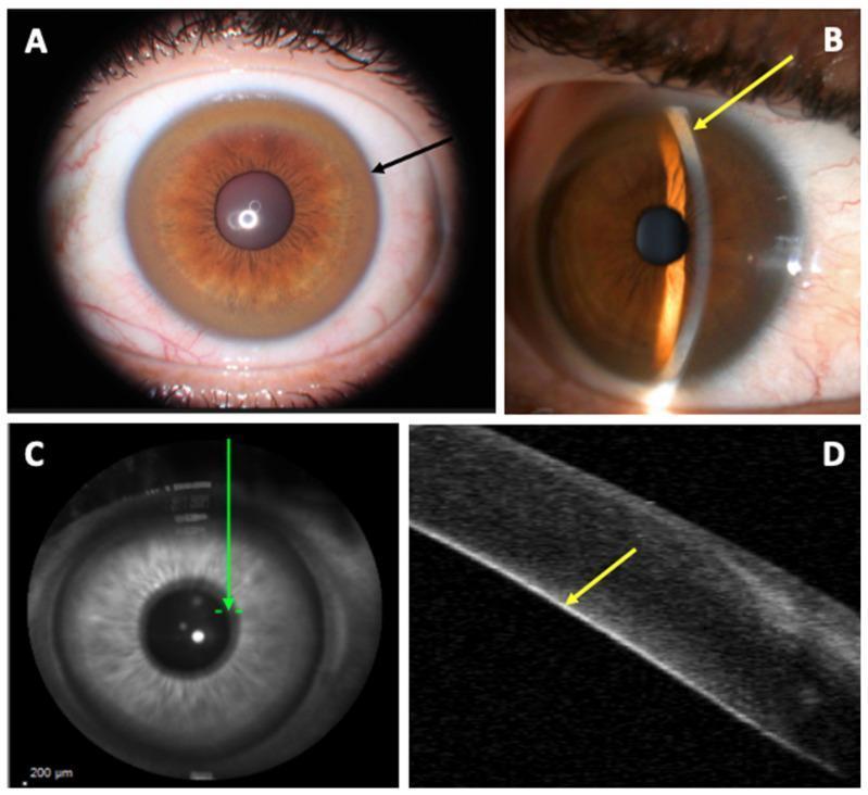
This composite educational graphic illustrates clinical and diagnostic findings of the Kayser–Fleischer ring (KFR), a pathognomonic sign of Wilson disease. Panel A is a clinical photograph from a slit-lamp examination showing a diffuse, circumferential golden-brown pigment ring (black arrow) in the corneal limbic area. Panel B utilizes a fine-slit lamp beam to demonstrate the precise localization of copper deposits at the posterior aspect of the cornea (yellow arrow). Panel C provides a localized infrared corneal B-scan view, while Panel D presents an Anterior Segment Optical Coherence Tomography (AS-OCT) cross-sectional image. The AS-OCT scan reveals a distinct, linear hyper-reflective layer at the level of the Descemet’s membrane (yellow arrow), corresponding exactly to the clinical site of copper deposition. This visual comparison highlights how diagnostic imaging can verify clinical observations of ocular copper accumulation in patients with copper metabolism disorders.
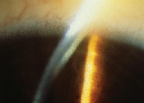
This clinical photograph shows a close-up slit-lamp examination of the eye, specifically focusing on the anterior segment and cornea. A prominent Kayser-Fleischer ring is visible as a golden-brown or greenish-yellow circumferential band located at the corneal periphery. This pigment deposition occurs within Descemet's membrane near the limbus. The slit-lamp beam provides cross-sectional illumination, highlighting the depth of the copper deposition and the relative transparency of the central corneal stroma. Subtle limbal vascularization is observed in the peripheral sclera. This finding is a pathognomonic sign of Wilson's disease (hepatolenticular degeneration), reflecting systemic copper overload and abnormal metabolism of ceruloplasmin. The image serves as a high-yield diagnostic reference for ophthalmology and internal medicine, illustrating the classic ocular manifestation of this metabolic disorder.
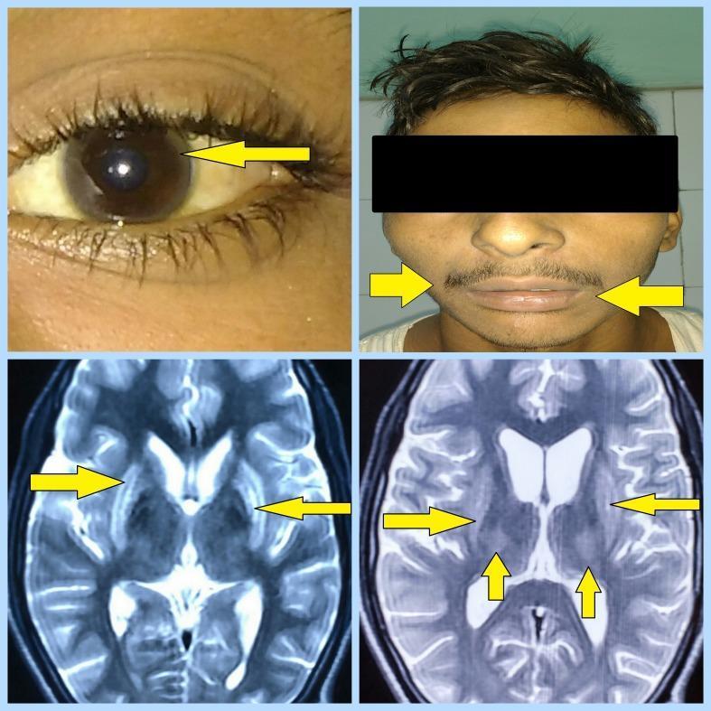
This composite figure illustrates the clinical and radiological hallmarks of Wilson's disease. The top-left clinical photograph shows a close-up of an eye with a yellow arrow pointing to a Kayser-Fleischer ring, characterized by a brownish-gold copper deposition at the corneal limbus. The top-right photograph depicts a patient with a 'mask-like facies' (hypomimia), noted by arrows pointing toward the mouth and facial muscles. The bottom panels consist of two axial MRI brain images (T2-weighted/FLAIR sequences). Yellow arrows indicate bilateral, symmetrical hyperintensities within the basal ganglia, specifically involving the lentiform nuclei (globus pallidus and putamen) and the thalami. These findings are highly characteristic of copper accumulation in the central nervous system and are critical for the diagnostic evaluation of hepatolenticular degeneration. The visual evidence correlates with neurological manifestations such as tremors and spasticity in pediatric or young adult patients.
Treatment of Wilson Disease
Treatment Overview by Phase
| Phase | Preferred Treatment |
|---|---|
| Acute liver failure (fulminant) | Plasmapheresis + liver transplantation |
| Initial treatment - hepatic | Trientine or combined trientine + zinc |
| Initial treatment - neurological | Tetrathiomolybdate (clinical trial) or trientine ± zinc |
| Presymptomatic | Zinc or trientine |
| Maintenance | Zinc or trientine |
| Pregnancy | Zinc (preferred) |
- Yamada's Textbook of Gastroenterology, 7th ed., Table 94.3
1. Copper Chelating Agents
D-Penicillamine (Cuprimine)
- The original first-line agent introduced by J.M. Walshe in 1956
- A sulfhydryl-containing amino acid (D-isomer of cysteine with 2 methyl groups) that greatly increases urinary copper excretion
- Also inhibits collagen cross-linking and has mild immunosuppressive properties
- Dose: 1-2 g/day in 3-4 divided doses, taken at least 30 minutes before or 2 hours after meals; start at one-quarter to one-half the target dose and increase over several weeks
- Children: ~20 mg/kg/day rounded to nearest 250 mg
- Pyridoxine (vitamin B6) 25 mg/day must be co-administered because penicillamine has an anti-pyridoxine effect
- Early: fever, rash, proteinuria (within 7-10 days) - manage with glucocorticoids or switch agents
- Skin: elastosis perforans serpiginosa, pemphigus, various rashes
- Hematologic: leukopenia, thrombocytopenia, aplastic anemia (rare but may not reverse on stopping)
- Renal: nephrotic syndrome, Goodpasture syndrome
- Others: myasthenia syndrome, SLE-like syndrome, hypothyroidism, loss of taste
- Neurologic paradox: ~10-50% of neurologically symptomatic patients worsen initially after starting penicillamine - many recover with cautious continued use; starting low and titrating slowly helps avoid this
- Sleisenger and Fordtran's GI and Liver Disease; Harrison's Principles of Internal Medicine 22E
Trientine (Triethylenetetramine Dihydrochloride / Syprine)
-
Now the preferred first-line chelator at many centers due to better tolerability
-
Chelates copper through 4 constituent nitrogens in a planar ring (lacks sulfhydryl groups unlike penicillamine)
-
Increases urinary copper excretion; also may reduce intestinal copper absorption
-
Less potent than penicillamine, but the difference is not clinically significant
-
Dose (adults): 10-20 mg/kg/day or 1-2 g/day in 2-3 divided doses, 1 hour before or 2 hours after meals (some patients tolerate single daily dosing, which aids adherence)
-
Adverse effects: minimal - occasional gastritis, iron deficiency (chelates dietary iron), rare bone marrow suppression
-
Neurologic deterioration after starting trientine is rare (much rarer than with penicillamine)
-
Side effects of penicillamine do not recur if switched to trientine
-
Sleisenger and Fordtran's GI and Liver Disease; Yamada's Textbook of Gastroenterology
Tetrathiomolybdate (TM)
-
Forms stable tripartite complexes: TM-albumin-copper, trapping free circulating copper
-
Reduces both intestinal copper absorption and circulating free copper
-
Faster acting than other chelators - restores copper balance in weeks vs. months
-
Bis-choline tetrathiomolybdate (ALXN1840) is the newer, stable formulation in Phase 2/3 trials - particularly preferred for neurologic Wilson disease because it does not cause paradoxical neurologic worsening
-
Adverse effects: bone marrow suppression and hepatotoxicity are noteworthy; risk of copper deficiency with prolonged use
-
Still considered investigational/clinical trial use for most guidelines
-
Harrison's 22E; Sleisenger and Fordtran's GI and Liver Disease
2. Zinc Therapy (Reduction of Copper Absorption)
-
Zinc acetate (Galzin): induces metallothionein synthesis in intestinal epithelial cells, which binds dietary copper and blocks its absorption
-
Dose: typically 50 mg elemental zinc three times daily (150 mg/day), taken 1 hour before meals
-
Onset slow: 4-6 months to restore proper copper balance when used as monotherapy
-
Ideal for:
- Presymptomatic patients
- Maintenance therapy after initial de-coppering
- Pregnancy (preferred over chelators due to teratogenic concerns)
-
Adverse effects: minimal - gastric irritation (most common), otherwise well tolerated
-
Monitoring: 24-hour urinary copper can still be used to monitor zinc therapy because urinary copper reflects total body copper load even without cupriuresis
-
Harrison's 22E; Sleisenger and Fordtran's GI and Liver Disease
3. Liver Transplantation (LT)
- Reserved for:
- Acute liver failure (fulminant Wilson disease) - this is the primary indication
- Advanced liver disease unresponsive to medical therapy
- Delayed diagnosis or poor medication compliance leading to irreversible hepatic damage
- LT corrects the metabolic defect entirely (transplanted liver has normal ATP7B)
- Outcomes: 1-year survival 80-90%, excellent beyond 1 year
- LT is not indicated for neurological Wilson disease alone; neurological disease may or may not improve after LT
- Plasmapheresis / plasma exchange may act as a bridge to transplantation in acute liver failure
4. Dietary Measures
- Low-copper diet is recommended as adjunct therapy (not sufficient alone)
- Foods to avoid: organ meats, shellfish, nuts, chocolate, mushrooms
- If drinking water has high copper content: test the water and install a copper-removing filtration device
- Vegetarians require specific dietary counseling (many plant foods are copper-rich)
5. Special Situations
Neurological Wilson Disease
- Trientine is preferred over penicillamine (lower risk of paradoxical worsening)
- Tetrathiomolybdate (in clinical trials) is considered optimal
- Combination trientine + zinc is also used
- Starting at low doses and titrating slowly reduces neurologic deterioration risk
Pregnancy
- Treatment must never be stopped - untreated Wilson disease increases spontaneous miscarriage risk; postpartum hepatic decompensation occurs if treatment is stopped
- Zinc is preferred - safest profile for the fetus (teratogenic concerns with penicillamine; trientine safety unknown)
- If on penicillamine or trientine: reduce dose by ~25-50% (especially if cesarean section planned)
- Breastfeeding is not recommended (all anticopper medications enter breast milk)
- A 2024 systematic review confirmed adverse pregnancy outcomes and treatment effects in Wilson disease (PMID 39206599).
Presymptomatic Patients (Diagnosed on Screening)
- Zinc monotherapy is effective and preferred
- Alternative: low-dose trientine
- These patients generally have the best long-term prognosis
6. Monitoring on Treatment
- Baseline and initial months: CBC, urinalysis, renal and liver function tests weekly for first month (especially in advanced liver disease)
- After stabilization: every 1-3 months for first year, then every 6-12 months
- Urinary copper: used to monitor chelation efficacy; target during chelation ~200-500 µg/24h; during maintenance with zinc, lower values expected
- Serum ceruloplasmin and serum copper: followed over time
- Adequate treatment reduces urine copper, normalizes liver enzymes, and stabilizes neurological status
7. Emerging Therapies
-
Gene therapy: Adeno-associated virus (AAV8) mediated ATP7B gene introduction into hepatocytes has been successful in mouse models; transduction of only 20% of hepatocytes normalized copper homeostasis. Clinical trials are underway.
-
Methanobactin: A bacterial copper-binding peptide (from Methylocystis trichosporium) with very high affinity for copper; shown to reverse acute liver failure in rat models
-
MAP kinase pathway inhibitors: To rescue misfolded ATP7B mutants (e.g., H1069Q, R778L) retained in the endoplasmic reticulum
-
Liver X receptor agonists: Under investigation to enhance hepatocyte copper handling
-
Antioxidants (e.g., vitamin E/alpha-tocopherol): Adjunctive role supported by anecdotal data in hepatic decompensation
-
Harrison's Principles of Internal Medicine 22E; Sleisenger and Fordtran's GI and Liver Disease
Key Take-Away Points
- Wilson disease requires life-long treatment - stopping therapy risks fatal relapse
- Trientine has replaced penicillamine as first-line chelator at most centers due to its better tolerability
- Zinc is the preferred agent for presymptomatic disease, maintenance, and pregnancy
- Neurological presentations require careful selection - tetrathiomolybdate or trientine preferred to avoid paradoxical worsening
- Liver transplantation is curative for the metabolic defect and is the treatment for fulminant liver failure
- Gene therapy is on the horizon with promising preclinical results and active clinical trials