
Explain ascites condition
Reading File
Now let me search the medical library for ascites content in parallel with a PubMed search.
Finding Sources
Searching PubMed
ascites pathophysiology management
Good. Now let me read the most comprehensive textbook sections on ascites in parallel.
Reading File
Reading File
Reading File
Now let me get the detailed pathophysiology section from Sleisenger & Fordtran and the laboratory/SAAG section from Tietz.
Reading File
Reading File
Now let me get more detailed pathophysiology and clinical features from the main GI textbook block.
Finding Sources
Finding Sources
Let me now read the comprehensive ascites chapter from Sleisenger & Fordtran (block 23) and the Harrison's SAAG algorithm image.
Reading File
Reading File
Now let me fetch the Harrison's SAAG algorithm image and get the detailed pathophysiology + SAAG classification table.
Loading Image
Reading File
I now have comprehensive material from multiple authoritative textbooks. Let me compile a thorough, well-structured answer.
Ascites
Ascites is the pathological accumulation of free fluid within the peritoneal (abdominal) cavity. Under normal conditions, the peritoneal cavity contains less than 150 mL of fluid. When this volume increases to detectable levels (>500 mL on physical exam, or any amount on imaging), it constitutes ascites.
Cirrhosis accounts for approximately 84% of all cases. It carries a significant prognosis: median survival after onset is about 2 years for nonmalignant ascites and only 4 months for malignant ascites.
- Tietz Textbook of Laboratory Medicine, 7th Ed.
- Harrison's Principles of Internal Medicine 22E
Causes
Portal hypertension-related (high SAAG):
- Cirrhosis (most common)
- Congestive heart failure / constrictive pericarditis
- Budd-Chiari syndrome (hepatic vein thrombosis)
- Inferior vena cava obstruction
- Portal vein occlusion
Hypoalbuminemia (low oncotic pressure):
- Nephrotic syndrome
- Protein-losing enteropathy
- Malnutrition
Miscellaneous / non-portal causes (low SAAG):
-
Peritoneal carcinomatosis (colon, gastric, pancreatic, ovarian, breast, endometrial)
-
Tuberculous peritonitis
-
Pancreatitis / pancreatic ascites
-
Myxedema (hypothyroidism)
-
Meigs syndrome
-
Chylous ascites (lymphatic disruption)
-
End-stage renal disease
-
Mulholland & Greenfield's Surgery, 7e
Pathophysiology
The development of ascites in cirrhosis involves several simultaneously operating mechanisms:
1. Portal Hypertension
Increased hydrostatic pressure in the portal venous system (due to sinusoidal fibrosis and capillarization of the space of Disse in cirrhosis) drives fluid out of the hepatic and splanchnic vasculature into the peritoneal space. As cirrhosis advances, sinusoidal fenestrations are lost, so the hepatic lymph produced is reduced and splanchnic lymph predominates - hence ascitic fluid in advanced cirrhosis tends to be low in protein.
2. Splanchnic Vasodilation and Nitric Oxide
The primary event is peripheral and splanchnic vasodilation, driven largely by nitric oxide (NO). Bacterial products (endotoxins, bacterial DNA from the gut) enter the systemic circulation via portal-systemic shunts and reduced reticuloendothelial clearance, stimulating excess NO synthesis. Endothelins and other vasoactive mediators also play a role.
3. Effective Arterial Underfilling
Splanchnic vasodilation causes a decrease in central blood volume (the circulation is "overfilled" in absolute terms but "underfilled" effectively). This activates compensatory neurohumoral systems:
- Renin-angiotensin-aldosterone system (RAAS) - leads to renal sodium and water retention
- Sympathetic nervous system activation
- ADH (vasopressin) - promotes water retention
4. Sodium and Water Retention
Sodium retention, driven by aldosterone activation, leads to fluid overload. Because of elevated portal hydrostatic pressure and reduced intravascular oncotic pressure (from hypoalbuminemia), the retained fluid leaks preferentially into the peritoneal "third space."
5. Hypoalbuminemia
Reduced hepatic albumin synthesis lowers plasma oncotic pressure, further tipping the Starling forces in favour of fluid extravasation into the peritoneum.
"The net effect of these forces is shrinkage of the central blood volume, which decreases renal perfusion and leads to sodium retention through activation of the renin-angiotensin system." - Tietz Textbook of Laboratory Medicine
- Tietz Textbook of Laboratory Medicine, 7th Ed.
- Brenner and Rector's The Kidney
- Mulholland & Greenfield's Surgery, 7e
Clinical Features
Symptoms:
- Abdominal distension and discomfort
- Early satiety, nausea
- Dyspnea (from diaphragm elevation and pleural effusion - usually right-sided)
- Fatigue and reduced mobility
Physical examination findings:
| Sign | Approximate Fluid Volume Required |
|---|---|
| Bulging flanks | ~1,000 mL |
| Shifting dullness on percussion | ~1,500 mL |
| Fluid wave (puddle sign) | ~5,000-10,000 mL |
Other findings: umbilical and inguinal hernias from increased intra-abdominal pressure, signs of underlying cirrhosis (jaundice, spider angiomata, palmar erythema, caput medusae).
- Mulholland & Greenfield's Surgery, 7e
- ROSEN's Emergency Medicine
Diagnosis
Imaging
Ultrasound is the most sensitive bedside tool - it can detect as little as 100 mL of ascites and also assesses for liver morphology, portal vein flow, and hepatocellular carcinoma.
Diagnostic Paracentesis
This is the key diagnostic step. Ascitic fluid is analysed for:
- Albumin (to calculate SAAG)
- Total protein
- Cell count + differential (neutrophils >250/mL diagnose SBP)
- Cultures (inoculated into blood culture bottles at bedside)
- Optional: glucose, LDH, amylase, triglycerides, cytology, TB culture
Gross appearance:
- Straw-coloured/clear: typical cirrhotic ascites
- Turbid: infection or malignant cells
- Milky/white: chylous ascites (triglycerides >200 mg/dL)
- Dark brown: biliary perforation
- Bloody: trauma, malignancy, hepatocellular carcinoma
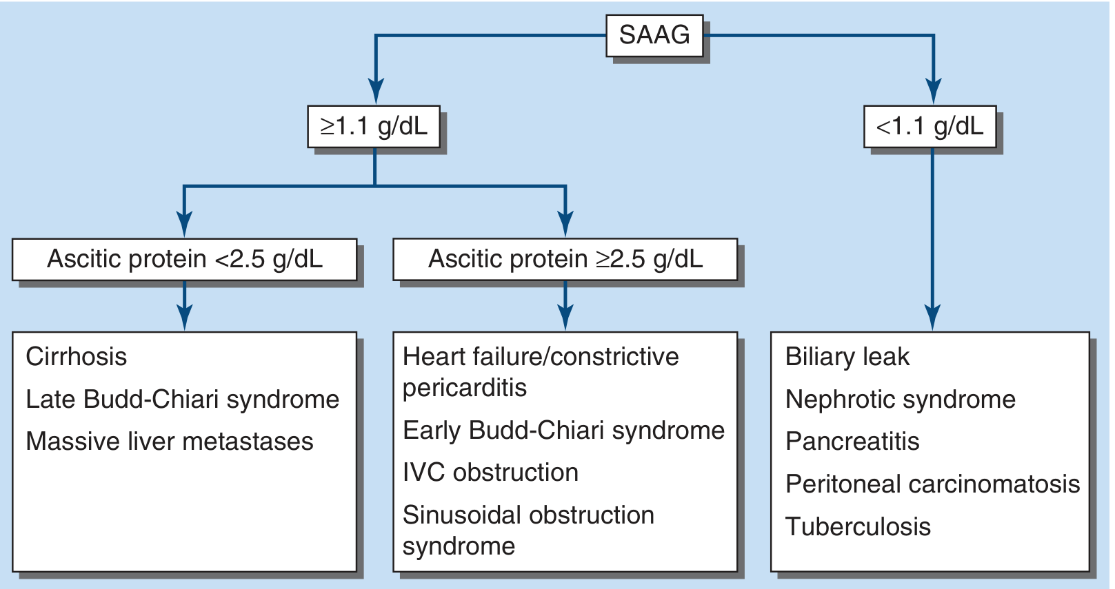
Serum-Ascites Albumin Gradient (SAAG)
SAAG = serum albumin - ascitic fluid albumin
This is the single most reliable test for categorising ascites:
| SAAG | Interpretation |
|---|---|
| ≥1.1 g/dL | Portal hypertension-related ascites (97% accuracy) |
| <1.1 g/dL | Non-portal hypertension cause |
-
High SAAG + ascitic protein <2.5 g/dL: cirrhosis, late Budd-Chiari, massive liver metastases
-
High SAAG + ascitic protein ≥2.5 g/dL: heart failure, constrictive pericarditis, early Budd-Chiari, IVC obstruction
-
Low SAAG: peritoneal carcinomatosis, tuberculosis, pancreatitis, nephrotic syndrome, biliary leak
-
Harrison's Principles of Internal Medicine 22E
-
Sleisenger and Fordtran's GI and Liver Disease
Complications
1. Spontaneous Bacterial Peritonitis (SBP)
The most feared acute complication. Defined as peritoneal infection (typically Gram-negative enteric organisms) without mechanical bowel disruption. Diagnosed when ascitic neutrophils exceed 250/mL. Presents with abdominal pain, fever, or leukocytosis - but can be clinically silent. Carries high mortality.
2. Hepatorenal Syndrome (HRS)
Functional kidney injury from hemodynamic abnormalities driven by end-stage liver disease and severe portal hypertension. The kidneys are structurally normal but fail due to extreme renal vasoconstriction. High mortality.
3. Hepatic Hydrothorax
Migration of ascitic fluid through diaphragmatic defects into the pleural space, usually right-sided.
4. Hyponatremia
Dilutional hyponatremia is common due to impaired free water excretion (ADH effect). Serum sodium <130 mEq/L indicates severe disease.
5. Umbilical Hernia Rupture
Can be a surgical emergency in tense ascites.
Management
First-Line: Salt Restriction + Diuretics
- Sodium restriction: 2 g/day (88 mEq). Stricter restriction is not recommended as it compromises nutrition.
- Spironolactone (aldosterone antagonist): start 100 mg/day, titrate every 3-4 days up to 400 mg/day. More effective than loop diuretics alone because RAAS activation is the dominant mechanism.
- Furosemide: start 40 mg/day, up to 160 mg/day. Given concurrently with spironolactone for tense ascites, or added if hyperkalemia or inadequate response.
- Target weight loss: 1 kg in the first week, then up to 2 kg/week (but no more than 0.5 kg/day without peripheral edema, or 1 kg/day with edema - to avoid intravascular depletion).
Large-Volume Paracentesis (LVP)
For refractory ascites (10-20% of patients) or symptomatic tense ascites:
- Remove all or most fluid
- For removal of >5 L: administer IV albumin at 6-8 g per litre removed to prevent paracentesis-induced circulatory dysfunction (PICD) - hemodynamic instability due to splanchnic vasodilation
- For <4-5 L removed, albumin replacement is optional
Transjugular Intrahepatic Portosystemic Shunt (TIPS)
- Reduces portal pressure mechanically
- More effective than LVP for preventing recurrent ascites
- Higher risk of hepatic encephalopathy with uncovered stents
- PTFE-covered TIPS can improve survival in patients requiring LVP >twice/month
Albumin Infusion (Long-term)
Long-term human albumin (40 g twice weekly x2 weeks, then 40 g weekly) added to diuretics significantly reduces recurrent ascites and improves 18-month survival.
Peritoneovenous Shunt / Automated Pump
For patients not candidates for TIPS or transplant. Automated pumps that drain ascitic fluid to the bladder (alfapump) reduce need for LVP and improve quality of life but carry infection risk.
Avoid These Medications in Cirrhotic Ascites:
- NSAIDs (promote sodium retention, precipitate renal failure)
- ACE inhibitors and ARBs (cause hypotension and renal failure)
- Aminoglycosides
Liver Transplantation
Definitive treatment in eligible patients. Refractory ascites is a Model for End-stage Liver Disease (MELD) score indication.
- Goldman-Cecil Medicine
- ROSEN's Emergency Medicine
- Mulholland & Greenfield's Surgery, 7e
Prognosis
Prognosis of cirrhotic ascites is linked to renal sodium excretion:
- Urine Na <10 mEq/day: median survival 1.5 years
- Urine Na ≥10 mEq/day: median survival 4.5 years
Other prognostic factors include arterial pressure, serum sodium, and serum creatinine.
A 2025 AGA Clinical Practice Update published in Gastroenterology provides the latest expert guidance on managing ascites, volume overload, and hyponatremia in cirrhosis (PMID: 41114681).
Sources: Sleisenger & Fordtran's GI and Liver Disease | Harrison's Principles of Internal Medicine 22E | Tietz Textbook of Laboratory Medicine, 7th Ed. | Goldman-Cecil Medicine | Mulholland & Greenfield's Surgery, 7e | ROSEN's Emergency Medicine | 2025 AGA Practice Update (PMID 41114681)
This is a shared conversation. Sign in to Orris to start your own chat.