
Give me notes of joints and types of joint basic arthrology as a 1st year bams student
Reading File
Now let me search the medical library for joints and arthrology content, targeting anatomy textbooks suitable for 1st year BAMS students.
Finding Sources
Finding Sources
Finding Sources
Finding Sources
Reading File
Reading File
Reading File
Reading File
Reading File
Reading File
Now let me fetch the key diagrams to include in the notes:
Loading Image
Loading Image
Finding Sources
Reading File
Finding Sources
Reading File
Loading Image
I now have all the material needed for comprehensive notes. Let me compile them.
Joints and Basic Arthrology - Notes for 1st Year BAMS
1. Definition of a Joint (Arthrology)
A joint (articulation) is the point where two or more bones come together. Arthrology is the branch of anatomy that studies joints. Joints allow the skeleton to move while also providing stability. Every joint is a unique structure fitted to its anatomic location and the type of motion required.
2. Classification of Joints
Joints are classified in two main ways:
- Structural classification - based on the tissue connecting the bones
- Functional classification - based on the degree of movement permitted
3. Structural Classification
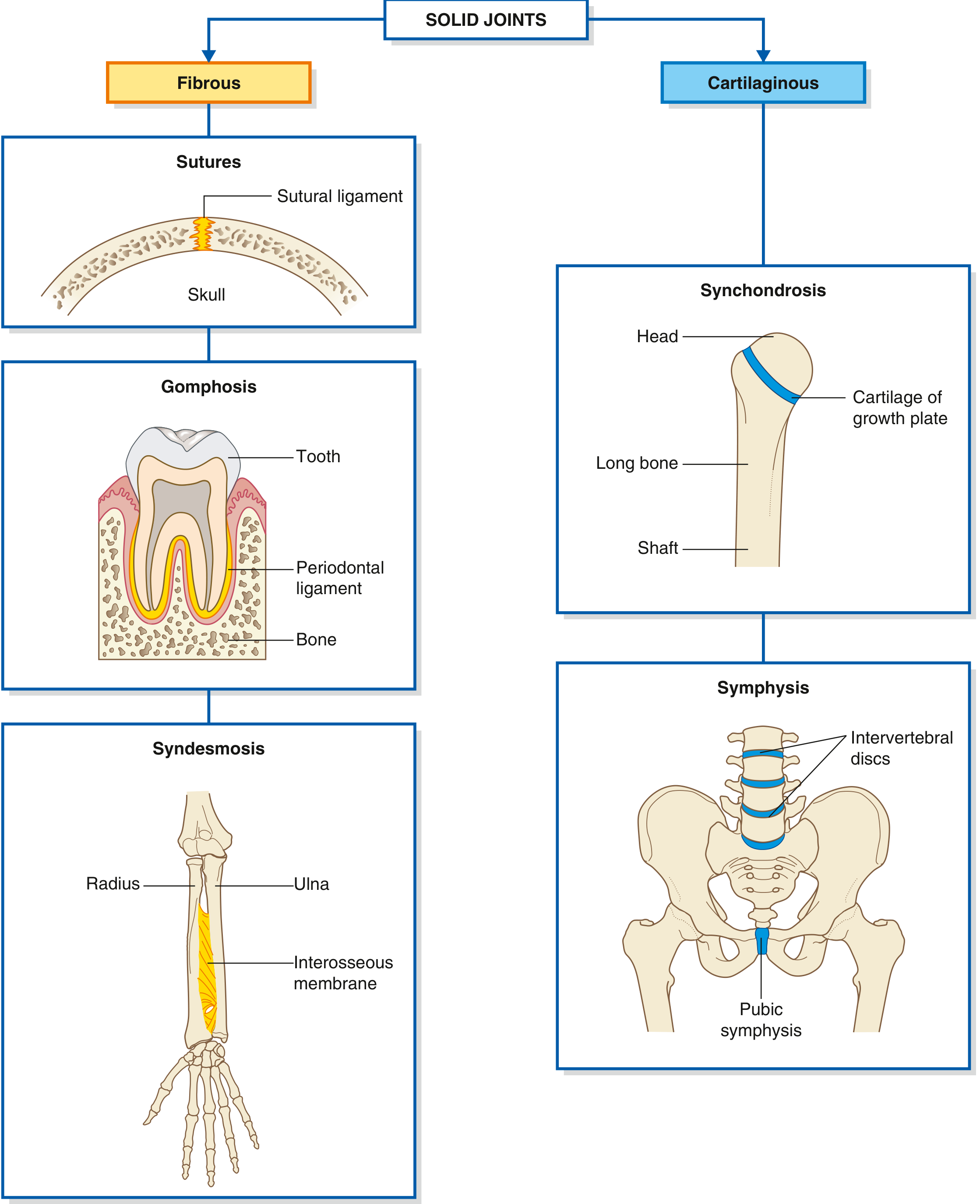
3A. FIBROUS JOINTS (Synarthroses)
- Bones are joined by dense connective tissue (collagen fibers)
- No joint cavity
- Movement: minimal to none
| Subtype | Description | Example |
|---|---|---|
| Sutures | Thin sutural ligament joins skull bones; eventually ossify | Coronal, sagittal, lambdoid sutures of skull |
| Syndesmoses | Ligament-like connective tissue bridges two bones | Interosseous membrane between radius and ulna; tibiofibular joint |
| Gomphoses | Periodontal ligament holds tooth root in bony socket | Tooth in jaw (only example in body) |
3B. CARTILAGINOUS JOINTS (Amphiarthroses)
- Bones are joined by hyaline cartilage or fibrocartilage
- No joint cavity
- Movement: slightly mobile
| Subtype | Tissue | Example |
|---|---|---|
| Primary cartilaginous (Synchondroses) | Hyaline cartilage | Epiphyseal (growth) plate of long bones; 1st rib - sternum joint |
| Secondary cartilaginous (Symphyses) | Fibrocartilage | Pubic symphysis; intervertebral discs |
Memory tip: Synchondroses are temporary - they eventually ossify. Symphyses are permanent.
3C. SYNOVIAL JOINTS (Diarthroses)
- Bones are separated by a synovial cavity (joint space)
- Freely moveable - the most important functional joints
- Also called true joints or diarthroses
4. Structure of a Synovial Joint
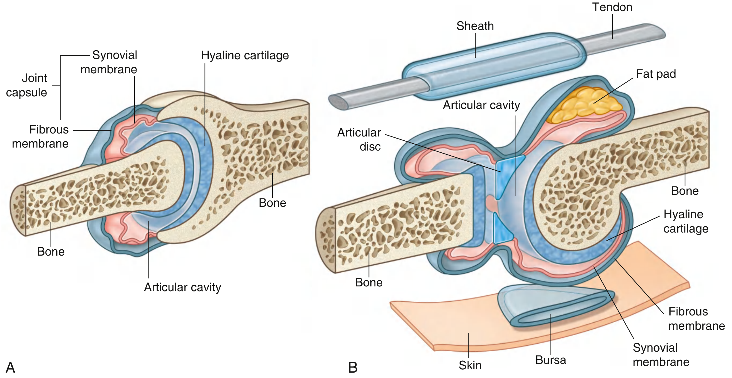
A typical synovial joint has these essential components:
Mandatory (Essential) Components:
- Articular surfaces - the bone ends that come together
- Articular cartilage - hyaline cartilage covers the bone ends; reduces friction; provides shock absorption; avascular (nourished by synovial fluid)
- Joint capsule - surrounds the joint completely; has two layers:
- Outer fibrous layer - tough dense connective tissue; provides strength and stability
- Inner synovial membrane - produces synovial fluid; can regenerate; lines all non-cartilaginous surfaces
- Synovial fluid - viscous fluid inside the cavity; composed of water, plasma filtrate, hyaluronic acid, lubricin (glycoprotein), and phospholipids; acts as lubricant and nourishes articular cartilage
- Synovial cavity - the actual joint space; only a few mm wide
Optional (Accessory) Components (not in all joints):
- Ligaments - intra- and extra-capsular; primary stabilizers
- Articular discs / Menisci - fibrocartilage structures that improve congruency and absorb force (e.g., menisci in knee, disc in TMJ)
- Glenoid labrum - deepens the socket at shoulder and hip
- Fat pads - cushioning (e.g., in knee)
- Bursae - fluid-filled sacs near joints that reduce friction between tendons/bones
- Tendon sheaths - synovial sheaths around tendons
5. Types of Synovial Joints
Synovial joints are further classified by shape of articular surfaces and number of axes of motion.
5A. Based on Shape and Movement:
| Type | Movement Allowed | Axes | Example |
|---|---|---|---|
| Plane (Gliding) | Sliding/gliding only | Non-axial | Acromioclavicular joint, intercarpal joints |
| Hinge (Ginglymus) | Flexion and extension only | Uniaxial (transverse) | Elbow (humero-ulnar), interphalangeal joints |
| Pivot (Trochoid) | Rotation only | Uniaxial (longitudinal) | Atlanto-axial joint, proximal radio-ulnar joint |
| Bicondylar | Flexion/extension mainly; limited rotation | Biaxial | Knee joint, temporomandibular joint |
| Condylar (Ellipsoid) | Flexion, extension, abduction, adduction, circumduction | Biaxial | Wrist (radiocarpal) joint, metacarpophalangeal joints |
| Saddle (Sellar) | Flexion, extension, abduction, adduction, circumduction | Biaxial | Carpometacarpal joint of thumb (1st CMC) |
| Ball and Socket (Spheroidal) | Flexion, extension, abduction, adduction, circumduction, rotation | Multiaxial | Hip joint, shoulder (glenohumeral) joint |
5B. Based on Number of Axes:
| Term | Axes | Movement |
|---|---|---|
| Uniaxial | 1 axis | Movement in 1 plane only |
| Biaxial | 2 axes | Movement in 2 planes |
| Multiaxial (Polyaxial) | 3+ axes | Movement in all planes |
| Non-axial | No axis | Gliding only |
6. Functional Classification Summary Table
| Functional Type | Structural Type | Mobility | Example |
|---|---|---|---|
| Synarthrosis | Fibrous | Immovable | Skull sutures |
| Amphiarthrosis | Cartilaginous | Slightly moveable | Pubic symphysis, IVDs |
| Diarthrosis | Synovial | Freely moveable | Hip, knee, shoulder |
| Amphiarthrosis (stiff) | Synovial | Very limited movement | Sacroiliac joint |
7. Movements at Synovial Joints
| Movement | Description |
|---|---|
| Flexion | Decreasing the angle at a joint |
| Extension | Increasing the angle at a joint |
| Abduction | Moving away from the midline |
| Adduction | Moving toward the midline |
| Rotation | Turning around a longitudinal axis (medial/internal or lateral/external) |
| Circumduction | Circular movement combining flexion, extension, abduction, adduction |
| Pronation | Rotating the forearm to turn the palm downward/backward |
| Supination | Rotating the forearm to turn the palm upward/forward |
8. Clinical Correlations (Important for BAMS)
| Term | Meaning |
|---|---|
| Ankylosis | Abnormal bony fixation of a true joint (complete stiffness) |
| Arthrodesis | Surgically induced joint ossification (to achieve stability at expense of mobility) |
| Synovectomy | Surgical removal of synovial membrane (done in chronic polyarthritis) |
| Pseudarthrosis | False joint formed due to abnormal fracture healing |
| Osteoarthritis | Degenerative joint disease - loss of water and proteoglycan in cartilage, eventually leading to cartilage wear, osteophyte formation |
9. Quick Revision: Mnemonic
"FCSB" = Fibrous, Cartilaginous, Synovial = Big to Small movement
- Fibrous → Fixed (no movement)
- Cartilaginous → Controlled (slight movement)
- Synovial → Sliding freely (full movement)
Types of Synovial Joints mnemonic: "Please Handle Big Cats Properly, Silly"
- Plane
- Hinge
- Bicondylar
- Condylar (Ellipsoid)
- Pivot
- Saddle
- Spherical (Ball and Socket)
Sources
- Gray's Anatomy for Students - Classification and types of joints
- General Anatomy and Musculoskeletal System (THIEME Atlas of Anatomy) - Joint structure, true vs false joints, intra-articular structures
- Firestein & Kelley's Textbook of Rheumatology - Classification table: fibrous, cartilaginous, synovial joints
This is a shared conversation. Sign in to Orris to start your own chat.