
Why do implants fail part 1
Which type of implant are you asking about?
"dental implant failure" risk factors
why do dental implants fail causes osseointegration peri-implantitis
https://pmc.ncbi.nlm.nih.gov/articles/PMC9674049
dental implant failure peri-implantitis bone loss diagram
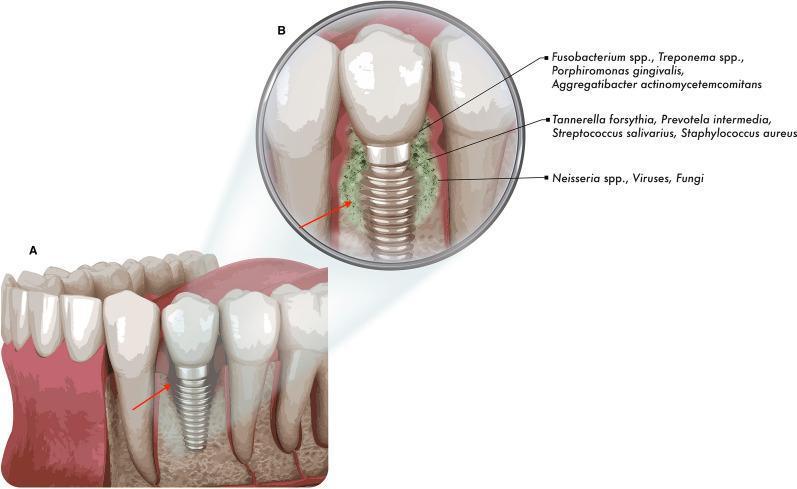
A medical anatomical diagram illustrating the pathophysiology of peri-implantitis and associated bone loss. Section A shows a cross-sectional view of the mandible with a dental implant integrated between natural teeth. A red arrow indicates localized alveolar bone resorption around the implant's threaded body. Section B provides a magnified view of the infectious process. It depicts the exposed implant surface following bone loss, which is heavily colonized by a complex microbial biofilm (represented in green). Labels identify specific pathogens involved in the biofilm community, including anaerobic bacteria such as Fusobacterium spp., Treponema spp., Porphyromonas gingivalis, Tannerella forsythia, and Aggregatibacter actinomycetemcomitans, as well as opportunistic fungi and viruses. The diagram highlights how progressive bone destruction provides an environment for bacterial colonization and the subsequent formation of a multi-species biofilm, which further exacerbates tissue inflammation and implant failure. This visual is designed for dental education, focusing on implantology and oral microbiology.
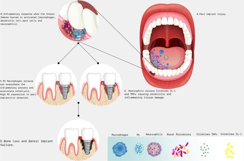
This medical infographic illustrates the pathophysiology and progression of peri-implantitis within the oral cavity. It features a central anatomical view of an open mouth showing microbiota distribution on the tongue and a localized peri-implant injury site. Surrounding diagrams detail the immune response and bone degradation sequence. Section B highlights the activation of the innate immune system, identifying macrophages, dendritic cells, mast cells, and neutrophils at the infection site. Inset C depicts M1 macrophage activity exacerbating inflammation and accelerating osteolysis. Inset D shows neutrophils releasing pro-inflammatory cytokines, specifically IL-1 and TNF-alpha, which drive osteolytic damage. The progression culminates in Section E, illustrating significant alveolar bone loss, gingival recession, and dental implant failure. A detailed legend identifies key components: Macrophages, M1 phenotype, Neutrophils, Buccal Microbiota, TNF-alpha, and IL-1. This educational diagram is intended for dental professionals and students to understand the host-microbe interactions leading to peri-implant tissue destruction.
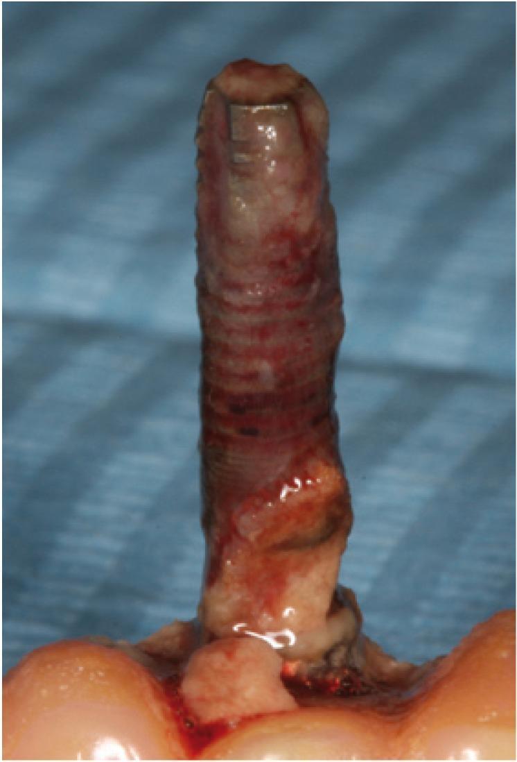
This clinical photograph displays a failed dental implant within an intraoral setting, illustrating severe peri-implantitis and implant failure. The visual focus is on a vertically oriented titanium implant fixture that exhibits extensive exposure of its threaded surface, indicating profound marginal bone loss and loss of osseointegration. The surrounding soft tissues are pathologically altered, appearing erythematous (dark red) and edematous, suggesting active inflammation and mucositis. At the base of the implant, there is evidence of gingival recession and irregular tissue attachment. The overall presentation serves as a teaching case for complications arising from the lack of supportive periodontal therapy (SPT), highlighting the progression from peri-implant mucositis to significant peri-implant bone destruction and ultimate fixture failure.
Why Do Dental Implants Fail? (Part 1)
Overview: Early vs. Late Failure
| Early Failure | Late Failure | |
|---|---|---|
| Timing | Before final prosthesis placement (within osseointegration phase) | 1-3+ years after implant placement |
| Core mechanism | Failure of bone-implant integration ("rejection") | Loss of an already-integrated implant |
| Main drivers | Biological non-acceptance, surgical trauma, systemic disease | Peri-implantitis, biomechanical overload, parafunctional habits |
1. Failure of Osseointegration
- Poor bone quality/quantity - Maxillary (upper jaw) bone is predominantly cancellous (spongy) and has lower density than mandibular bone, making osseointegration harder. Areas with bone loss from prior tooth extraction or resorption may lack sufficient volume.
- Lack of primary stability - The implant must be initially stable at placement to allow bone ingrowth without micromotion. Excessive movement (>150 µm) during healing disrupts the process and leads to fibrous encapsulation instead.
- Surgical trauma - Overheating bone during drilling (>47°C) causes osteonecrosis at the implant interface. Poor drilling technique, excessive force, or using dull burs are common culprits.
2. Peri-Implantitis (the #1 Cause of Late Failure)
- Bacterial biofilm (plaque) accumulates on the exposed implant surface
- Key pathogens include Porphyromonas gingivalis, Tannerella forsythia, Treponema spp., and Fusobacterium spp.
- The immune response (macrophages, neutrophils) releases pro-inflammatory cytokines - IL-1, TNF-alpha - driving osteolysis
- Progressive bone destruction exposes more implant surface, worsening the cycle
- History of periodontitis (strongest risk factor)
- Poor oral hygiene
- Smoking
- Diabetes mellitus
- Retained subgingival cement from the restoration
3. Smoking
- Increased peripheral vascular resistance and platelet aggregation reduce blood flow to the healing bone
- Impaired oxygen delivery slows fibroblast activity, collagen synthesis, and macrophage function
- Nicotine has a direct toxic effect on osteoblasts
- Higher infection susceptibility due to impaired immune response
4. Systemic Diseases
Diabetes Mellitus
- Microvascular and macrovascular changes impair blood supply to bone
- Impaired wound healing and increased infection susceptibility
- Hyperglycemia inhibits osteoblast function
- More likely to affect implants in the maxilla (cancellous bone) than the denser mandibular cortical bone
- Uncontrolled diabetes is a contraindication; well-controlled diabetes is a relative risk
Osteoporosis
- Reduced bone mineral density impairs osseointegration
- Bisphosphonate therapy (used to treat osteoporosis) poses a separate risk - medication-related osteonecrosis of the jaw (MRONJ) can be triggered by implant surgery
- Failure rate is approximately 10% higher in osteoporosis patients
Cardiovascular Disease
- Impairs tissue oxygenation, directly hindering the wound healing and osseointegration cascade
- Some antihypertensives (e.g., calcium channel blockers) cause gingival overgrowth, complicating hygiene
Immunosuppression (AIDS, corticosteroid therapy)
- Reduces immune surveillance, allowing opportunistic infection
- Corticosteroids also directly suppress bone formation
5. Bruxism (Teeth Grinding / Parafunction)
- Micromotion beyond the critical threshold (~150 µm) during the healing phase prevents osseointegration
- After osseointegration, excessive cyclical loading causes fatigue fracture of the implant fixture or abutment screw
- Lateral (non-axial) forces are particularly destructive since implants are designed for axial load
6. Bone Quality and Site Factors
| Type | Description | Failure Risk |
|---|---|---|
| Type I | Dense cortical bone | Lowest |
| Type II | Thick cortical + dense trabecular | Low |
| Type III | Thin cortical + dense trabecular | Moderate |
| Type IV | Very thin cortical + sparse trabecular | Highest |
- Posterior maxilla (upper back jaw) is the highest-risk site - Type III/IV bone, low blood supply, proximity to the maxillary sinus
- Anterior mandible is the lowest-risk site - dense cortical bone, excellent blood supply
7. Medications
- Bisphosphonates (e.g., alendronate, zoledronic acid) - IV bisphosphonates pose the highest MRONJ risk; oral bisphosphonates are a relative risk requiring careful evaluation
- Corticosteroids - Suppress bone metabolism and immune defense
- Antidepressants (SSRIs) - A 2024 meta-analysis (Harutyunyan et al., Int J Oral Maxillofac Implants 2024) found a statistically significant association between SSRI use and increased implant failure, likely due to effects on bone turnover (serotonin receptors are expressed on osteoblasts and osteoclasts)
- Anticoagulants/immunosuppressants - Impair healing
8. Clinical photograph of a failed implant
Summary Table
| Category | Cause | Early/Late |
|---|---|---|
| Biological | Failed osseointegration (fibrous encapsulation) | Early |
| Infection | Peri-implantitis (biofilm, IL-1/TNF-α driven osteolysis) | Late |
| Patient habit | Smoking (impaired vascularity/healing) | Both |
| Parafunctional | Bruxism (excessive micromotion/loading) | Both |
| Systemic | Diabetes, osteoporosis, cardiovascular disease, immunosuppression | Both |
| Medication | Bisphosphonates (MRONJ), SSRIs, corticosteroids | Both |
| Bone factors | Poor bone quality (Type IV), maxillary posterior site | Early |
| Surgical | Overheating (>47°C), poor primary stability, placement errors | Early |
| Mechanical | Overloading, traumatic occlusion, poor prosthetic design | Late |
Abdel salam article
Abdel Salam dental implant failure
Abdel Salam dental implant failure article
Abdelsalam implant failure
"Abdel Salam" OR "Abdelsalam" implant failure dental article journal
-
Slight name variation - Could the author's name be spelled differently? For example: Abdelsalam, Abd El-Salam, Abdelssalaam, or a first name like Mohamed Abdel Salam?
-
Different topic - Is this the "Part 1" article you are referring to, or is it a separate reference your instructor/textbook cited?
-
Gray literature / local journal - The article may be in a regional Arabic-language or Egyptian dental journal not indexed on PubMed (e.g., Egyptian Dental Journal, Alexandria Dental Journal).
-
Different field - Could it be about orthopedic implants, breast implants, or another specialty?
- The full author name
- The journal name or year of publication
- The topic covered in the article (e.g., risk factors, classification, surgical technique)
- Whether it was a handout, lecture slide, or textbook reference
How can i send the article file to you
- PDF (.pdf) - most common for journal articles
- Word document (.doc or .docx)
- Images (.jpg, .png) - if you have a scanned copy or screenshot