
SBO full details
"small bowel obstruction" management
small bowel obstruction CT scan abdominal X-ray
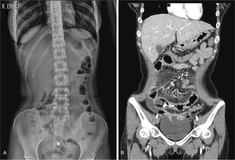
A two-panel composite showing diagnostic abdominal imaging for a small bowel obstruction. Image A is a frontal erect abdominal radiograph (X-ray) demonstrating the 'stepladder sign,' characterized by multiple dilated loops of small bowel with air-fluid levels, indicating mechanical obstruction. A radiopaque, cylindrical video capsule endoscopy (VCE) device is visible as a foreign body retained in the pelvic region. Image B is a coronal view from a Computed Tomography (CT) scan of the same patient. The CT demonstrates multifocal mural thickening and luminal narrowing (stenosis) within the ileum, consistent with Crohn's disease complications. The retained endoscopy capsule is identified as a high-attenuation object lodged proximal to a site of ileal stenosis. Proximal small bowel dilation is observed, corroborating the radiographic findings of obstruction. This comparison illustrates the complication of capsule retention in patients with inflammatory bowel disease and intestinal strictures.
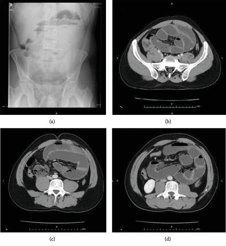
This composite of diagnostic images illustrates findings characteristic of a mechanical intestinal obstruction. Panel (a) is an upright plain abdominal X-ray (radiograph) demonstrating multiple dilated loops of small bowel with prominent air-fluid levels, visible as sharp horizontal interfaces between radiolucent gas and radiopaque fluid. Panels (b), (c), and (d) are axial (transverse) slices of a contrast-enhanced abdominal computerized tomography (CT) scan. These images show significantly dilated loops of the small intestine with associated wall thickening. The CT images reveal the clustering of the small bowel loops and help delineate the transition zone or specific location of the pathology. Together, these imaging modalities provide evidence for small bowel ileus, often caused by conditions such as abdominal cocoon syndrome (ACS), adhesions, or internal herniation. The visual data is intended for medical education regarding the radiological diagnosis of acute mechanical intestinal obstruction and the assessment of bowel wall edema and luminal dilatation.
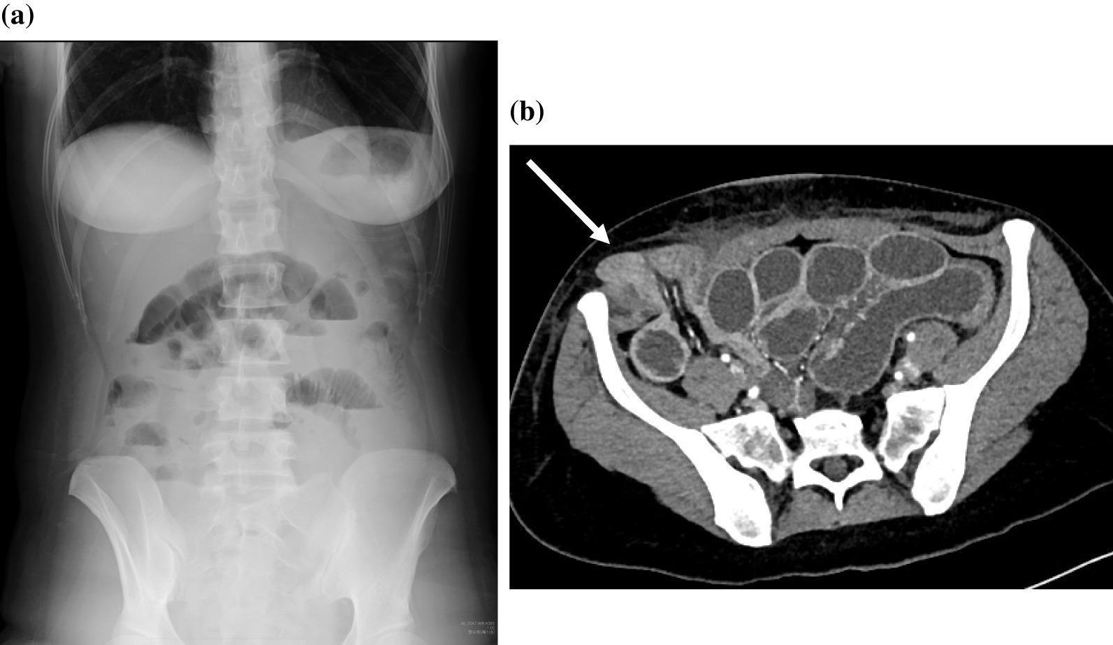
This composite diagnostic image illustrates a case of small bowel obstruction secondary to a trocar site hernia. (a) Anteroposterior erect abdominal radiograph (X-ray) demonstrating multiple dilated loops of small bowel with prominent air-fluid levels. These horizontal interfaces indicate the pathologic accumulation of gas and liquid, a classic sign of mechanical bowel obstruction. (b) Axial contrast-enhanced computed tomography (CT) scan of the lower abdomen and pelvis. The image shows multiple dilated, fluid-filled small bowel loops within the abdominal cavity. A white arrow specifically indicates a segment of small bowel that has herniated through a defect in the abdominal wall fascia at a previous 12-mm lateral trocar site. The herniated loop is visible in the subcutaneous space, superficial to the fascial plane, confirming a trocar site hernia (TSH). The imaging highlights the progression from clinical signs of ileus on plain film to a definitive anatomical diagnosis of strangulated or obstructed hernia on CT, emphasizing postoperative complications of laparoscopic surgery.
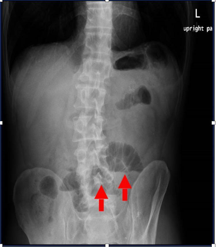
This diagnostic image is an upright posterior-anterior (PA) abdominal radiograph demonstrating signs of an evolving small bowel obstruction. The X-ray shows multiple dilated loops of small bowel in the mid and lower abdominal regions, identified by their radiolucent gas-filled appearance and central distribution. Red arrows specifically highlight dilated loops in the lower quadrants. The valvulae conniventes (plicae circulares) are visible within these segments, distinguishing them from large bowel. Bony landmarks including the lumbar spine and pelvic girdle appear intact and in normal alignment. Soft tissue shadows of solid organs are present but partially obscured by the increased bowel gas. This imaging is clinically significant for evaluating acute abdominal pain and mechanical obstruction, providing a visual baseline before further cross-sectional imaging (CT scan) or surgical intervention.
Small Bowel Obstruction (SBO) - Full Details
Definition
- Rosen's Emergency Medicine, p. 4509
- Schwartz's Principles of Surgery, p. 1255
Classification
1. By Mechanism
| Type | Description |
|---|---|
| Mechanical | Physical barrier to intestinal transit |
| Functional (Adynamic Ileus) | Disrupted peristalsis without a physical blockage; common post-operatively |
2. By Anatomical Relationship to Bowel Wall
| Category | Examples |
|---|---|
| Extrinsic (most common) | Adhesions, hernias, carcinomatosis, volvulus |
| Intramural | Crohn's strictures, primary tumors, hematoma, radiation stricture |
| Intraluminal | Gallstone ileus, bezoars, foreign bodies, meconium |
3. By Degree
- Partial obstruction - some gas/fluid can pass distally; slower progression; strangulation less likely
- Complete obstruction - no gas or fluid passes beyond obstruction point
4. Simple vs. Closed-Loop
-
Simple obstruction - blocked at a single point
-
Closed-loop obstruction - blocked at two points (e.g., volvulus, internal hernia in mesenteric defect); blood supply compromised proximally and distally - can rapidly progress to strangulation and infarction, with twice the mortality of simple SBO
-
Rosen's Emergency Medicine, p. 4534
Etiology & Epidemiology
| Cause | Frequency |
|---|---|
| Postoperative adhesions | ~60-75% of all SBO |
| Tumors (extrinsic compression, carcinomatosis, primary) | ~20% |
| Hernias (inguinal, femoral, internal) | ~10% |
| Crohn's disease strictures | Less common |
| Radiation-induced stricture | Less common |
| Gallstone ileus | Rare |
| Volvulus / intussusception | Rare |
| Congenital (malrotation, webs, duplications) | Rare in adults |
| SMA syndrome | Very rare |
- Schwartz's Principles of Surgery, pp. 1255-1256
Pathophysiology
- Gas and fluid accumulate proximal to the obstruction - gas is mostly swallowed air; fluid is swallowed liquids plus GI secretions (obstruction stimulates intestinal epithelial water secretion)
- Intestinal motility increases initially to overcome the obstruction - causes colicky pain and early diarrhea
- Bowel distends - intraluminal and intramural pressures rise
- Motility eventually decreases - fewer contractions
- Luminal flora changes - small bowel (normally near-sterile) develops bacterial overgrowth; bacterial translocation to regional lymph nodes occurs
- If intramural pressure becomes critical - microvascular perfusion is impaired → ischemia → necrosis (strangulated obstruction)
- Schwartz's Principles of Surgery, p. 1256
Clinical Presentation
Symptoms
| Symptom | Notes |
|---|---|
| Colicky abdominal pain | Periodic, crampy; from bowel hypermotility against obstruction |
| Nausea and vomiting | More prominent with proximal SBO; feculent vomiting suggests established/distal obstruction |
| Abdominal distension | Maximal with distal ileal obstruction; may be absent in proximal SBO |
| Obstipation | Inability to pass gas or stool; passage of flatus/stool >6-12 h after symptom onset suggests partial obstruction |
Signs
- Early: Hyperactive, high-pitched ("tinkling") bowel sounds
- Late: Absent or minimal bowel sounds
- Abdominal distension
- Peritoneal signs (guarding, rigidity, rebound) - raise concern for strangulation or perforation
Red Flags for Strangulation
- Pain disproportionate to abdominal findings
- Tachycardia
- Fever
- Localized tenderness rather than diffuse crampy pain
- Leukocytosis (progressive)
- Elevated lactate
No single history or physical exam finding can reliably diagnose or exclude SBO or strangulation.
- Schwartz's Principles of Surgery, p. 1256
Investigations
Laboratory
- CBC: Mild leukocytosis common; marked leukocytosis suggests strangulation or perforation
- BMP/electrolytes: Volume depletion; hypokalemia, hypochloremia, metabolic alkalosis
- Lactate: Elevated in ischemia/strangulation
- Serum amylase: Can be mildly elevated (non-specific)
- ABG: In unstable patients; to assess acid-base status
- Type and Screen: Pre-operative preparation
Imaging
Plain Abdominal X-ray (AXR)
- Supine view: Multiple loops of dilated small bowel (central location, valvulae conniventes/plicae circulares traversing the full diameter) with paucity of colonic gas
- Upright/decubitus view: Multiple air-fluid levels ("step-ladder sign")
- String of pearls sign: Small gas bubbles trapped along superior margin of fluid-filled loops on upright films
- Gasless abdomen: Can occur when patient swallows no air - multiple fluid-filled loops may resemble abdominal masses
- Distinguishing small from large bowel: small bowel is central, has valvulae conniventes (tightly spaced, full-width folds); colon is peripheral with haustral folds that don't span full diameter
CT Abdomen/Pelvis (Gold Standard)
- Most sensitive and specific imaging for SBO
- Identifies transition zone (point of obstruction)
- Determines etiology (adhesion band, hernia, mass)
- Detects strangulation (mesenteric fat stranding, bowel wall thickening/pneumatosis, portal venous gas, ascites, vascular engorgement)
- Can detect closed-loop obstruction
Water-Soluble Contrast Studies (Gastrografin/Oral Contrast)
- Used in stable patients with suspected adhesive SBO for both diagnosis and therapy
- Therapeutic effect: Water-soluble contrast exerts osmotic effect, drawing water into bowel lumen and stimulating peristalsis - shown to reduce time to resolution and reduce need for surgery
- Contrast reaching the colon within 24 hours has high sensitivity for predicting successful non-operative resolution (~97%)
- A 2025 meta-analysis (PMID 40341790) confirmed benefit of water-soluble contrast agents in adhesional SBO
MRI / Ultrasound
-
Less commonly used; MRI useful in pregnancy
-
Ultrasound: can detect dilated loops and peristalsis at bedside
-
Yamada's Textbook of Gastroenterology, p. 2632
Management
Initial Resuscitation (All Patients)
- IV fluid resuscitation - vigorous isotonic crystalloid (NS or Lactated Ringer's); guided by urine output (target >0.5 mL/kg/hr)
- Nasogastric (NG) decompression - relieves vomiting, prevents aspiration, reduces ongoing distension
- Electrolyte correction - especially K⁺ (after establishing urine output) and Cl⁻
- Urinary catheter - to monitor urine output
- Blood work and monitoring as above
- NPO (nil by mouth)
Colloid solutions (albumin, hetastarch) have little role in resuscitation of SBO patients.
- Maingot's Abdominal Operations, pp. 620-621
Non-Operative Management
- No peritonitis
- No progressive leukocytosis
- No impaired bowel wall perfusion on imaging
- Uncomplicated (simple, partial) obstruction
- Bowel rest (NPO)
- NG decompression
- IV fluids and electrolyte replacement
- Pain control
- Water-soluble contrast (Gastrografin) challenge - therapeutic and diagnostic
Surgical Management
Absolute Indications for Emergency Surgery
- Peritonitis (signs of perforation)
- Strangulation or ischemia (suspected or confirmed)
- Closed-loop obstruction
- Incarcerated/irreducible hernia
- Complete SBO failing non-operative management
- Hemodynamic instability
Operative Approach
- Traditional standard, especially for complex, multiple adhesions or suspected strangulation
- Lysis of adhesions (adhesiolysis) - freeing the obstructing band(s)
- Bowel resection if necrotic/ischemic segment found
- Assessment of bowel viability after relief of obstruction (colour, peristalsis, Doppler); resect non-viable segments
- Increasingly used; best for proximal SBO likely due to a single band in patients without massive distension
- Associated with significantly lower rates of complications, surgical site infections, and shorter hospital stay (4 vs 10 days)
- Conversion rate 17-33% (distended bowel limits visualization)
- Risk of iatrogenic bowel injury is a concern but comparable to open in pooled analyses
- Schwartz's Principles of Surgery, p. 1258
- Fluid resuscitation to hemodynamic stability
- Electrolyte correction and adequate urine output
- Broad-spectrum antibiotics within 1 hour of incision (prophylaxis)
Special Scenarios
Gallstone Ileus
- Gallstone passes through a cholecystoduodenal fistula and impacts the terminal ileum
- Rigler's triad on AXR: air in biliary tree (pneumobilia), small bowel obstruction, ectopic calcified gallstone
- Treated surgically: enterolithotomy ± cholecystectomy
Crohn's Disease
- Stricturoplasty preferred over resection when possible to preserve bowel length
- May require resection of fibrotic strictures unresponsive to medical therapy
Malignant SBO
- Often requires palliation rather than curative surgery
- Stenting (via endoscopy or radiology), bypass procedures, or gastrostomy for decompression in unresectable cases
SBO in Pregnancy
- High mortality for both mother and fetus if delayed
- CT should NOT be withheld due to radiation fear if clinical suspicion is high
- A 2024 systematic review (PMID 38600568) found conservative management feasible in select pregnant patients with SBO
Virgin Abdomen (No Prior Surgery)
- SBO in a patient without prior abdominal surgery should always prompt workup for hernia, malignancy, volvulus, or Crohn's disease
- Lower threshold for surgery
Outcomes
| Situation | Perioperative Mortality |
|---|---|
| Non-strangulating SBO - surgical | <5% |
| Strangulated SBO - surgical | Significantly higher (exact varies) |
| Overall modern mortality | <8% |
-
Risk of recurrent obstruction: 5.5% at 1 yr, 11.3% at 3 yr, 13.5% at 5 yr
-
Risk of reoperation: 3.7% at 1 yr, 4.8% at 3 yr, 5.8% at 5 yr
-
Most patients managed conservatively do NOT require readmission - <20% readmitted over 5 years
-
Schwartz's Principles of Surgery, p. 1259
Prevention of Adhesive SBO
-
Good surgical technique: careful tissue handling, minimize peritoneal trauma, minimize use of foreign material
-
Laparoscopic approach preferred - open surgery associated with a 4-fold increase in SBO risk vs. laparoscopy within 5 years (Swedish National Inpatient Register data)
-
Adhesion barriers (e.g., Seprafilm - sodium hyaluronate/carboxymethylcellulose): reduces adhesion formation and may reduce reoperation rate for SBO (but did not reduce overall SBO rate in large trials; increases septic complications when placed around anastomoses)
-
Schwartz's Principles of Surgery, pp. 1259-1260
Summary Table
| Feature | Detail |
|---|---|
| Most common cause | Adhesions (60-75%) |
| Classic presentation | Colicky pain + vomiting + distension + obstipation |
| Key investigation | CT abdomen/pelvis |
| Emergency surgery indicators | Peritonitis, strangulation, closed-loop, hernia |
| Conservative success rate | ~70-80% of adhesive SBO |
| Mortality (non-strangulating) | <5% surgical; <8% overall |
| Key prognostic test | Gastrografin challenge (contrast in colon <24h = likely resolves) |
- A 2025 meta-analysis (PMID 40341790) confirmed water-soluble contrast agents reduce need for surgery in adhesional SBO
- A 2024 systematic review (PMID 37938387) identified CT-based prognostic factors that predict which patients will resolve without surgery