
thrombosis
What aspect of thrombosis are you interested in?
causes and consequences of thrombosis
thrombosis Virchow's triad pathophysiology diagram
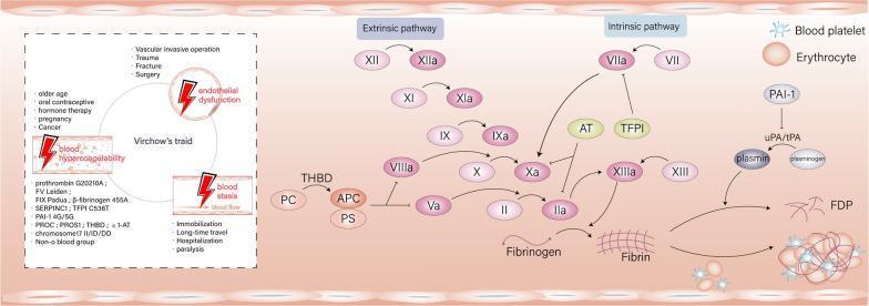
This medical infographic illustrates the complex pathophysiology of thrombosis, integrating Virchow’s triad with the coagulation and fibrinolytic cascades. On the left, a schematic of Virchow’s triad identifies three primary contributors to thrombosis: vascular endothelial dysfunction (associated with surgery and trauma), blood hypercoagulability (linked to genetic mutations like Factor V Leiden and prothrombin G20210A, or acquired states like pregnancy and cancer), and blood stasis (linked to immobilization). Each element is marked with a red lightning bolt indicating a trigger point for thrombus formation. The right side depicts the biochemical signaling pathways of coagulation. It details the intrinsic pathway (Factors XII, XI, IX, VIII) and extrinsic pathway (Factor VII) converging on the common pathway at Factor Xa. Key regulatory components are shown, including anticoagulants like Antithrombin (AT), Tissue Factor Pathway Inhibitor (TFPI), and the Protein C system (PC, APC, PS) with Thrombomodulin (THBD). The cascade culminates in the conversion of prothrombin (II) to thrombin (IIa), which catalyzes fibrinogen into a fibrin mesh. The final stage illustrates the fibrinolytic system where plasmin converts fibrin into fibrin degradation products (FDP), and the structural role of blood platelets and erythrocytes in forming a mature clot.
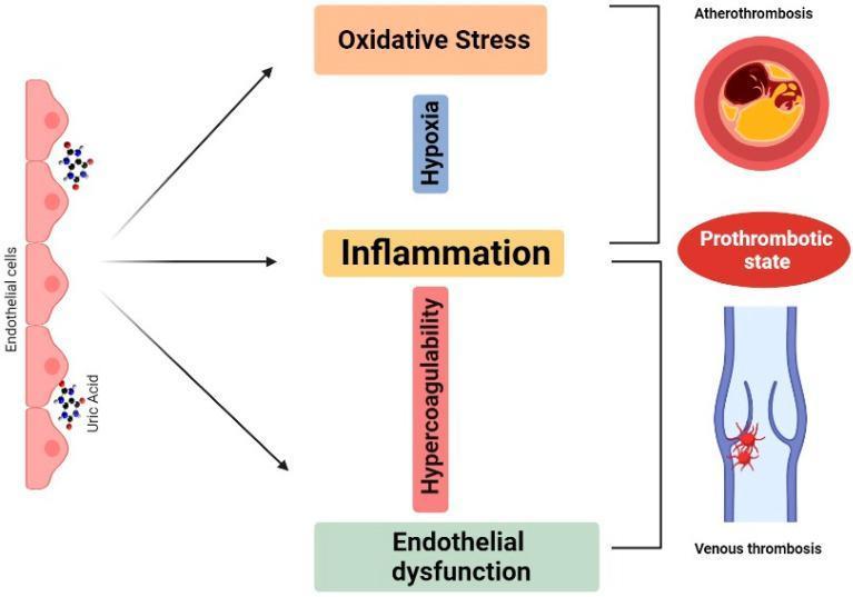
A pathophysiology diagram illustrating the proposed mechanism linking uric acid to thrombosis. The flowchart progresses from left to right. On the far left, an illustration of the vascular endothelium shows uric acid molecules being absorbed by endothelial cells. This process triggers three central intermediary pathways: 'Oxidative Stress', 'Inflammation', and 'Endothelial dysfunction'. These pathways are further modulated by factors labeled 'Hypoxia' and 'Hypercoagulability'. On the right, the diagram culminates in three clinical outcomes: 'Atherothrombosis' (represented by a cross-section of an artery with an occlusive plaque), a 'Prothrombotic state', and 'Venous thrombosis' (depicted by a venous valve with a developing red thrombus). The visual layout uses arrows and brackets to establish a causal relationship between hyperuricemia-induced cellular stress and various forms of vascular clotting. This educational diagram is intended for medical students and clinicians studying cardiovascular pathophysiology and the metabolic drivers of thrombotic disease.
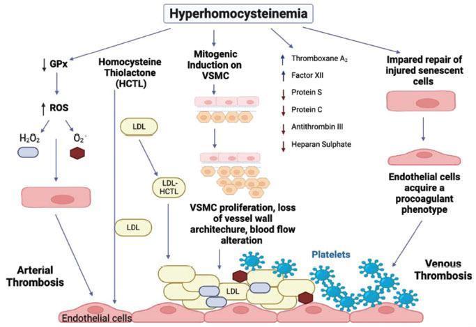
A pathophysiology diagram illustrating the mechanisms by which hyperhomocysteinemia induces endothelial damage, leading to arterial and venous thrombosis. The diagram flows from the central header 'Hyperhomocysteinemia' into five distinct pathways. The left pathway details oxidative stress, showing decreased Glutathione Peroxidase (GPx) and increased Reactive Oxygen Species (ROS), such as H2O2 and O2-, leading to arterial thrombosis. A second pathway shows Homocysteine Thiolactone (HCTL) interacting with LDL. The central 'Mitogenic Induction' pathway depicts vascular smooth muscle cell (VSMC) proliferation and loss of vessel wall architecture. A fourth pathway lists hematologic alterations, specifically increased Thromboxane A2 and Factor XII, alongside decreased Protein S, Protein C, Antithrombin III, and Heparan Sulphate. The right pathway illustrates impaired repair of injured senescent cells and the acquisition of a procoagulant phenotype. The bottom of the diagram shows a damaged endothelial layer with adherent LDL and platelets, visually differentiating the mechanisms that culminate in arterial versus venous thrombotic events.
Thrombosis: Causes and Consequences
What is Thrombosis?
CAUSES: Virchow's Triad
1. Endothelial Injury
- Physical injury or surgery
- Infectious agents (bacterial, viral)
- Abnormal blood flow
- Inflammatory cytokines
- Metabolic abnormalities: hypercholesterolaemia, homocysteinaemia
- Cigarette smoke toxins
- Hypertension
- Downregulating anticoagulants: thrombomodulin, protein C receptor, tissue factor pathway inhibitor (TFPI)
- Upregulating tissue factor (initiates the coagulation cascade)
- Increasing plasminogen activator inhibitors (PAI), which suppress fibrinolysis
2. Abnormal Blood Flow (Stasis or Turbulence)
- Turbulence promotes arterial/cardiac thrombosis by causing endothelial injury and forming countercurrents and pockets of stasis
- Stasis is the major factor in venous thrombosis
- Platelets and activated clotting factors to accumulate (instead of being diluted/washed away)
- Inhibition of fresh anticoagulant factor inflow
- Endothelial cell hypoxia, promoting a pro-thrombotic phenotype
- Atrial fibrillation (stasis in atrial appendage)
- Aortic aneurysms
- Atherosclerotic plaques (turbulence)
- Prolonged immobility or bed rest (venous stasis)
- Long-haul flights
3. Hypercoagulability
Primary (Genetic) Hypercoagulable States
| Risk Level | Condition |
|---|---|
| Common (>1% of US population) | Factor V Leiden mutation (Arg506Glu) - ~3-fold increased VTE risk |
| Prothrombin G20210A mutation | |
| Elevated factor VIII, IX, XI, or fibrinogen | |
| Rare | Antithrombin deficiency |
| Protein C deficiency | |
| Protein S deficiency | |
| Very Rare | Fibrinolysis defects |
| Homozygous homocystinuria |
Secondary (Acquired) Hypercoagulable States
| Risk Level | Condition |
|---|---|
| High Risk | Prolonged bed rest / immobilisation |
| Myocardial infarction | |
| Atrial fibrillation | |
| Surgery, fracture, burns | |
| Cancer | |
| Prosthetic cardiac valves | |
| DIC (disseminated intravascular coagulation) | |
| Heparin-induced thrombocytopenia (HIT) | |
| Antiphospholipid syndrome | |
| Elevated Risk | Cardiomyopathy |
| Nephrotic syndrome | |
| Pregnancy / postpartum | |
| Oral contraceptive use | |
| Sickle cell anaemia | |
| Smoking |
- Autoimmune conditions: SLE, rheumatoid arthritis
- Infections: Bacterial/viral (SARS-CoV-2, HIV, hepatitis C) - activate platelet TLRs
- Hyperhomocysteinaemia (both arterial and venous thrombosis)
- CD40L/CD40 axis dysregulation (links thrombosis and inflammation)
CONSEQUENCES of Thrombosis
A. Fate of a Thrombus (Four Possible Outcomes)
- Propagation - the thrombus grows and causes more obstruction
- Embolisation - parts detach and travel distally (most dangerous outcome)
- Dissolution - fibrinolysis breaks it down (therapeutic thrombolytics exploit this window)
- Organisation and recanalisation - older thrombi are invaded by endothelial cells, smooth muscle cells, and fibroblasts; capillary channels eventually re-establish limited blood flow
B. Venous Thrombosis Consequences
- Superficial (saphenous system, varicosities): cause local pain, congestion, oedema; rarely embolise but predispose to skin infections and varicose ulcers
- Deep Vein Thrombosis (DVT) - larger leg veins at or above the knee: the main danger is pulmonary embolism (PE)
- PE can cause sudden death (massive PE occluding pulmonary trunk), acute right heart strain, or infarction
- Post-thrombotic syndrome: chronic venous insufficiency, pain, oedema, skin changes
C. Arterial and Cardiac Thrombosis Consequences
| Site | Consequence |
|---|---|
| Coronary artery | Myocardial infarction (MI) |
| Cerebral artery | Ischaemic stroke |
| Renal artery | Renal infarction / renovascular hypertension |
| Mesenteric artery | Bowel ischaemia / infarction |
| Peripheral arteries | Acute limb ischaemia |
| Left ventricle (mural thrombus) | Systemic arterial embolism (stroke, splenic/renal infarct) |
| Left atrium (e.g., in AF) | Stroke |
Key Clinical Summary
| Feature | Venous Thrombosis | Arterial Thrombosis |
|---|---|---|
| Major cause | Stasis + hypercoagulability | Endothelial injury (atherosclerosis) |
| Composition | Red (fibrin + RBCs) | White (platelet-rich) |
| Main consequence | PE, post-thrombotic syndrome | Infarction (MI, stroke, limb ischaemia) |
| Key risk factors | Immobility, cancer, pregnancy, genetic thrombophilia | Hypertension, smoking, dyslipidaemia, diabetes |