I now have comprehensive content from multiple authoritative textbooks. Let me compile the complete, detailed note on shock.
SHOCK - A Comprehensive Clinical Note
1. DEFINITION
Shock is a state of circulatory failure that impairs tissue perfusion and leads to cellular hypoxia. More precisely, it is defined as inadequate perfusion of tissues resulting in insufficient delivery of oxygen and nutrients to meet cellular metabolic demands. At the outset, the cellular injury is reversible; however, prolonged shock eventually leads to irreversible tissue injury and is often fatal.
"A manifestation of the rude unhinging of the machinery of life." - Gross, 1872
(Sabiston Textbook of Surgery, p. 563)
Shock may complicate severe hemorrhage, extensive trauma or burns, myocardial infarction, pulmonary embolism, and microbial sepsis.
2. HISTORICAL BACKGROUND
- The term "shock" first appeared in a 1743 translation of Henri Francois Le Dran's French treatise on battlefield wounds, used to designate the act of collision.
- In 1815, Guthrie used it to describe physiologic instability in Gunshot Wounds of the Extremities.
- In 1830, Herman attempted IV rehydration with water for cholera - one of the first documented attempts at intravascular volume replacement.
- In 1872, Gross defined shock as "a manifestation of the rude unhinging of the machinery of life."
- In 1899, George Crile proposed that profound decline in blood pressure accounted for all symptoms of shock, leading to the routine clinical measurement of BP.
(Sabiston Textbook of Surgery, p. 563)
3. CLASSIFICATION / TYPES OF SHOCK
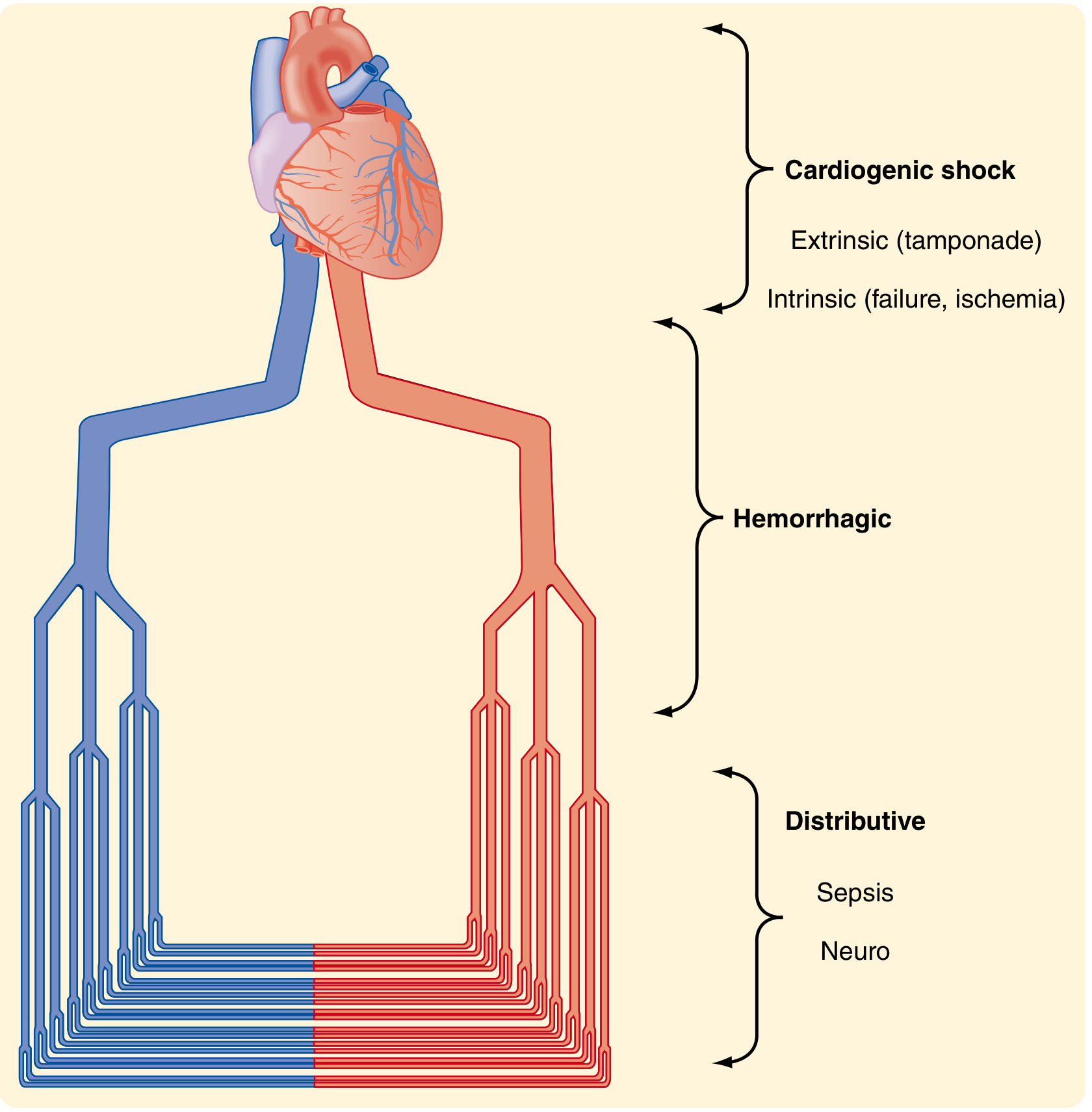
Figure: Types of shock showing cardiogenic (extrinsic and intrinsic), hemorrhagic, and distributive (sepsis, neurogenic) categories at different levels of the cardiovascular system. (Sabiston Textbook of Surgery)
Shock can be conceptualized as occurring at three anatomical levels:
- At the heart - cardiogenic abnormalities (intrinsic or extrinsic)
- At large/medium vessels - hemorrhage and loss of blood volume
- At small vessels - neurologic dysfunction or sepsis causing vasodilatation and maldistribution
A. Cardiogenic Shock
- Results from low cardiac output due to myocardial pump failure
- Intrinsic causes: Myocardial infarction, ventricular rupture, arrhythmia, myocardial contusion/laceration
- Extrinsic causes: Cardiac tamponade, tension pneumothorax, hemothorax
- Obstructive causes: Pulmonary embolism (outflow obstruction)
- Hemodynamics: Low CO, high SVR, high PCWP
B. Hypovolemic Shock
- Results from low cardiac output due to loss of blood or plasma volume
- Causes: Hemorrhage (most common), fluid loss from severe burns, vomiting, diarrhea, third-spacing
- Subdivided into:
- Hemorrhagic shock (most studied; caused by blood loss)
- Non-hemorrhagic hypovolemic shock (plasma loss, GI losses)
- Hemodynamics: Low CO, high SVR, low PCWP
C. Distributive (Vasodilatory) Shock
The defining feature is peripheral vasodilation with relative maldistribution of blood flow. This category includes:
1. Septic Shock
- Most common form of distributive shock
- Caused by overwhelming microbial infections (gram-positive bacteria most common, followed by gram-negative bacteria and fungi)
- Sepsis: life-threatening organ dysfunction caused by a dysregulated host response to infection
- Septic shock: a subset of sepsis with profound circulatory, cellular, and metabolic abnormalities
- Hemodynamics: High CO (hyperdynamic), low SVR, warm periphery
2. Neurogenic Shock
- Loss of sympathetic vascular tone due to spinal cord injury or anesthesia
- Acute vasodilation leads to hypotension and tissue hypoperfusion
- Hemodynamics: Low CO (due to bradycardia), very low SVR
- Classic triad: Hypotension + Bradycardia + Warm extremities
3. Anaphylactic Shock
- IgE-mediated hypersensitivity reaction
- Triggered by drugs (penicillin), foods (peanuts), insect stings
- Systemic vasodilation and increased vascular permeability
- Features: Urticaria, bronchospasm, angioedema, hypotension
(Robbins, Cotran & Kumar Pathologic Basis of Disease, p. 134; Sabiston Textbook of Surgery, p. 563)
| Type | CO | SVR | PCWP | Clinical Example |
|---|
| Cardiogenic | ↓ | ↑ | ↑ | MI, tamponade |
| Hypovolemic | ↓ | ↑ | ↓ | Hemorrhage, burns |
| Septic | ↑ (early) | ↓ | Low/normal | Bacterial sepsis |
| Neurogenic | ↓ | ↓↓ | ↓ | Spinal cord injury |
| Anaphylactic | ↓ | ↓↓ | ↓ | Drug reaction |
(CO = cardiac output, SVR = systemic vascular resistance, PCWP = pulmonary capillary wedge pressure)
4. ATLS CLASSIFICATION OF HEMORRHAGIC SHOCK
The ATLS course classifies hemorrhagic shock into four classes based on the volume of blood lost:
| CLASS I | CLASS II | CLASS III | CLASS IV |
|---|
| Blood loss (%) | 0-15 | 15-30 | 30-40 | >40 |
| CNS status | Slightly anxious | Mildly anxious | Anxious/confused | Confused/lethargic |
| Pulse (beats/min) | <100 | >100 | >120 | >140 |
| Blood pressure | Normal | Normal | Decreased | Decreased |
| Pulse pressure | Normal | Decreased | Decreased | Decreased |
| Respiratory rate | 14-20/min | 20-30/min | 30-40/min | >35/min |
| Urine output (mL/h) | >30 | 20-30 | 5-15 | Negligible |
| Fluid replacement | Crystalloid | Crystalloid | Crystalloid + blood | Crystalloid + blood |
(Sabiston Textbook of Surgery, p. 567)
Note: These classes were not rigorously validated; patients (especially the young, elderly, those on beta-blockers, or those with TBI) often do not follow this pattern.
5. PATHOPHYSIOLOGY
5.1 General Mechanism
Regardless of the cause, the core problem is:
Oxygen delivery (DO₂) fails to meet oxygen consumption (VO₂) at the cellular level
This results in:
- Shift from aerobic to anaerobic glycolysis
- Excessive production of lactic acid → metabolic acidosis
- Failure of Na⁺/K⁺ ATPase pumps → cellular swelling
- Lysosomal enzyme release → cellular necrosis
- Eventual irreversible cell death if not corrected
5.2 Pathogenesis of Septic Shock
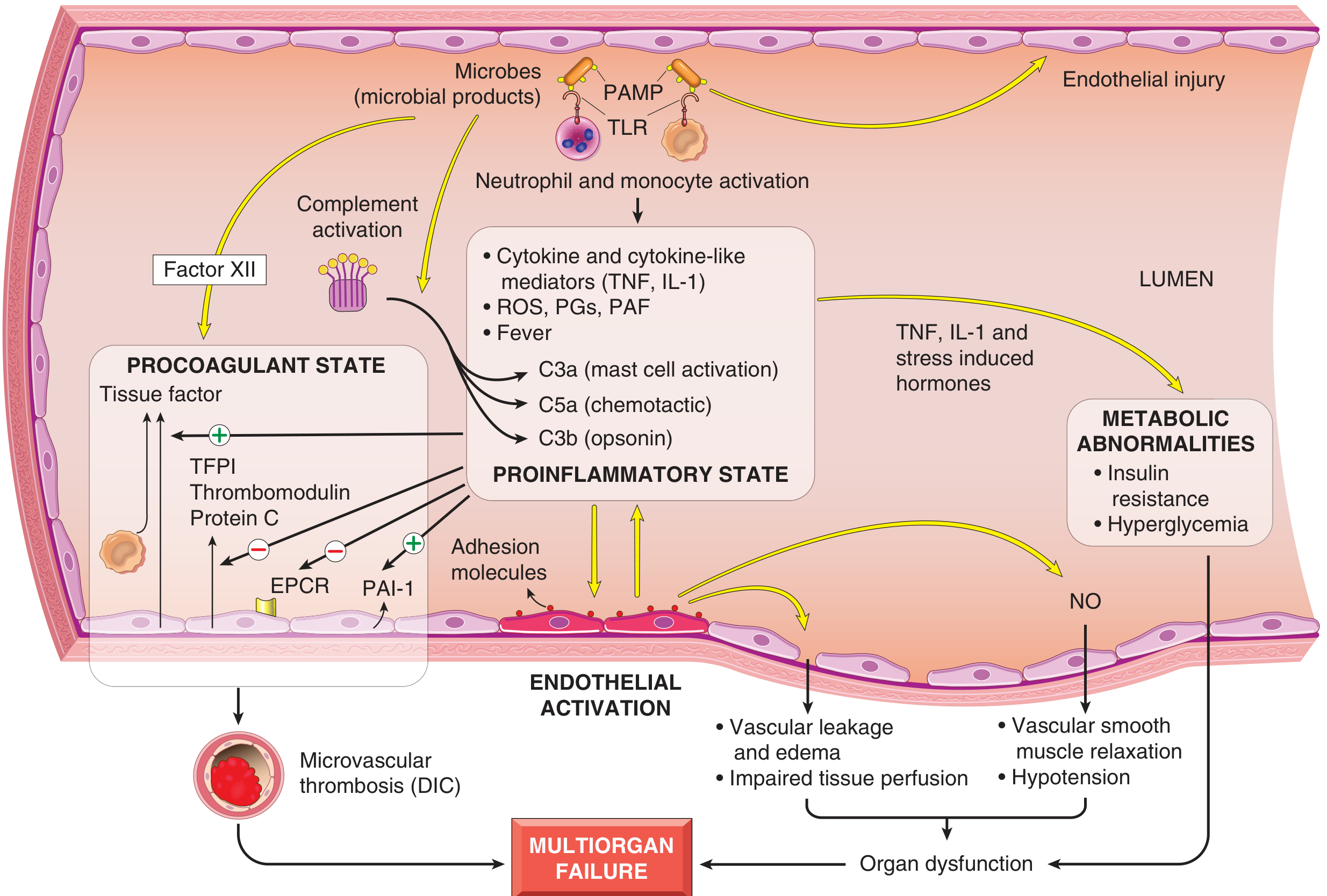
Figure: Microbial products activate endothelial cells and cellular elements of innate immunity, triggering cascades that lead to proinflammatory state, procoagulant state, metabolic abnormalities, endothelial activation, and ultimately multiorgan failure. (Robbins, Cotran & Kumar Pathologic Basis of Disease)
The key pathogenic mechanisms include:
a) Inflammatory & Counter-inflammatory Responses
- Microbial constituents (PAMPs) activate Toll-like receptors (TLRs), G-protein-coupled receptors, and C-type lectin receptors on innate immune cells
- Activated macrophages, neutrophils, and dendritic cells release:
- Cytokines: TNF, IL-1, IL-12, IL-18, type I interferons
- Reactive oxygen species (ROS)
- Lipid mediators: prostaglandins, platelet-activating factor (PAF)
- Complement cascade is activated → produces C3a (mast cell activation), C5a (chemotaxis), C3b (opsonin)
- Elevated acute phase reactants: C-reactive protein, procalcitonin
- With time, the hyperinflammatory state triggers counter-regulatory immunosuppression involving both innate and adaptive immune cells
b) Endothelial Activation and Injury
- Inflammatory cytokines loosen tight junctions → vascular leakage and protein-rich edema
- Activated endothelium upregulates NO and other vasoactive mediators (C3a, C5a, PAF) → smooth muscle relaxation → systemic hypotension
- Microvascular dysfunction: increased capillaries with intermittent flow, heterogeneous flow, loss of autoregulation → oxygen supply-demand mismatch
c) Induction of a Procoagulant State
- Proinflammatory cytokines increase tissue factor production by monocytes/endothelial cells
- Decrease anticoagulant factors: tissue factor pathway inhibitor (TFPI), thrombomodulin, protein C
- Dampen fibrinolysis by increasing PAI-1 expression
- Result: systemic thrombin activation and fibrin-rich thrombi in small vessels → further compromise tissue perfusion
- In up to 50% of septic patients: Disseminated Intravascular Coagulation (DIC) - consumption of clotting factors and platelets leads to paradoxical bleeding
d) Metabolic Abnormalities
- Insulin resistance and hyperglycemia: driven by TNF, IL-1, catecholamines, glucocorticoids, growth hormone
- Proinflammatory cytokines suppress insulin release and promote insulin resistance
- Sepsis is associated with rises in blood glucose, triglycerides, and lactate
- Hyperglycemia decreases neutrophil bactericidal function and increases endothelial adhesion molecule expression
- Adrenal insufficiency may follow initial glucocorticoid surge, sometimes from Waterhouse-Friderichsen syndrome (adrenal necrosis from DIC)
- Cellular hypoxia + diminished oxidative phosphorylation → lactic acidosis
(Robbins, Cotran & Kumar Pathologic Basis of Disease, p. 134-137)
6. COMPENSATORY MECHANISMS (Stages of Shock)
Shock is a progressive disorder that - unless the underlying problem is corrected - evolves through three stages:
Stage 1: Initial Nonprogressive (Compensated) Stage
- Reflex compensatory mechanisms are activated
- Vital organ perfusion is maintained
- Neurohumoral responses activated:
- Baroreceptor reflexes
- Release of catecholamines and ADH (antidiuretic hormone)
- Activation of renin-angiotensin-aldosterone axis
- Generalized sympathetic stimulation
- Net effects:
- Tachycardia
- Peripheral vasoconstriction (skin becomes cool, pale, clammy)
- Renal fluid conservation (oliguria)
- Blood shunting away from skin to heart and brain (coronary and cerebral vessels less sensitive to sympathetic signals)
- Note: In septic shock, initial cutaneous vasodilation may produce warm, flushed skin
Stage 2: Progressive (Decompensated) Stage
- Underlying cause not corrected → widespread tissue hypoxia
- Intracellular aerobic respiration replaced by anaerobic glycolysis → excessive lactic acid production
- Metabolic lactic acidosis blunts the vasomotor response
- Arterioles dilate → blood pools in the microcirculation
- Peripheral pooling worsens cardiac output and risks DIC from endothelial anoxic injury
- Vital organs affected and begin to fail
Stage 3: Irreversible Stage
- Widespread cell injury → lysosomal enzyme leakage, further aggravating shock
- Myocardial contractile function worsens (partly from increased NO synthesis)
- Ischemic bowel → intestinal flora enters circulation → superimposed bacteremic shock
- Renal failure from ischemic kidney injury
- Despite best interventions, the downward spiral culminates in death
(Robbins, Cotran & Kumar Pathologic Basis of Disease, p. 136)
7. CLINICAL FEATURES
General Features
- Hypotension (SBP <90 mmHg or MAP <65 mmHg)
- Tachycardia (>100 beats/min; not always reliable in isolation)
- Tachypnea
- Altered mental status (anxiety, confusion, obtundation)
- Oliguria (<0.5 mL/kg/h, indicating reduced renal perfusion)
- Metabolic acidosis with elevated lactate (>2 mmol/L)
Type-Specific Features
| Feature | Cardiogenic | Hypovolemic | Septic | Neurogenic |
|---|
| Skin | Cold, clammy | Cold, clammy | Warm (early) | Warm, dry |
| HR | Tachycardia | Tachycardia | Tachycardia | Bradycardia |
| JVP/CVP | Elevated | Low | Low/normal | Low |
| Lung sounds | Crackles/rales | Clear | Clear | Clear |
| Fever | Absent | Absent | Present | Absent |
| Response to fluids | Poor/worsens | Good | Partial | Variable |
Markers of Shock Severity
- Serum lactate: best marker; >4 mmol/L = severe; measures anaerobic metabolism
- Base deficit: correlates well with lactate; >8 mmol/L associated with ~25% mortality
- pH: lactic acidosis reflects severity
- ScvO₂ (central venous oxygen saturation): <70% indicates inadequate oxygen delivery
- Procalcitonin & CRP: markers of sepsis
(Sabiston Textbook of Surgery, p. 568-570)
8. MORPHOLOGICAL CHANGES (Pathology)
The cellular and tissue effects are essentially those of hypoxic injury caused by a combination of hypoperfusion and microvascular thrombosis.
Organs Affected:
| Organ | Morphological Changes |
|---|
| Brain | Ischemic encephalopathy, hypoxic neuronal injury |
| Heart | Subendocardial hemorrhage and necrosis (myocyte loss) |
| Kidneys | Acute tubular necrosis (ATN) - most vulnerable segment is the straight portion of the proximal tubule; "shock kidney" → acute kidney injury |
| Adrenals | Cortical cell lipid depletion (increased steroid synthesis); Waterhouse-Friderichsen syndrome (with DIC) |
| GI tract | Mucosal hemorrhage, erosions, stress ulcers (Curling's ulcers); bacterial translocation |
| Liver | Centrilobular necrosis (zone 3) due to relative anoxia |
| Lungs | In sepsis/trauma: Diffuse Alveolar Damage (DAD) - "Shock Lung" or ARDS (Acute Respiratory Distress Syndrome) |
| Glomeruli | Fibrin thrombi in capillaries (in DIC-associated shock) |
- Neuronal and cardiomyocyte loss is irreversible; other affected tissues can recover completely if patient survives
- Lungs are relatively resistant to hypoxic injury in pure hypovolemic shock (hemorrhage alone) - but are vulnerable in sepsis or trauma
(Robbins, Cotran & Kumar Pathologic Basis of Disease, p. 136-137)
9. INVESTIGATIONS / DIAGNOSIS
Bedside Assessment
- Blood pressure (systolic, diastolic, pulse pressure, MAP)
- Heart rate, respiratory rate, pulse oximetry
- Mental status, urine output (catheter)
- Temperature
Laboratory
- Full blood count: Hb, Hct (hemorrhagic shock); WBC (sepsis)
- Serum lactate: elevated (>2 mmol/L in shock; >4 mmol/L = severe)
- Arterial blood gas (ABG): metabolic acidosis (↓ pH, ↓ HCO₃, ↑ base deficit)
- Electrolytes, urea, creatinine: acute kidney injury
- Coagulation profile (PT, aPTT, D-dimers, fibrinogen): DIC screen
- Blood cultures x 2: in suspected septic shock (ideally before antibiotics)
- Procalcitonin, CRP: infection markers
- Troponin, BNP: cardiogenic shock
- Cross-match/Group & Screen: for blood transfusion
Imaging
- ECG: MI, arrhythmia (cardiogenic shock)
- Chest X-ray: pneumothorax, hemothorax, pulmonary edema, infiltrates (ARDS)
- FAST ultrasound (Focused Assessment with Sonography in Trauma): fluid in peritoneum/pericardium
- Echo (bedside): cardiac function, tamponade, volume status (IVC collapsibility)
- CT scan: source of hemorrhage, aortic dissection, PE (CT-PA)
Monitoring (ICU)
- Central venous catheter: CVP, ScvO₂
- Arterial line: continuous arterial BP, ABG access
- Pulmonary artery catheter (Swan-Ganz): CO, PCWP, SVR (in complex cases)
- Point-of-care lactate clearance (target >10% every 2 hours)
10. MANAGEMENT
General Principles
Management follows the ABCDE approach while simultaneously addressing the underlying cause. The priorities are:
- Airway and Breathing: Supplemental oxygen (high-flow), intubation if GCS ≤8 or respiratory failure
- Circulation: Two large-bore IV lines (16G or larger), fluid resuscitation
- Control of bleeding (in hemorrhagic shock): The most critical step is identifying and stopping the source
- Monitoring: Continuous BP, pulse oximetry, ECG, urine output
- End-point resuscitation goals: Normalize lactate, base deficit, urine output, MAP ≥65 mmHg
Resuscitation Endpoints
- MAP ≥65 mmHg (or ≥80 mmHg in neurogenic shock due to spinal cord perfusion needs)
- Urine output >0.5 mL/kg/h
- Serum lactate normalization (<2 mmol/L)
- ScvO₂ >70%
- Correction of base deficit within 48 hours (persistent elevation = ongoing shock)
(Sabiston Textbook of Surgery, p. 568)
A. Hemorrhagic Shock Management
Permissive Hypotension (Damage Control Resuscitation)
- Maintain SBP 80-90 mmHg (or MAP ~50 mmHg) in penetrating trauma until surgical hemorrhage control
- Prevents "popping the clot" before definitive surgical control
- Not applied in traumatic brain injury (requires higher MAP for cerebral perfusion)
Fluids
- Initial: Crystalloid (Normal saline or Lactated Ringer's) - Class I and II
- Class III/IV: Crystalloid + blood products
- Massive transfusion protocol (MTP): Packed red blood cells : Fresh frozen plasma : Platelets in 1:1:1 ratio
- Avoid excessive crystalloid (hyperchloremic acidosis, abdominal compartment syndrome)
Vasopressors: Generally avoided until blood volume replaced; norepinephrine if refractory hypotension
Surgical Control: Damage control surgery - stop hemorrhage, contamination, and definitive repair in a staged approach
B. Septic Shock Management (Surviving Sepsis Campaign Bundles)
"Hour-1 Bundle" (within 1 hour of recognition):
- Measure serum lactate (re-measure if >2 mmol/L)
- Obtain blood cultures before antibiotics
- Administer broad-spectrum antibiotics
- Administer 30 mL/kg crystalloid for hypotension or lactate ≥4 mmol/L
- Apply vasopressors if patient remains hypotensive to maintain MAP ≥65 mmHg
Antibiotics:
- Within 1 hour of septic shock recognition
- Broad-spectrum empirical coverage (e.g., piperacillin-tazobactam + aminoglycoside)
- De-escalate based on culture results
Fluid Resuscitation:
- 30 mL/kg crystalloid (Lactated Ringer's preferred over normal saline)
- Reassess with dynamic indicators of fluid responsiveness (pulse pressure variation, IVC collapsibility)
Vasopressors:
- Norepinephrine: first-line vasopressor for septic shock (titrate to MAP ≥65 mmHg)
- Vasopressin (0.03 units/min): second-line, added to reduce norepinephrine requirements
- Dopamine: alternative in select patients (bradycardia + low cardiac output)
- Dobutamine: if myocardial dysfunction (low CO with high filling pressures)
Corticosteroids:
- Hydrocortisone 200 mg/day IV if refractory to vasopressors (vasopressor-dependent septic shock)
- Fludrocortisone 50 mcg/day (oral) may be added
Source Control: Surgical drainage, debridement, or removal of infected device within 6-12 hours
C. Cardiogenic Shock Management
- Treat the underlying cause: Reperfusion therapy for MI (primary PCI), pericardiocentesis for tamponade, thrombolysis/embolectomy for PE
- Fluids: cautious (small boluses) - avoid fluid overload (worsens pulmonary edema)
- Vasopressors: Norepinephrine (first-line), Dopamine
- Inotropes: Dobutamine (positive inotrope to improve CO)
- Intra-aortic balloon pump (IABP): increases diastolic coronary perfusion, reduces afterload
- Mechanical circulatory support: Impella device, ECMO (extracorporeal membrane oxygenation) in refractory cases
D. Neurogenic Shock Management
- Immobilize spine (spinal precautions)
- Aggressive fluid resuscitation: restoration of intravascular volume
- Vasopressors: Norepinephrine preferred (phenylephrine for pure vasoconstriction)
- Atropine/cardiac pacing: for associated bradycardia
- Target MAP ≥85 mmHg (for first 7 days post-spinal injury) to maintain spinal cord perfusion
- Avoid secondary spinal cord injury
E. Anaphylactic Shock Management
- Epinephrine (adrenaline) 0.5 mg IM (anterolateral thigh): first-line, immediately
- Remove/neutralize trigger
- Airway: early intubation if laryngeal edema
- IV fluids: large-volume crystalloid resuscitation
- Antihistamines: Chlorphenamine (H1), Ranitidine/Famotidine (H2)
- Corticosteroids: Hydrocortisone 200 mg IV (slow; reduces biphasic reactions)
- Bronchodilators: Salbutamol nebulization for bronchospasm
11. COMPLICATIONS
| Complication | Notes |
|---|
| Acute Kidney Injury (AKI) | Most common; ATN from ischemic hypoperfusion |
| ARDS (Acute Respiratory Distress Syndrome) | "Shock lung"; DAS/diffuse alveolar damage from sepsis or trauma |
| Disseminated Intravascular Coagulation (DIC) | Especially in septic shock; consumption coagulopathy |
| Multiorgan Dysfunction Syndrome (MODS) | Sequential failure of 2+ organs |
| Stress Ulcers (Curling's ulcers) | GI mucosal ischemia; prophylaxis with PPIs |
| Acute Hepatic Failure | Centrilobular ischemic necrosis |
| Ischemic Gut / Ileus | Gut ischemia, bacterial translocation, secondary bacteremia |
| Cardiomyopathy | Stress (Takotsubo) cardiomyopathy; sepsis-induced myocardial depression |
| Adrenal Insufficiency | Relative adrenal insufficiency or Waterhouse-Friderichsen syndrome |
| Death | From irreversible multiorgan failure |
12. SPECIAL CONSIDERATIONS
Hypothermia in Shock ("Lethal Triad")
- Hemorrhagic shock patients are prone to hypothermia
- Lethal Triad of Trauma: Hypothermia + Acidosis + Coagulopathy - mutually reinforcing
- Even a few degrees drop in core temperature causes 40% inefficiency in coagulation enzymes
- Cold affects coagulation by decreasing enzyme activity, enhancing fibrinolysis, inhibiting thromboxane B2 (platelet dysfunction)
- Prevention: Warm all IV fluids; warming blankets; heated humidified ventilation
(Sabiston Textbook of Surgery, p. 572)
Lactate as a Biomarker
- Not merely a waste product - may be a "pseudohormone" affecting cellular redox state
- Lactate levels depend on equilibrium between production AND elimination (liver metabolizes lactate)
- Elevated in:
- Anaerobic conditions (tissue hypoxia)
- High adrenergic states (stress response)
- Liver failure (decreased clearance)
- Used to guide resuscitation: target lactate clearance of >10-20% every 2 hours
- Lactate normalization within 6 hours is a key resuscitation target
Fluid Choice Considerations
- Lactated Ringer's: preferred over normal saline (avoids hyperchloremic acidosis)
- Balanced crystalloids: reduce AKI risk vs. normal saline (SMART trial)
- Albumin: may be used in septic shock after adequate crystalloid loading
- Colloids (HES - hydroxyethyl starch): Avoid in sepsis (increased AKI risk and mortality)
13. PROGNOSIS
| Shock Type | Mortality (approximate) |
|---|
| Hypovolemic (treated) | 10-25% |
| Cardiogenic (after MI) | 40-60% |
| Septic shock | 20-40% |
| Septic shock + MODS | Up to 80% |
| Neurogenic | Variable (depends on injury level) |
| Anaphylactic (treated promptly) | <1% |
Adverse prognostic markers:
- Persistent elevated lactate (failure to clear)
- Persistent base deficit >6-8 mmol/L after 24-48 hours
- Development of ≥3 organ dysfunction (MODS)
- Age extremes
- Multidrug-resistant organisms (sepsis)
- Delayed recognition and treatment
SUMMARY TABLE
| Aspect | Key Points |
|---|
| Definition | Circulatory failure → inadequate tissue perfusion → cellular hypoxia |
| Types | Cardiogenic, Hypovolemic (hemorrhagic), Distributive (septic, neurogenic, anaphylactic) |
| Stages | Compensated (nonprogressive) → Decompensated (progressive) → Irreversible |
| Biochemistry | Anaerobic glycolysis → lactic acidosis → cell death |
| Sepsis mediators | TLRs + PAMPs → TNF, IL-1 → DIC, NO production, endothelial injury |
| Key investigations | Lactate, ABG, blood cultures, echo, CBC, coagulation |
| Resuscitation goals | MAP ≥65, urine >0.5 mL/kg/h, lactate normalization |
| Vasopressor of choice | Norepinephrine (all distributive/cardiogenic shock) |
| Sepsis: 1st-line vasoactive | Norepinephrine → add vasopressin → add epinephrine if needed |
| Anaphylaxis: 1st-line | Epinephrine 0.5 mg IM |
| Hemorrhage 1st-line | Source control + 1:1:1 MTP (pRBCs:FFP:platelets) |
| Key complication | AKI, ARDS, DIC, MODS |
Sources:
- Robbins, Cotran & Kumar Pathologic Basis of Disease (10th ed.), Chapter 4 - Hemodynamic Disorders, Thromboembolic Disease, and Shock
- Robbins & Kumar Basic Pathology (11th ed.), Chapter 3
- Sabiston Textbook of Surgery - Chapter 33: Shock
- Goodman & Gilman's Pharmacological Basis of Therapeutics
- Goldman-Cecil Medicine
- Ganong's Review of Medical Physiology