attachment
attachment

Explain the ms related

Reading File
Finding Sources
Searching Images

multiple sclerosis MRI brain lesions demyelination

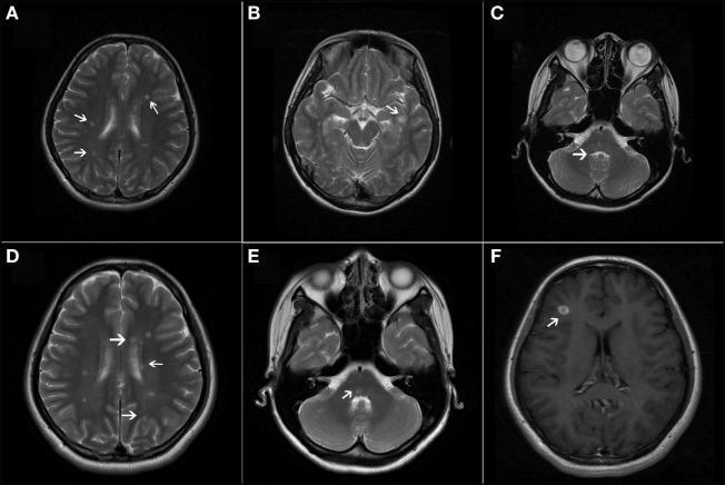
This composite figure displays multiple axial brain MRI sequences highlighting characteristic lesions associated with a demyelinating disease, such as Multiple Sclerosis (MS). Panels A through E are T2-weighted axial images showing multiple, ovoid-shaped, hyperintense lesions (white arrows) distributed in classic locations: the bilateral periventricular regions (A, D), the juxtacortical area of the left inferior temporal lobe (B), and the infratentorial compartment, specifically the pons and cerebellum (C, E). Panel F is a T1-weighted axial image with gadolinium contrast enhancement, demonstrating a single actively enhancing lesion in the right frontal lobe's juxtacortical region. The presence of both chronic T2-hyperintense lesions and an acute gadolinium-enhancing lesion illustrates the concept of dissemination in space and time, fundamental to the McDonald diagnostic criteria for MS. These visual findings support the identification of inflammatory demyelination within the central nervous system across different neuroanatomical regions.

This composite figure displays multiple axial brain MRI sequences highlighting characteristic lesions associated with a demyelinating disease, such as Multiple Sclerosis (MS). Panels A through E are T2-weighted axial images showing multiple, ovoid-shaped, hyperintense lesions (white arrows) distributed in classic locations: the bilateral periventricular regions (A, D), the juxtacortical area of the left inferior temporal lobe (B), and the infratentorial compartment, specifically the pons and cerebellum (C, E). Panel F is a T1-weighted axial image with gadolinium contrast enhancement, demonstrating a single actively enhancing lesion in the right frontal lobe's juxtacortical region. The presence of both chronic T2-hyperintense lesions and an acute gadolinium-enhancing lesion illustrates the concept of dissemination in space and time, fundamental to the McDonald diagnostic criteria for MS. These visual findings support the identification of inflammatory demyelination within the central nervous system across different neuroanatomical regions.

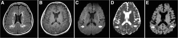
This composite of axial brain MRI scans illustrates the neuroimaging characteristics of Multiple Sclerosis (MS) lesions. Image A (T2-FLAIR) demonstrates multiple hyperintense demyelinating lesions within the bilateral periventricular white matter. Image B (Contrast-Enhanced T1WI) shows that these lesions lack enhancement, indicating no active blood-brain barrier disruption. Images C, D, and E utilize functional imaging to assess lesion chronicity. Two specific posterior periventricular lesions (marked with arrows) appear hypointense on Diffusion-Weighted Imaging (DWI, Image C), hyperintense on the Apparent Diffusion Coefficient (ADC) map (Image D), and hypointense on the exponentially weighted ADC (eADC) map (Image E). This imaging pattern—specifically the hyperintensity on ADC—represents 'facilitated diffusion,' which is characteristic of Type IVB chronic lesions where severe tissue destruction and increased extracellular space allow for greater water mobility. These findings are clinically relevant for differentiating chronic, inactive MS plaques from acute, inflammatory demyelination.

This composite of axial brain MRI scans illustrates the neuroimaging characteristics of Multiple Sclerosis (MS) lesions. Image A (T2-FLAIR) demonstrates multiple hyperintense demyelinating lesions within the bilateral periventricular white matter. Image B (Contrast-Enhanced T1WI) shows that these lesions lack enhancement, indicating no active blood-brain barrier disruption. Images C, D, and E utilize functional imaging to assess lesion chronicity. Two specific posterior periventricular lesions (marked with arrows) appear hypointense on Diffusion-Weighted Imaging (DWI, Image C), hyperintense on the Apparent Diffusion Coefficient (ADC) map (Image D), and hypointense on the exponentially weighted ADC (eADC) map (Image E). This imaging pattern—specifically the hyperintensity on ADC—represents 'facilitated diffusion,' which is characteristic of Type IVB chronic lesions where severe tissue destruction and increased extracellular space allow for greater water mobility. These findings are clinically relevant for differentiating chronic, inactive MS plaques from acute, inflammatory demyelination.

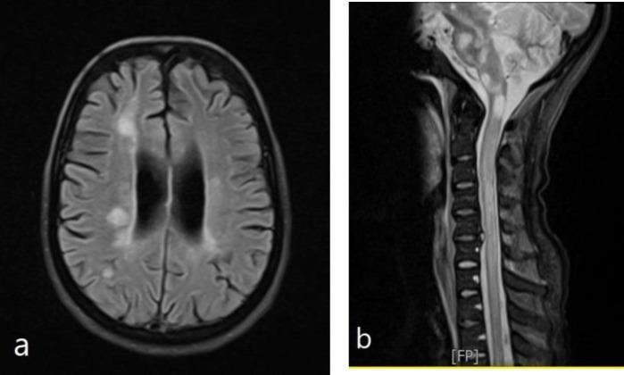
This diagnostic image composite presents MRI findings typical of a demyelinating disease, such as Multiple Sclerosis (MS). Panel A shows an axial FLAIR (Fluid-Attenuated Inversion Recovery) sequence of the brain at the level of the lateral ventricles. It demonstrates multiple hyperintense lesions in the periventricular and juxtacortical white matter. Several lesions are oriented perpendicular to the ventricles, characteristic of Dawson's fingers. Panel B displays a sagittal T2-weighted MRI of the cervical spine, revealing multiple intramedullary hyperintense lesions. These spinal cord lesions are seen at several levels, including the upper cervical (C3-C5) and lower cervical/upper thoracic junction (C7-T1). The lesions appear as distinct, hyperintense plaques within the spinal cord parenchyma. Together, these images illustrate dissemination in space, a key diagnostic criterion for central nervous system demyelination. The material is intended for intermediate to advanced medical learners focusing on neurology and neuroradiology.

This diagnostic image composite presents MRI findings typical of a demyelinating disease, such as Multiple Sclerosis (MS). Panel A shows an axial FLAIR (Fluid-Attenuated Inversion Recovery) sequence of the brain at the level of the lateral ventricles. It demonstrates multiple hyperintense lesions in the periventricular and juxtacortical white matter. Several lesions are oriented perpendicular to the ventricles, characteristic of Dawson's fingers. Panel B displays a sagittal T2-weighted MRI of the cervical spine, revealing multiple intramedullary hyperintense lesions. These spinal cord lesions are seen at several levels, including the upper cervical (C3-C5) and lower cervical/upper thoracic junction (C7-T1). The lesions appear as distinct, hyperintense plaques within the spinal cord parenchyma. Together, these images illustrate dissemination in space, a key diagnostic criterion for central nervous system demyelination. The material is intended for intermediate to advanced medical learners focusing on neurology and neuroradiology.

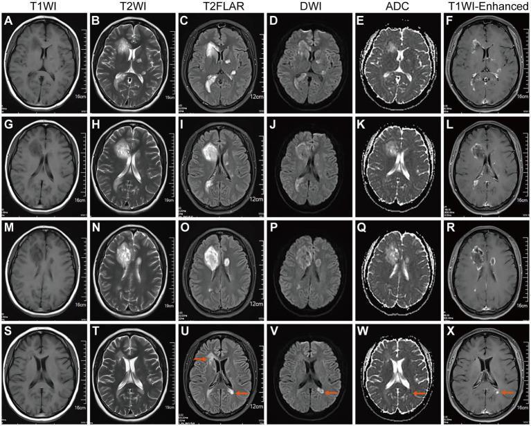
This diagnostic image panel displays axial brain MRI scans from a patient with Tumefactive Demyelinating Lesions (TDLs) in the context of Multiple Sclerosis, comparing findings from February 2023 (A-R) and May 2023 (S-X) following steroid therapy. The February scans show multiple large, hyperintense periventricular lesions on T2WI (B, H, N) and T2-FLAIR (C, I, O). Corresponding Diffusion-Weighted Imaging (DWI) (D, J, P) and Apparent Diffusion Coefficient (ADC) maps (E, K, Q) demonstrate restricted diffusion at lesion margins, characteristic of active demyelination. Contrast-enhanced T1WI (F, L, R) shows prominent ring-shaped and open-ring enhancement, indicating blood-brain barrier breakdown. By May 2023 (S-X), follow-up imaging reveals a significant reduction in the size and signal intensity of the initial periventricular lesions, some of which appear liquefied or chronic. However, a new active lesion is identified near the left lateral ventricle horn (marked by red arrows), demonstrating high signal on T2-FLAIR (U), restricted diffusion on DWI (V), and focal contrast enhancement on T1WI-C (X), signifying ongoing disease activity despite treatment.

This diagnostic image panel displays axial brain MRI scans from a patient with Tumefactive Demyelinating Lesions (TDLs) in the context of Multiple Sclerosis, comparing findings from February 2023 (A-R) and May 2023 (S-X) following steroid therapy. The February scans show multiple large, hyperintense periventricular lesions on T2WI (B, H, N) and T2-FLAIR (C, I, O). Corresponding Diffusion-Weighted Imaging (DWI) (D, J, P) and Apparent Diffusion Coefficient (ADC) maps (E, K, Q) demonstrate restricted diffusion at lesion margins, characteristic of active demyelination. Contrast-enhanced T1WI (F, L, R) shows prominent ring-shaped and open-ring enhancement, indicating blood-brain barrier breakdown. By May 2023 (S-X), follow-up imaging reveals a significant reduction in the size and signal intensity of the initial periventricular lesions, some of which appear liquefied or chronic. However, a new active lesion is identified near the left lateral ventricle horn (marked by red arrows), demonstrating high signal on T2-FLAIR (U), restricted diffusion on DWI (V), and focal contrast enhancement on T1WI-C (X), signifying ongoing disease activity despite treatment.

Reading File
Finding Sources
Finding Sources
Finding Sources
Finding Sources
Finding Sources
Reading File
Reading File
Reading File
I now have comprehensive content. Let me compile the full answer covering all aspects of the Unit 17 syllabus.

Multiple Sclerosis (MS) - Unit 17 Complete Notes

Source: Adams and Victor's Principles of Neurology, 12th Ed.; Bradley and Daroff's Neurology in Clinical Practice

1. Definition & Historical Background

Multiple Sclerosis (MS) is a chronic inflammatory demyelinating disease of the central nervous system (CNS), characterized by multifocal demyelinated plaques in the brain and spinal cord. It was originally called "disseminated sclerosis" by British physicians and "sclérose en plaques" by the French. J.M. Charcot at the Salpêtrière is credited with the first systematic clinicopathologic description.

2. Epidemiology

  • Most common cause of non-traumatic neurological disability in young adults
  • Peak onset: 20-40 years; can present from adolescence to late middle age
  • Female:Male ratio = 2:1 (females predominate)
  • Higher prevalence at greater distances from the equator (latitude gradient)
  • Leading to unemployment within 10 years of disease in 50-80% of patients
  • Prevalence of major depression: ~50% lifetime; bipolar disorder: 2x general population

3. Etiology

The exact cause remains unknown. MS is considered multifactorial, involving:

Genetic Factors

  • Concordance in identical twins: 25-30% (confirming a genetic, not purely genetic etiology)
  • Dizygotic twins: ~5% - similar to other siblings
  • HLA-DR2 (DRB1*1501) is the strongest genetic association
  • Other susceptibility loci: IL-7R, IL-2R alpha chain genes

Environmental Factors

  • Vitamin D deficiency - low sun exposure in northern latitudes
  • Epstein-Barr virus (EBV) infection - near-universal prior EBV infection in MS patients
  • Cigarette smoking increases risk 1.5x
  • Childhood obesity
  • Migration studies: people who migrate before age 15 acquire the risk of their new country; those who migrate after 15 retain original country risk - suggesting a critical exposure period in childhood/adolescence

Immunological Mechanism

  • T-cell mediated autoimmunity is central
  • Autoreactive CD4+ Th1 and Th17 cells breach the blood-brain barrier
  • These attack myelin basic protein (MBP), proteolipid protein (PLP), myelin oligodendrocyte glycoprotein (MOG)
  • Results in oligodendrocyte destruction → demyelination
  • B cells and antibodies also contribute (especially anti-MOG, anti-AQP4 variants)
  • Inflammation → demyelination → axonal injury (in progressive stages)

4. Pathology / Pathophysiology

The MS Plaque

  • Characteristic lesion = demyelinated plaque (sclerosis = scar)
  • Predilection sites: periventricular white matter, optic nerves, brainstem, spinal cord (lateral and posterior columns), cerebellum
  • Dawson's fingers - periventricular plaques oriented perpendicular to ventricles along medullary veins (classic on MRI)

Stages of a Plaque

StagePathology
Acute (active)Lymphocytic infiltration, macrophages stripping myelin, edema, blood-brain barrier (BBB) breakdown
SubacuteOligodendrocyte loss, partial remyelination (shadow plaques)
Chronic (inactive)Astrocytic gliosis, axonal loss, atrophy

Why Symptoms Occur

  • Demyelination → slowed/blocked conduction along axons
  • During attacks: inflammation and edema worsen conduction block
  • Recovery: remyelination (thin myelin) restores partial function
  • Uhthoff's phenomenon: worsening of symptoms with rise in body temperature (fever, exercise, hot bath) - because heat further impairs conduction in demyelinated axons
  • Progression: axonal transection and neurodegeneration cause permanent disability

5. Classification of MS

(Adams and Victor Table 35-1)

A. Relapsing-Remitting MS (RRMS) - Most common (~85% at onset)

  • Episodes ("relapses/attacks") of acute neurological dysfunction lasting days to weeks
  • Followed by partial or complete recovery ("remissions")
  • Between attacks, no disease progression

B. Secondary Progressive MS (SPMS)

  • After years of RRMS, the course transitions to gradual progressive worsening with or without occasional relapses
  • Approximately 50% of untreated RRMS converts to SPMS within 10 years; 90% within 25 years

C. Primary Progressive MS (PPMS) - ~10-15% of cases

  • Gradual neurological decline from onset, without distinct relapses or remissions
  • More common in males; older age of onset (~40s)
  • Typically manifests as progressive myelopathy (spastic paraparesis)

D. Progressive-Relapsing MS (PRMS)

  • Progressive from onset but WITH clear acute relapses superimposed

E. Acute MS Variants

  • Marburg disease: fulminant, rapidly progressive, fatal variant
  • Tumefactive MS: large (>2 cm) pseudotumoral lesions mimicking brain tumor
  • Balo's concentric sclerosis: alternating rings of demyelination and preserved myelin
  • Schilder disease (Diffuse cerebral sclerosis): large bilateral hemispheric lesions

6. Clinical Signs and Symptoms

MS can affect any part of the CNS, and the clinical picture is highly variable. Key features by system:

Motor

  • Spastic paraparesis/weakness (most common in spinal cord involvement)
  • Upper motor neuron signs: hyperreflexia, extensor plantar response (Babinski sign), spasticity
  • Fatigue (most common and disabling symptom overall)

Sensory

  • Paresthesias (tingling, numbness) - often an early symptom
  • Loss of vibration and joint position sense (dorsal column involvement)
  • Lhermitte's sign: electric shock sensation radiating down the spine/limbs on neck flexion - indicates cervical cord involvement (highly suggestive of MS but not pathognomonic)

Visual

  • Optic neuritis - most common first manifestation
    • Unilateral painful loss of vision
    • Central scotoma
    • Relative afferent pupillary defect (Marcus Gunn pupil)
    • Recovery is typical but recurrence is common
  • Internuclear ophthalmoplegia (INO): impaired adduction of one eye with nystagmus of the abducting eye (due to MLF lesion) - bilateral INO is virtually pathognomonic of MS in a young adult
  • Diplopia (CN VI or MLF lesion)
  • Nystagmus

Cerebellar/Brainstem

  • Charcot's triad: Intention tremor + Nystagmus + Scanning (staccato) speech - classic (though not commonly seen together)
  • Ataxia, dysmetria, dysarthria
  • Vertigo (due to brainstem lesions)

Bladder/Bowel/Sexual

  • Bladder dysfunction (very common): urgency, frequency, hesitancy, incontinence
  • Constipation
  • Erectile dysfunction in males

Cognitive and Psychiatric

  • Cognitive impairment in ~40-50% of patients (attention, memory, processing speed)
  • Depression (25-50%)
  • Emotional lability, pseudobulbar affect
  • Fatigue (present in >80%)

Paroxysmal Symptoms

  • Trigeminal neuralgia (facial pain)
  • Paroxysmal dysarthria and ataxia
  • Tonic spasms (painful, brief muscle contractions)
  • Paresthesias - episodic

7. Investigations

MRI Brain and Spine (most important diagnostic tool)

  • T2/FLAIR: hyperintense white matter lesions
    • Periventricular ("Dawson's fingers")
    • Juxtacortical
    • Infratentorial (brainstem, cerebellum)
    • Spinal cord
  • T1 gadolinium enhancement: active lesions (BBB breakdown) - indicates acute inflammation
  • T1 hypointense "black holes": chronic axonal loss
  • Fulfills McDonald 2017 criteria for Dissemination in Space (DIS) and Dissemination in Time (DIT)
MS MRI - Periventricular lesions and Dawson's fingers
MRI showing periventricular hyperintense lesions (Dawson's fingers) on FLAIR, and multiple spinal cord lesions on T2
MS MRI - Active and chronic lesions
T2 and gadolinium-enhanced T1 images showing dissemination in space and time - the basis of McDonald criteria

Cerebrospinal Fluid (CSF)

  • Oligoclonal IgG bands (OCBs) - present in >90% of MS patients (absent in serum = intrathecal synthesis)
  • Elevated IgG index
  • Mild pleocytosis (lymphocytes, typically <50 cells/mm³)
  • Mildly elevated protein
  • Normal glucose

Evoked Potentials

  • Visual Evoked Potentials (VEP): prolonged P100 latency - indicates demyelination of optic nerve even subclinically
  • Somatosensory evoked potentials (SSEP): delayed
  • Brainstem Auditory Evoked Potentials (BAEP)
  • Useful to detect subclinical lesions and confirm DIS

Blood Tests (to exclude differentials)

  • ANA, anti-dsDNA, APS antibodies (to exclude SLE/lupus sclerosis)
  • Anti-AQP4 (NMO-IgG) and anti-MOG antibodies - to exclude NMOSD
  • B12, folate, HIV, VDRL
  • Thyroid function

8. McDonald Diagnostic Criteria (2017)

Diagnosis requires demonstration of Dissemination in Space (DIS) + Dissemination in Time (DIT):
  • DIS: Lesions in ≥2 of 4 characteristic CNS regions (periventricular, juxtacortical, infratentorial, spinal cord)
  • DIT: New T2 lesion on follow-up MRI, OR simultaneous presence of enhancing and non-enhancing lesions, OR CSF-specific OCBs

9. Differential Diagnosis

ConditionKey Distinguishing Features
NMOSD (Devic's disease)Anti-AQP4+, longitudinally extensive spinal cord lesion >3 vertebral segments, area postrema syndrome
Acute disseminated encephalomyelitis (ADEM)Post-infectious/vaccination, monophasic, diffuse lesions, children, bilateral OPN
SLE/VasculitisANA+, systemic features, anti-dsDNA
NeurosarcoidosisACE elevated, hilar lymphadenopathy, granulomas
Vitamin B12 deficiency (SCD)Posterior column + pyramidal, low B12, macrocytosis
CNS lymphomaPeriventricular lesions but no OCBs, mass effect
Cervical spondylotic myelopathyProgressive myelopathy, spondylotic changes on spine MRI
Lyme diseaseExposure history, serology
HIV myelopathy/vacuolar myelopathyHIV+, CD4 low

10. Medical Management

A. Treatment of Acute Relapses

  • IV Methylprednisolone 1g/day for 3-5 days - shortens relapse duration but does not affect long-term outcome
  • Oral prednisolone is an alternative
  • Plasmapheresis for severe steroid-unresponsive attacks

B. Disease-Modifying Therapies (DMTs)

1st Line / Moderate Efficacy

DrugMechanismRoute
Interferon beta-1a (Avonex, Rebif)Anti-inflammatory, immunomodulatoryIM/SC
Interferon beta-1b (Betaseron)Reduces relapse rate ~30%SC
Glatiramer acetate (Copaxone)Mimics MBP, tolerizes T cellsSC
TeriflunomideInhibits pyrimidine synthesis, reduces T/B cell proliferationOral
Dimethyl fumarate (Tecfidera)Activates Nrf2 pathway, anti-inflammatoryOral

High Efficacy

DrugMechanismNote
Natalizumab (Tysabri)Anti-alpha-4 integrin, blocks lymphocyte trafficking across BBBRisk of PML (JC virus)
Alemtuzumab (Lemtrada)Anti-CD52, depletes T and B cellsAnnual infusion, immune reconstitution
Ocrelizumab (Ocrevus)Anti-CD20, depletes B cellsOnly approved for both RRMS and PPMS
CladribineDNA alkylator, depletes lymphocytesOral

C. Symptomatic Treatment

SymptomTreatment
SpasticityBaclofen (oral or intrathecal), Tizanidine, Diazepam
FatigueAmantadine, Modafinil, aerobic exercise
Bladder urgency/frequencyOxybutynin, Solifenacin, intermittent self-catheterization
Neuropathic painGabapentin, Pregabalin, Carbamazepine (for trigeminal neuralgia)
TremorClonazepam, Propranolol
DepressionSSRIs (Fluoxetine, Sertraline)
Pseudobulbar affectDextromethorphan + Quinidine (Nuedexta)
Erectile dysfunctionSildenafil
Paroxysmal symptomsCarbamazepine, Gabapentin

D. Rehabilitation (important for Physiotherapy students)

  • Physiotherapy: exercise programs, stretching for spasticity, balance/gait training, fall prevention
  • Occupational therapy: adaptive equipment, energy conservation techniques
  • Speech therapy: dysarthria, dysphagia management
  • Psychological support: CBT for depression, fatigue management programs
  • Multidisciplinary MS care team

11. Complications

ComplicationDetails
Progressive disabilityAccumulation of neurological deficits; 50% need walking aid within 15 years in untreated RRMS
Urinary tract infectionsFrom neurogenic bladder dysfunction and catheterization
Pressure sores/skin breakdownIn immobile patients
Deep vein thrombosis (DVT)Reduced mobility
Aspiration pneumoniaFrom dysphagia in advanced disease
Cognitive decline/dementiaSubcortical dementia in advanced disease
Falls and fracturesAtaxia, spasticity, weakness
Depression and suicideLifetime suicide risk elevated 7.5x vs general population
OsteoporosisReduced mobility + steroid use
ContracturesSpastic paralysis without adequate physiotherapy
Secondary progressive courseConversion from RRMS → SPMS with irreversible disability

Quick Summary Table

FeatureDetail
TypeInflammatory demyelinating, autoimmune
Peak age20-40 years
SexF > M (2:1)
PathologyMultifocal CNS demyelination plaques
Common onsetOptic neuritis, sensory symptoms, motor weakness
Hallmark signsINO, Lhermitte's, Uhthoff's, Charcot's triad
DiagnosisMcDonald criteria (MRI + CSF OCBs)
Best imagingMRI (T2/FLAIR + gadolinium)
Relapse treatmentIV Methylprednisolone
Disease modificationInterferons, Natalizumab, Ocrelizumab
Most common typeRelapsing-Remitting (85%)
Sources: Adams and Victor's Principles of Neurology 12th Ed., Bradley and Daroff's Neurology in Clinical Practice

Case study of this

Here is a detailed, realistic case study of Multiple Sclerosis covering all the Unit 17 topics (etiology, pathophysiology, classification, clinical signs & symptoms, investigations, differential diagnosis, medical management, and complications) — structured in the format used in clinical neurology teaching.

CASE STUDY: Multiple Sclerosis


PATIENT PROFILE

NameMrs. Aisha Mir (fictional)
Age28 years
SexFemale
OccupationSchool teacher
ResidenceSrinagar, Kashmir (high altitude, northern latitude)
Marital StatusMarried, 1 child
Referred byGeneral Physician → Neurology OPD

CHIEF COMPLAINTS

  1. Sudden painless blurring of vision in the right eye - 3 weeks ago (resolved spontaneously)
  2. Tingling and numbness in both legs - present for 2 weeks
  3. Weakness in both lower limbs - progressive over 10 days
  4. Electric shock sensation down the spine on bending the neck - 1 week
  5. Urinary urgency and occasional incontinence - 5 days

HISTORY OF PRESENT ILLNESS

Mrs. Aisha Mir, a 28-year-old school teacher from Srinagar, presented to the Neurology OPD with a 3-week history of sudden, painless blurring of vision in her right eye associated with mild pain on eye movement. She noticed a "washed out" appearance of colors. This episode resolved completely over 2 weeks without any treatment, and she dismissed it.
Two weeks prior to the current visit, she developed tingling and numbness starting in the feet and ascending to the thighs bilaterally. Over the next week, she noticed progressive weakness in both legs, making it difficult to climb stairs and walk long distances. She describes her legs as feeling "stiff and heavy."
One week ago, she noticed that on bending her neck forward, she experienced a sudden electric shock-like sensation radiating down her spine into both legs - lasting a second but frightening her enough to seek medical care.
She also reports urinary urgency with difficulty holding urine and two episodes of incontinence in the last 5 days.
She denies fever, headache, seizures, vomiting, or recent vaccinations/infections.

PAST HISTORY

  • 5 years ago: An episode of transient diplopia (double vision) lasting 3 weeks that resolved spontaneously - never investigated
  • No history of diabetes, hypertension, tuberculosis, epilepsy
  • No prior hospitalizations
Clinical Note: The history of transient diplopia 5 years ago represents a prior demyelinating episode - highly significant in retrospect.

FAMILY HISTORY

  • No first-degree relative with MS or autoimmune disease
  • Mother has Hashimoto's thyroiditis (autoimmune background in family)

PERSONAL HISTORY

  • Non-smoker, non-alcoholic
  • Diet: predominantly vegetarian, limited sun exposure (lives in high-latitude, frequently cloudy Kashmir)
  • No recent travel
  • Menstrual history: regular; symptoms worsened after a hot bath last week

DRUG HISTORY

  • No current medications
  • No history of drug allergy

GENERAL EXAMINATION

  • Conscious, cooperative, well-oriented to time, place, and person
  • Vitals: BP 118/76 mmHg, Pulse 78/min, Temp 37°C, SpO2 98%
  • No pallor, icterus, cyanosis, clubbing, lymphadenopathy, or edema
  • No skin rash, oral ulcers, or joint swelling

NEUROLOGICAL EXAMINATION

Higher Mental Functions

  • Mini-Mental State Examination (MMSE): 27/30 (mild inattention, slightly slow processing)
  • Mood: mildly anxious, appropriate affect

Cranial Nerve Examination

NerveFinding
CN INormal
CN II (Right eye)Visual acuity 6/18 (reduced); Central scotoma on Amsler grid; Pale optic disc on fundoscopy (resolving optic neuritis)
CN II (Left eye)Normal 6/6
CN III/IV/VIInternuclear ophthalmoplegia (INO) on left gaze: impaired adduction of right eye, nystagmus of left abducting eye (MLF lesion - right side)
CN VNormal sensation
CN VIINormal
CN VIIINo hearing loss; mild positional nystagmus
CN IX, XNormal gag reflex
CN XI, XIINormal
Key Finding: Unilateral optic neuritis + INO in a young woman = MS until proven otherwise

Motor Examination (Lower Limbs)

FindingRightLeft
ToneIncreased (spasticity)Increased (spasticity)
Power (hip flexion)3/53/5
Power (knee extension)4/54/5
Power (ankle dorsiflexion)4/54/5
Reflexes (knee jerk)++++ (brisk)++++ (brisk)
Reflexes (ankle jerk)+++ (brisk)+++ (brisk)
Plantar responseExtensor (Babinski +ve)Extensor (Babinski +ve)
ClonusPresent at anklePresent at ankle
Upper limbs: essentially normal power, mildly brisk reflexes bilaterally

Sensory Examination

ModalityFinding
Pain & TemperatureMildly reduced below T6 level bilaterally
Vibration senseSignificantly reduced in both feet and knees
Joint position senseImpaired at toes bilaterally
Light touchReduced below mid-thoracic level
Sensory level at approximately T6 = cervicothoracic cord involvement

Cerebellar Examination

  • Mild bilateral dysmetria on finger-nose test
  • Heel-shin test mildly impaired bilaterally
  • Tandem gait impossible (falls to sides)
  • No dysarthria at this stage

Special Signs

  • Lhermitte's sign: POSITIVE - electric shock sensation down the spine on neck flexion
  • Uhthoff's phenomenon: POSITIVE - symptoms worsened after hot bath (history)
  • Romberg's test: POSITIVE - sways markedly, near-fall

Bladder

  • Palpable bladder on lower abdominal examination
  • Post-void residual urine on bladder scan: 120 mL (neurogenic bladder)

PROVISIONAL DIAGNOSIS

Relapsing-Remitting Multiple Sclerosis (RRMS)
Based on:
  • Young female, episodic CNS attacks with recovery
  • Current attack: spinal cord + optic nerve + brainstem involvement
  • Previous attacks: diplopia (5 yrs ago) + optic neuritis (3 wks ago)
  • Dissemination in SPACE: optic nerve + brainstem (INO) + spinal cord
  • Dissemination in TIME: prior episode (5 yrs) + current attack

INVESTIGATIONS

1. MRI Brain with Gadolinium (MOST IMPORTANT)

Report:
  • Multiple T2/FLAIR hyperintense lesions in periventricular white matter, oriented perpendicular to ventricles (Dawson's fingers)
  • Juxtacortical lesions in right frontal and parietal regions
  • Infratentorial lesion in dorsal pons (explains INO)
  • Gadolinium-enhancing lesion in right pericallosal region (active, current attack)
  • Non-enhancing chronic lesions in left periventricular region (prior attack)
Conclusion: Lesions satisfy McDonald 2017 criteria for both DIS and DIT

2. MRI Cervical Spine

Report:
  • T2 hyperintense intramedullary lesion at C3-C5 level (right lateral column)
  • Lesion < 3 vertebral segments in length
  • No cord expansion or swelling

3. Cerebrospinal Fluid (CSF) Analysis

ParameterResultReference
AppearanceClear, colorlessClear
Opening pressure140 mmH₂ONormal
WBC12 cells/mm³ (lymphocytes)<5
Protein52 mg/dL15-45 mg/dL
Glucose58 mg/dL (blood glucose 100)Normal ratio
Oligoclonal bands (OCBs)POSITIVE (5 bands in CSF, absent in serum)Abnormal
IgG Index0.72 (elevated)<0.66
Myelin Basic Protein (MBP)ElevatedNormal
OCBs in CSF absent in serum = intrathecal IgG synthesis = highly supportive of MS

4. Visual Evoked Potentials (VEP)

  • Right eye: P100 latency = 128 ms (prolonged; normal <115 ms) - confirms subclinical right optic nerve demyelination
  • Left eye: P100 latency = 109 ms (borderline) - suggests subclinical prior involvement

5. Blood Investigations (to exclude differentials)

TestResultSignificance
CBCNormalExcludes anemia, infection
ESR18 mm/hrNormal (against SLE/vasculitis)
ANAWeakly positive 1:40Low titer, non-specific
Anti-dsDNANegativeExcludes lupus
Anti-AQP4 (NMO-IgG)NegativeExcludes NMOSD
Anti-MOG antibodyNegativeExcludes MOG-AD
Serum B12320 pg/mL (normal)Excludes B12 deficiency
Thyroid functionTSH mildly elevated (3.8 mIU/L)Autoimmune thyroid disease (incidental)
HIV ELISANegativeExcludes HIV myelopathy
VDRLNegativeExcludes neurosyphilis
ACE levelNormalExcludes neurosarcoidosis
Chest X-rayNormalExcludes sarcoidosis, TB

DIAGNOSIS CONFIRMED

Relapsing-Remitting Multiple Sclerosis (RRMS)

McDonald 2017 Criteria Fulfilled:
CriterionEvidence
DISPeriventricular + Juxtacortical + Infratentorial + Spinal cord lesions on MRI
DITSimultaneous enhancing + non-enhancing lesions on MRI + CSF OCBs

DIFFERENTIAL DIAGNOSES (Considered and Excluded)

DiagnosisArguments ForArguments Against / How Excluded
NMOSD (Devic's)Female, optic neuritis, myelopathyAnti-AQP4 negative; cord lesion < 3 segments; no area postrema syndrome
MOG antibody diseaseFemale, optic neuritis, relapsingAnti-MOG negative; no cortical encephalitis
SLE/Lupus sclerosisYoung female, ANA weakly positiveAnti-dsDNA negative; no systemic features; no renal/skin/joint involvement
NeurosarcoidosisCan mimic MS on MRIACE normal; no systemic sarcoid features; no granulomas
ADEMDemyelinationNo recent infection/vaccination; polyphasic course (prior attacks); OCBs present
Spinal cord tumorMyelopathy with sensory levelMRI shows no mass/cord expansion; no extramedullary compression
Vitamin B12 deficiency (SCD)Posterior column + pyramidal signsB12 normal; no macrocytosis; episodic relapsing course inconsistent
Cervical spondylotic myelopathyCervical cord lesionAge 28, female; MRI no spondylotic changes; episodic history inconsistent

MANAGEMENT

Phase 1: Acute Relapse Treatment

IV Methylprednisolone 1 g/day IV in 250 mL normal saline over 3-4 hours × 5 days
  • Shortens duration and severity of current relapse
  • Does NOT improve long-term disability
  • Followed by oral prednisolone taper (optional)
Monitoring during steroids: Blood glucose, BP, electrolytes, sleep disturbance, mood changes

Phase 2: Disease-Modifying Therapy (DMT)

Decision: Start moderate-efficacy DMT given first confirmed MS diagnosis at young age
Choice: Interferon beta-1a (Avonex) 30 mcg IM once weekly
  • Reduces relapse rate by ~30%
  • Slows MRI lesion accumulation
  • Counsel about: flu-like side effects (pre-treat with paracetamol), injection site reactions, liver function monitoring every 6 months, thyroid function (important here given autoimmune thyroid disease)
Alternative if intolerant: Glatiramer acetate or Dimethyl fumarate (oral)
Escalation plan: If ≥2 relapses/year or new MRI lesions on first-line therapy → escalate to Natalizumab (after JC antibody testing) or Ocrelizumab

Phase 3: Symptomatic Management

SymptomDrug / InterventionDose
SpasticityTab. BaclofenStart 5 mg TDS, titrate to 25 mg TDS
Urinary urgency/incontinenceTab. Oxybutynin5 mg BD; clean intermittent catheterization
Neuropathic tingling/painTab. Gabapentin300 mg TDS (titrated)
FatigueTab. Amantadine100 mg BD (morning + afternoon)
Depression (if develops)Tab. Sertraline50 mg OD
Vitamin D deficiencyCholecalciferol2000 IU/day (given northern latitude)

Phase 4: Rehabilitation (Physiotherapy - Key for BPT students)

Goals:
  • Improve functional mobility and independence
  • Reduce spasticity and prevent contractures
  • Improve balance and reduce fall risk
  • Manage fatigue
Physiotherapy Program:
ComponentDetails
StretchingDaily passive and active stretching of hamstrings, hip flexors, gastrocnemius to manage spasticity
StrengtheningGraduated resistance training for hip flexors, quadriceps (avoid overexertion/heat)
Gait trainingParallel bars → walking frame → cane progression
Balance trainingFrenkel's exercises, proprioceptive training on wobble board
Aquatic therapyHydrotherapy in cool water (avoid warm water - Uhthoff's phenomenon)
Fatigue managementEnergy conservation techniques, rest periods, pacing
Breathing exercisesPrevent chest infections in later stages
Bladder retrainingTimed voiding schedule
Occupational Therapy:
  • Adaptive devices (grab rails, raised toilet seat, non-slip mats)
  • Home modification advice
  • Return-to-work planning

COUNSELLING

The neurologist and team explained to Mrs. Aisha:
  1. MS is not curable, but is manageable - majority of RRMS patients remain ambulatory for decades
  2. DMT reduces relapse rate and slows disability progression - must be taken regularly
  3. Avoid heat - no hot baths, manage fever promptly (Uhthoff's phenomenon)
  4. Pregnancy - MS relapses decrease during pregnancy (especially 3rd trimester) but increase post-partum; interferon beta must be stopped before conception
  5. Importance of vitamin D supplementation and maintaining active lifestyle
  6. Regular follow-up: MRI every 6-12 months, neurologist review every 3-6 months
  7. MS Society support group referral
  8. Psychological support offered

FOLLOW-UP at 4 Weeks

  • Patient reports significant improvement in leg strength (power improved to 4+/5 bilaterally)
  • Urinary urgency reduced with oxybutynin
  • Tingling partially resolved
  • Lhermitte's sign still present but less intense
  • Started walking with minimal support
  • Commenced physiotherapy 5 days/week
Repeat MRI at 3 months:
  • Gadolinium-enhancing lesion resolved (inactive)
  • No new lesions (treatment response)

COMPLICATIONS NOTED / ANTICIPATED

ComplicationStatus in this Patient
Neurogenic bladder / UTI riskActive - managed with oxybutynin + hygiene
DepressionCounselled; watchful monitoring
FatigueActive - amantadine + physiotherapy
FallsRisk present - home safety review done
Cognitive declineMildly present - monitored
ContracturesPrevented by physiotherapy stretching program
Secondary progression (SPMS)Risk in future; DMT initiated to delay
Pregnancy planningDiscussed - DMT adjustment needed
Vitamin D deficiencyCorrected with supplementation

LEARNING POINTS FROM THIS CASE

  1. Optic neuritis in a young woman is MS until proven otherwise - 50% develop MS within 15 years
  2. Bilateral INO in a young adult = pathognomonic of MS (virtually no other cause)
  3. Lhermitte's sign + Uhthoff's phenomenon are hallmark clinical features
  4. Dissemination in space AND time is the diagnostic cornerstone (McDonald criteria)
  5. A prior undiagnosed episode (diplopia 5 years ago) is frequently identified retrospectively - thorough history is essential
  6. CSF oligoclonal bands support diagnosis and allow earlier treatment initiation
  7. Anti-AQP4 and anti-MOG must always be checked to exclude NMOSD/MOGAD before labeling MS
  8. Physiotherapy is a core pillar of MS management - not supplementary
  9. Avoid heat during physiotherapy sessions - use cool environments
  10. MS is a lifelong condition requiring multidisciplinary care: Neurologist + Physiotherapist + Occupational Therapist + Urologist + Psychologist + Ophthalmologist

This case study is designed for 4th Year BPT students, Dept. of Neurology, GMC Srinagar, as per Unit 17 syllabus (Dr. Sheikh Hilal Ahmad).
This is a shared conversation. Sign in to Orris to start your own chat.