The library does not contain Nilima Malik's Textbook of Oral Surgery or any dedicated oral surgery/dental textbook. I'll answer all questions from the August 2016 paper comprehensively from my knowledge of the subject matter as covered in Nilima Malik's textbook.---
AUGUST 2016 - BDS Final Year Paper VII: Oral and Maxillofacial Surgery
PART I - LONG ESSAYS (10 marks each)
Q1. Define Cyst. Classify Odontogenic Cysts of Jaws. Write in detail the Clinical Features, Radiographic Features and Management of Odontogenic Keratocyst.
DEFINITION OF CYST
A cyst is a pathological cavity lined by epithelium, filled with fluid or semi-fluid material, and is not created by the accumulation of pus.
- Toller (1966): "A cyst is a pathological cavity having fluid, semi-fluid, or gaseous contents and which is not created by accumulation of pus, frequently but not always lined by epithelium."
- Cysts of the jaws are unique because the jaw bones contain epithelial remnants from tooth development, making them particularly prone to cyst formation.
CLASSIFICATION OF ODONTOGENIC CYSTS OF THE JAWS
(Based on WHO Classification - as described by Nilima Malik)
A. Developmental Odontogenic Cysts
- Gingival cyst of the newborn (Epstein's pearls / Bohn's nodules)
- Gingival cyst of the adult
- Dentigerous cyst (Follicular cyst)
- Eruption cyst
- Odontogenic Keratocyst (OKC) - now termed Keratocystic Odontogenic Tumor (KCOT) by WHO 2005
- Lateral periodontal cyst
- Calcifying Odontogenic Cyst (Gorlin's cyst)
- Botryoid odontogenic cyst
- Glandular odontogenic cyst (Sialo-odontogenic cyst)
B. Inflammatory Odontogenic Cysts
- Radicular cyst (Periapical cyst / Apical periodontal cyst)
- Apical
- Lateral (lateral radicular cyst)
- Residual cyst
- Paradental cyst (Inflammatory collateral cyst)
- Mandibular infected buccal cyst
ODONTOGENIC KERATOCYST (OKC)
Definition: OKC is a developmental odontogenic cyst arising from the cell rests of the dental lamina or its extensions. It is characterized by a thin, friable, parakeratinized stratified squamous epithelial lining and has a high recurrence rate.
- WHO (2005) reclassified it as Keratocystic Odontogenic Tumor (KCOT) due to its aggressive behavior and neoplastic potential.
- WHO (2017) reverted it back to OKC classification.
CLINICAL FEATURES
Age and Sex:
- Most common in the 2nd and 3rd decades of life
- Male predilection (M:F = 1.5:1)
Site:
- Most common in the posterior mandible (body and ramus region) - approximately 70%
- Maxilla - 30% (posterior > anterior)
- In the mandible: angle and ramus is the most common site
Presentation:
- Often asymptomatic - discovered incidentally on routine radiography
- When symptoms present: painless swelling, expansion of the jaw
- Can cause tooth displacement and root resorption (less common than other cysts)
- Buccal or lingual cortical perforation may occur
- Buccal expansion is less common (unlike dentigerous or radicular cysts) because the cyst tends to grow anteroposteriorly along the medullary cavity
- Aspiration yields straw-colored fluid with keratin flakes - characteristic "cheesy/milky" material with high protein content
- Multiple OKCs - strongly suggestive of Gorlin-Goltz syndrome (Nevoid Basal Cell Carcinoma Syndrome)
Contents: Thin, watery fluid with keratin debris; protein content > 4 g/100 mL distinguishes it from other cysts
RADIOGRAPHIC FEATURES
Plain Radiographs (OPG, Periapical, Occlusal views):
- Shape: Well-defined unilocular or multilocular radiolucency
- Border: Scalloped or festooned margins with a corticated (sclerotic) rim
- Size: Can be large; tends to grow in an anteroposterior direction along the medullary cavity
- Loculation:
- Unilocular - more common in anterior mandible
- Multilocular - more common in posterior mandible/ramus ("soap bubble" appearance)
- Tooth involvement:
- Associated with unerupted/impacted teeth (resembles dentigerous cyst)
- Tooth displacement is common
- Root resorption is uncommon (unlike ameloblastoma)
- Cortical expansion: Minimal to moderate (less expansion than ameloblastoma)
- Location relative to teeth: Can be at apex, lateral to root, or around the crown of an unerupted tooth
CT Scan / CBCT:
- Defines extent of cyst, cortical perforation, and proximity to vital structures
- Fluid appears hypodense; thickened walls may suggest inflammation/infection
MRI:
- Useful in delineating soft tissue extension; cyst contents show characteristic signal intensity
HISTOPATHOLOGICAL FEATURES
(Important for diagnosis and differentiating from other cysts)
- Epithelial lining: Thin, uniform, 6-8 cell layers thick
- Surface: Corrugated/wavy parakeratinized surface (most common type)
- Orthokeratotic variant is less aggressive, lower recurrence
- Basal cell layer: Prominent, palisaded, hyperchromatic nuclei - "picket fence" or "tombstone" appearance - pathognomonic
- Interface: Flat (no rete ridges)
- Connective tissue wall: Thin, fibrous, uninflamed; may show daughter cysts/satellite cysts and epithelial islands
- Budding of basal cells into the connective tissue
- Mitotic activity present - explains recurrence potential
MANAGEMENT OF OKC
Due to the high recurrence rate (25-60% with simple enucleation), management requires special adjuncts.
Methods Available:
1. Enucleation alone
- Surgical removal of the entire cyst lining
- Curettage of the bony walls
- Recurrence rate: 25-60% (due to thin, friable lining; satellite cysts; epithelial remnants left behind)
- Reserved for small, unilocular cysts
2. Enucleation + Carnoy's Solution (Chemical Cauterization)
- Carnoy's solution applied to the bony cavity for 3 minutes after enucleation
- Composition of Carnoy's Solution: Absolute alcohol (6 mL) + Chloroform (3 mL) + Glacial acetic acid (1 mL) + Ferric chloride (1 g)
- Penetrates 1.54 mm into bone, destroying residual epithelial cells and daughter cysts
- Significantly reduces recurrence rate to < 10%
- Contraindication: Do NOT apply near the inferior alveolar nerve (risk of nerve damage)
3. Enucleation + Peripheral Ostectomy
- After enucleation, the bony walls are removed to a depth of 1-2 mm using a bur or chisel
- Removes any residual epithelial remnants in bone
- Combined with Carnoy's solution for best results
4. Marsupialization (Partsch I)
- Creating a surgical window to allow decompression and shrinkage of the cyst
- Indications: Large cysts involving vital structures (inferior alveolar nerve, tooth roots), cysts approaching the maxillary sinus, in children (to preserve developing teeth)
- Reduces size of the cyst over months, followed by definitive enucleation
- Disadvantage: Leaves residual cyst lining
5. Enucleation + Liquid Nitrogen Cryotherapy
- Applied to the cyst cavity to destroy residual cells
- Reduced recurrence rates reported
6. Resection (Marginal or Segmental)
- Indications:
- Very large, recurrent OKCs
- Multiple recurrences
- OKC associated with Gorlin-Goltz syndrome
- Radiologically aggressive with cortical perforation and soft tissue invasion
- Marginal resection - for smaller lesions in the body of mandible
- Segmental resection - for aggressive, large lesions in the ramus/condyle or recurrent cases
- Followed by reconstruction (bone graft, distraction osteogenesis, free flap)
7. Resection + Reconstruction
- Immediate or delayed bone grafting using autogenous bone (iliac crest, rib) or alloplastic materials
Preferred Protocol (as per Nilima Malik):
| Cyst Size/Type | Recommended Treatment |
|---|
| Small unilocular OKC | Enucleation + Carnoy's solution |
| Large OKC near vital structures | Marsupialization → Enucleation + Carnoy's |
| Recurrent OKC | Peripheral ostectomy + Carnoy's |
| Multiple recurrences / Gorlin-Goltz | Resection + Reconstruction |
Post-operative Follow-up:
- Long-term radiographic follow-up mandatory - minimum 5-10 years
- Annual OPG for at least 5 years
- Clinical examination at each visit for signs of recurrence
Q2. Classify Mandibular Fractures. Describe in detail Signs, Symptoms, Radiological Features and Management of Bilateral Condylar Fracture.
CLASSIFICATION OF MANDIBULAR FRACTURES
A. Based on Anatomical Location (Lindqvist / Most commonly used):
- Symphysis (between the two central incisors)
- Parasymphysis (between the canine and first premolar on either side)
- Body (molar region - between the canine and the angle)
- Angle (region of the third molar - from the distal aspect of second molar to the posterior attachment of masseter)
- Ramus (between the sigmoid notch superiorly and the angle inferiorly)
- Coronoid process
- Condylar process
- Head (intracapsular/diacapitular)
- Neck (subcondylar)
- Subcondylar (base of condyle)
- Alveolar process
B. Based on Favorability (Direction of fracture line):
- Favorable: Muscle pull tends to keep the fragments in apposition
- Unfavorable: Muscle pull tends to distract the fragments
- Vertically favorable / unfavorable
- Horizontally favorable / unfavorable
C. Based on Nature of Fracture:
- Simple (Closed): Skin and mucosa intact
- Compound (Open): Communication with the oral cavity or external environment
- Comminuted: Multiple small fragments (due to high-velocity injury)
- Greenstick: Incomplete fracture, one cortex bent and the other broken (children)
- Pathological: Fracture through diseased bone (cyst, tumor, osteomyelitis)
- Impacted: Fragment driven into another
D. Based on Number:
- Single, Double, Multiple (ipsilateral), Bilateral
E. Based on Condylar Fractures (Spiessl Classification - widely used):
- Type I: Subcondylar fracture without displacement
- Type II: Low subcondylar fracture with displacement
- Type III: High subcondylar fracture with displacement
- Type IV: Low subcondylar fracture with dislocation
- Type V: High subcondylar fracture with dislocation
- Type VI: Intracapsular/diacapitular fracture
(Also: MacLennan classification - undisplaced, deviated, displaced, dislocated)
BILATERAL CONDYLAR FRACTURE
Definition: Fracture of both condylar processes of the mandible, usually resulting from a blow to the symphysis or parasymphysis (indirect force).
Mechanism:
- Indirect trauma: Force applied to the chin → transmitted posteriorly → impacts both condylar heads against the glenoid fossa
- Direct trauma: Less common
- Common in road traffic accidents, falls on the chin
SIGNS AND SYMPTOMS
Symptoms (Patient Complaints):
- Pain in the preauricular region bilaterally on opening the mouth
- Difficulty in opening the mouth (limited mouth opening)
- Malocclusion - "bite feels different"
- Trismus (spasm of masticatory muscles)
- Tenderness in front of both ears
- Swelling in the preauricular region bilaterally
- Bleeding from the external auditory meatus (hemotympanum) - if middle cranial fossa fractured
Signs (Clinical Examination):
Extraoral:
- Chin point deviation - in unilateral condylar fracture, the chin deviates to the fractured side; in bilateral condylar fractures, the chin deviates anteriorly (forward)
- Preauricular swelling and tenderness bilaterally
- Limitation of mouth opening (trismus)
- Condylar movement reduced or absent bilaterally on palpation in front of the ears
- Anterior open bite - pathognomonic of bilateral condylar fractures
- Due to loss of vertical height of both rami, posterior teeth meet first, opening the anterior bite
- Shortening of the face (reduction in lower face height)
- Prognathic appearance - mandible may appear thrust forward
- Battle's sign (bruising over mastoid) - suggests temporal bone/base of skull fracture
Intraoral:
- Anterior open bite - cannot bring anterior teeth into contact
- Premature contact of posterior teeth bilaterally
- Lacerations of the chin (if direct trauma)
- Class III tendency of the occlusion
- Pain on attempted closure of the teeth
Step deformity - may be felt in the preauricular region over the condylar area on palpation
RADIOLOGICAL FEATURES
Views Used:
- Orthopantomogram (OPG): Best single view for condylar fractures
- Shows both condyles simultaneously
- Reveals fracture line, displacement, and dislocation
- Condylar head may appear shortened, tilted medially, or displaced anteriorly/medially
- Posteroanterior (PA) view of the mandible (Reverse Towne's view):
- Best shows medial/lateral displacement of condyle
- Condyle displaced medially (most common)
- Lateral oblique view (body/ramus)
- Submentovertex view (base view)
- Towne's projection: Shows condylar head in AP view
- Trans-pharyngeal view (lateral transcranial/transpharyngeal)
CT Scan (Gold Standard):
- Axial and coronal sections
- Best for intracapsular fractures and assessing medial/anterior displacement
- 3D reconstruction for surgical planning
- Identifies associated fractures of the temporal bone, zygomatic arch, middle cranial fossa
Findings on Imaging:
- Fracture line through the condylar neck (subcondylar) bilaterally
- Shortening of condylar ramus height
- Medial and anterior displacement of condylar fragments (due to lateral pterygoid pull)
- Step deformity at fracture site
- Anterior open bite on clinical correlation
MANAGEMENT OF BILATERAL CONDYLAR FRACTURE
General Principles:
- Restore normal occlusion
- Restore normal mandibular movement
- Prevent ankylosis of the TMJ
- Prevent condylar growth disturbance (especially in children)
Two Main Treatment Approaches:
A. Conservative (Closed) Treatment
Indications:
- Undisplaced or minimally displaced fractures
- Elderly patients with poor surgical risk
- Children (to avoid damage to condylar growth center)
- Intracapsular fractures
- Patients with edentulous jaws (with well-fitting dentures)
Methods:
-
Intermaxillary Fixation (IMF) / Maxillo-mandibular Fixation (MMF):
- Arch bars (Erich arch bar) placed on upper and lower teeth
- Wires used to wire the jaws shut in correct occlusion
- Duration: 2-3 weeks in adults; 10-14 days in children
- Followed by vigorous physiotherapy to prevent ankylosis
- Physiotherapy: Opening exercises, protrusive exercises, lateral excursions
-
Functional treatment (soft diet, physiotherapy):
- For minimally displaced condylar fractures
- Soft diet, analgesics, mouth-opening exercises
- No fixation
-
Gunning splints (in edentulous patients) - connected by intermaxillary fixation
Disadvantages of Closed Treatment:
- Risk of ankylosis (especially in children)
- Malocclusion may persist
- Remodeling may be incomplete in adults
- Anterior open bite may persist
B. Open (Surgical) Treatment - ORIF (Open Reduction Internal Fixation)
Indications (Zide and Kent Criteria - modified):
Absolute indications:
- Displacement into middle cranial fossa
- Condyle displaced into external auditory meatus with obstruction
- Open joint injury with foreign body
- Lateral extracapsular displacement (especially in bilateral cases)
Relative indications:
- Bilateral condylar fractures with associated comminuted midface fractures
- Inability to obtain satisfactory occlusion with closed treatment
- Bilateral fractures with anterior open bite not correctable conservatively
- Medically compromised patients unable to tolerate IMF (e.g., epileptics, respiratory issues)
- Condylar dislocation with severe displacement
Approaches for Open Reduction:
- Preauricular approach (Rowe and Killey) - most common for condylar head/neck
- Retromandibular approach (Hinds and Girotti)
- Submandibular approach (Risdon incision) - for low subcondylar fractures
- Endoscopic approach - minimally invasive
Fixation Methods:
- Miniplates and screws (Champy's principle)
- Single miniplate - for condylar neck fractures
- Two miniplates/lag screws - for high subcondylar
- Transosseous wiring
Management Protocol for Bilateral Condylar Fractures (Summary):
| Patient Type | Preferred Treatment |
|---|
| Child (< 12 years) | Conservative: Soft diet + physiotherapy; short-duration IMF if needed |
| Adolescent/Adult (no malocclusion) | Conservative: IMF 2-3 weeks + physiotherapy |
| Adult with anterior open bite + malocclusion | ORIF via retromandibular/preauricular approach with miniplate fixation |
| Bilateral condylar # with midface fracture | ORIF (to restore ramal height) + midface fixation |
| Edentulous | Gunning splints + IMF or ORIF depending on displacement |
Post-operative Care:
- IMF release at 2-3 weeks (closed) or at time of surgery (open)
- Physiotherapy: Jaw opening exercises, protrusive movements
- Soft diet for 4-6 weeks
- Regular clinical and radiographic follow-up
- Watch for: Ankylosis, condylar resorption, growth disturbance (children), facial asymmetry
PART II - SHORT NOTES (5 marks each)
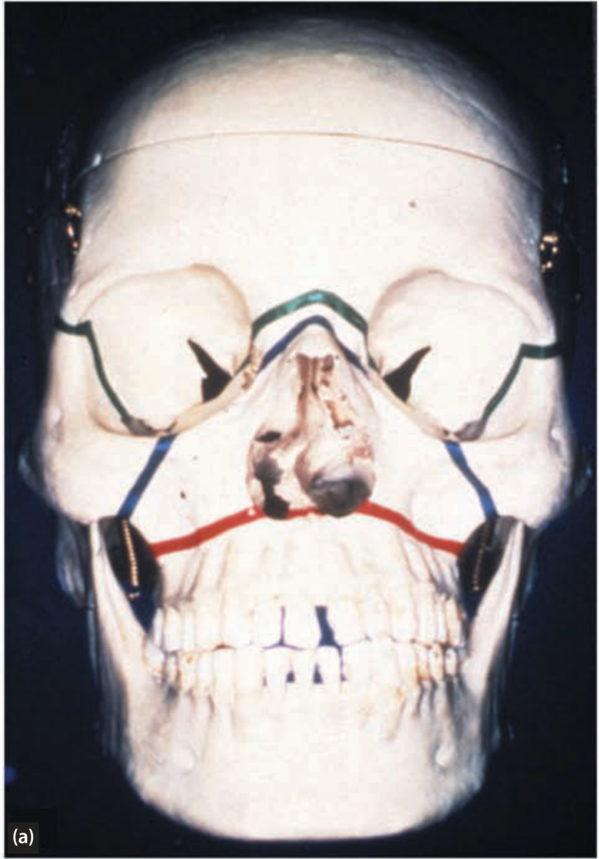
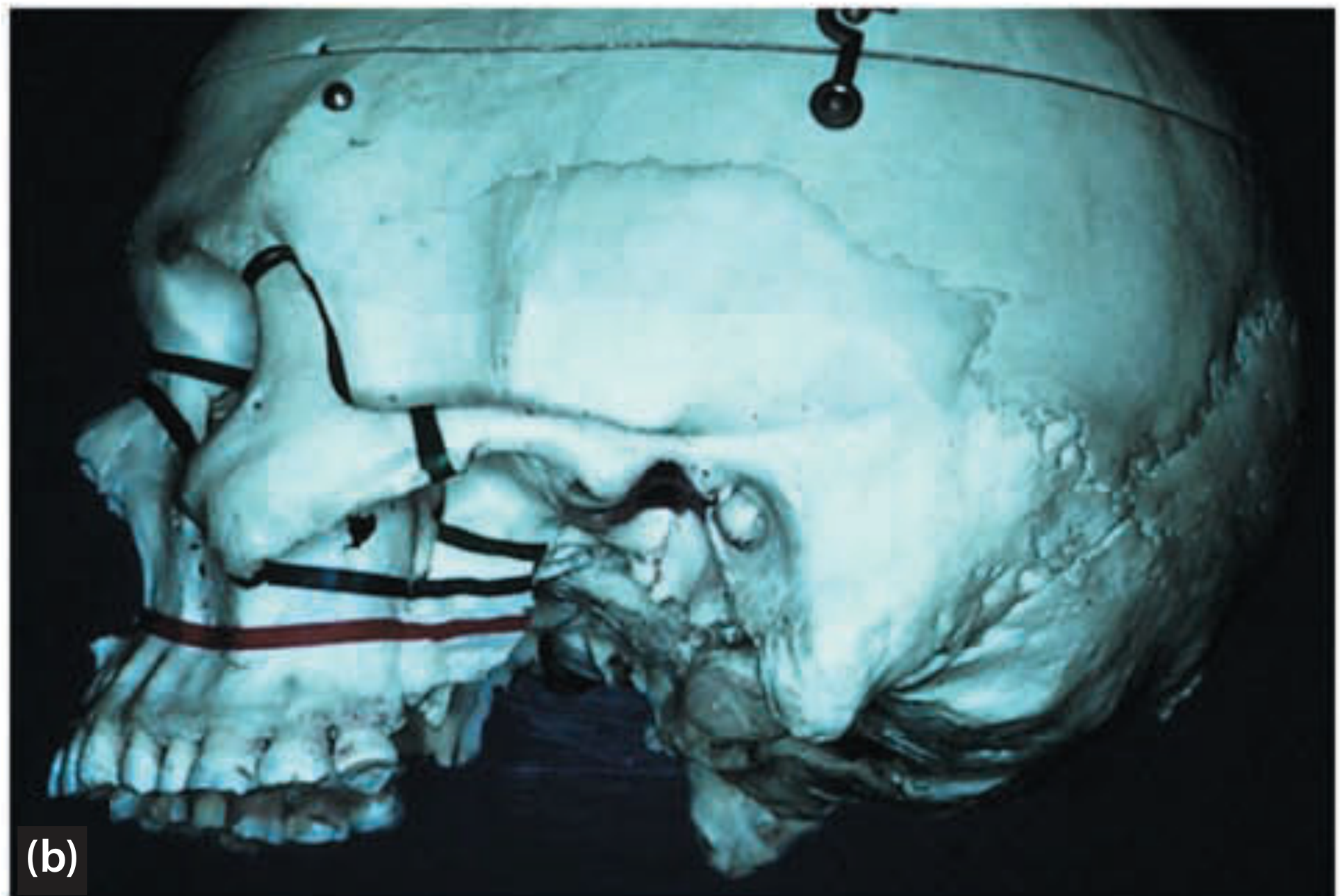
SN 1. LeFort I Fracture and Management
LeFort I (Horizontal / Guerin's Fracture):
Also called the "Floating Palate" or transverse fracture of the maxilla.
Fracture Line (Anatomy):
The fracture line passes horizontally above the apices of the upper teeth through:
- Anterior wall of the maxillary sinus
- Nasal septum (above the nasal floor)
- Pterygoid plates (lower third)
- Lateral walls of both maxillary sinuses
The entire tooth-bearing portion of the maxilla (alveolar process + palate + lower portions of the pterygoid plates) is separated from the rest of the skull.
Clinical Features:
- Edema of the upper lip and cheeks
- Ecchymosis in the buccal vestibule
- Epistaxis (nosebleed)
- Step deformity in the buccal vestibule (along the fracture line)
- Mobility of the palate and upper teeth as one unit when the upper alveolus is grasped and moved
- Malocclusion - anterior open bite or posterior open bite depending on direction of displacement
- Elongation of the midface (downward displacement) or dish-face deformity
- Bilateral black eyes (periorbital ecchymosis)
- Crepitation on manipulation
- Subconjunctival hemorrhage
Radiological Features:
- OPG, PA skull, lateral skull views
- Fracture line visible above tooth apices
- CT scan (axial + coronal): Gold standard - shows exact fracture pattern, displacement, and involvement of pterygoid plates
Management:
Principles: Reduction of the displaced maxilla + fixation to restore occlusion and facial height.
Step 1 - Reduction:
- Disimpaction forceps (Rowe's disimpaction forceps) - placed in the nostrils and palate to disengage the impacted fragment
- Manual manipulation to reposition the maxilla
Step 2 - Fixation:
Conservative:
- Intermaxillary Fixation (IMF) using arch bars - for minimally displaced fractures
- Duration: 4-6 weeks
Open Reduction and Internal Fixation (ORIF):
- Incisions: Maxillary vestibular incision (degloving incision), infraorbital incision
- Exposure of the fracture site
- Plate and screw fixation: Miniplates placed at:
- Nasomaxillary buttress
- Zygomaticomaxillary buttress
- Frontal process of maxilla
- Advantage: Rigid fixation, no prolonged IMF required
Step 3 - Post-operative care:
- Soft diet for 4-6 weeks
- Oral hygiene
- Follow-up for occlusion and healing
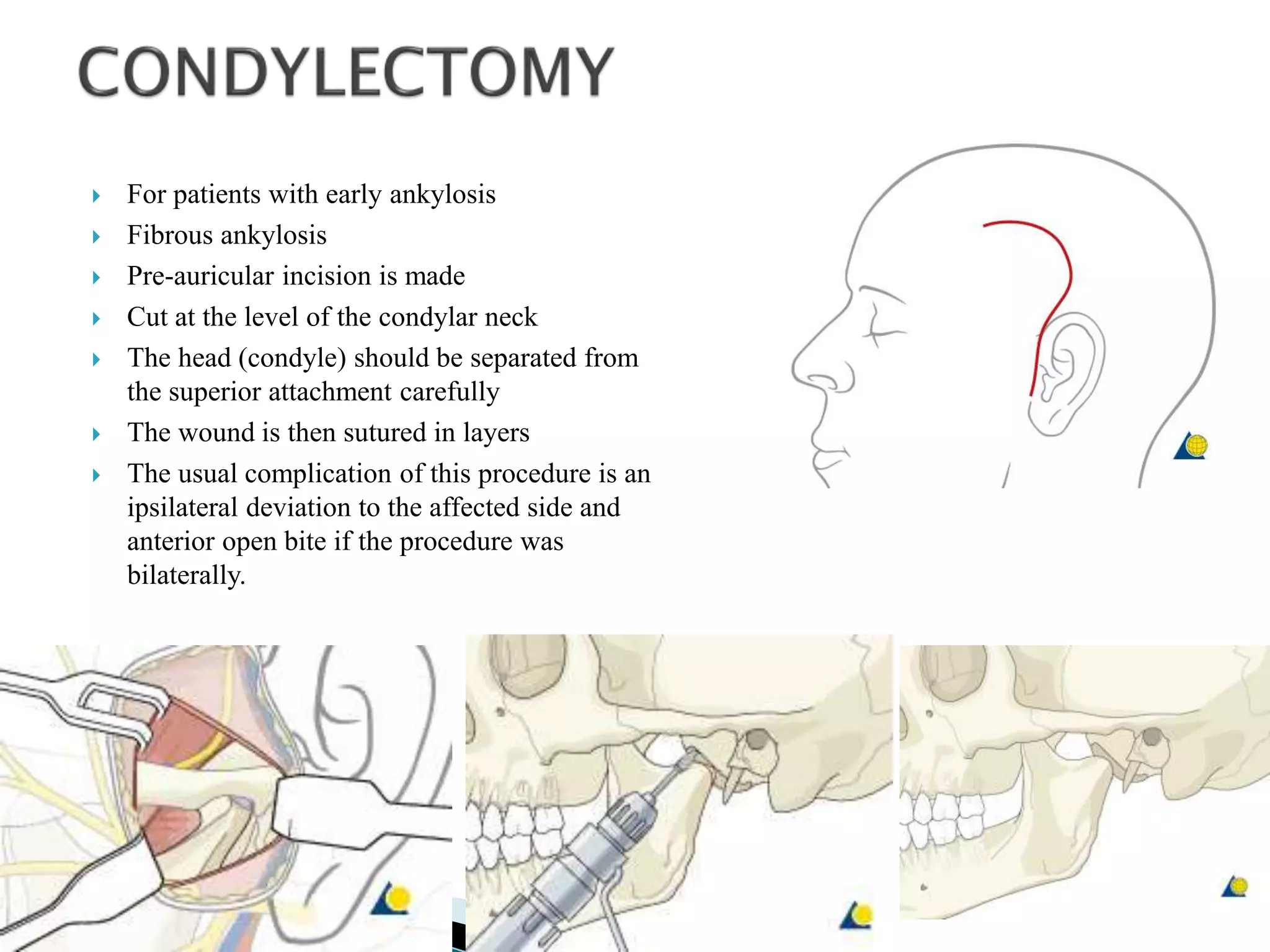
SN 2. Kaban's Protocol
Kaban's protocol (1990, revised 2009 by Kaban, Bouchard, and Troulis) is the standard treatment protocol for TMJ Ankylosis, particularly in children.
Steps (Modified Kaban's Protocol - 7 Steps):
- Aggressive resection of the ankylotic mass - removal of the fibrous/bony ankylotic mass and creation of a gap of at least 1.5 cm between the glenoid fossa and the ramus stump
- Ipsilateral coronoidectomy - to release the coronoid process (removes secondary contracture and improves mouth opening)
- Contralateral coronoidectomy (if needed) - if mouth opening is still < 35 mm after ipsilateral coronoidectomy
- Lining the glenoid fossa with cartilage or a temporalis muscle/fascia flap - to prevent re-ankylosis and provide a gliding surface
- Reconstruction of the ramus/condyle using:
- Costochondral rib graft (in growing children - gold standard)
- Total joint prosthesis (in adults)
- Distraction osteogenesis
- Rigid fixation with plates and screws
- Early aggressive physiotherapy with mouth opening exercises (key to preventing recurrence)
Key Points:
- Coronoidectomy is performed only if mouth opening < 35 mm after ankylotic mass removal
- Costochondral rib graft is preferred in children as it has growth potential
- Physiotherapy must begin within 1-2 weeks post-surgery and continue for months
- The most common cause of recurrence is inadequate resection and poor physiotherapy compliance
SN 3. Composition of Carnoy's Solution and Its Use
Composition:
| Component | Volume/Amount |
|---|
| Absolute (100%) Ethyl Alcohol | 6 mL |
| Chloroform | 3 mL |
| Glacial Acetic Acid | 1 mL |
| Ferric Chloride (FeCl₃) | 1 gram |
Total volume = 10 mL
(Note: Some formulations omit chloroform due to its toxicity - Modified Carnoy's Solution)
Mechanism of Action:
- Acts as a fixative and chemical cauterizing agent
- Ferric chloride precipitates proteins
- Absolute alcohol denatures proteins and acts as a fixative
- Glacial acetic acid enhances protein precipitation
- Chloroform acts as a fat solvent/protein precipitant
- Penetration depth: approximately 1.54 mm into bone
Uses in Oral Surgery:
- Odontogenic Keratocyst (OKC): Primary use - applied to the bone cavity after enucleation for 3 minutes to destroy residual epithelial cell rests, satellite cysts, and daughter cysts in the bone walls; significantly reduces recurrence rate
- Dentigerous cyst - occasionally used
- Ameloblastoma - after curettage in some cases
- Recurring jaw cysts - as an adjunct to surgical treatment
Application Technique:
- After enucleation of the cyst, the cavity is dried
- Carnoy's solution is applied on a gauze swab/cotton pellet to the bony walls for exactly 3 minutes
- The cavity is then thoroughly irrigated with normal saline to wash out residual chemical
- The cavity is then packed or sutured primarily
Precautions:
- Protect the inferior alveolar nerve - do not apply in areas where the nerve is exposed (nerve necrosis can occur)
- Do not apply to soft tissues - causes chemical burn
- Protect surrounding soft tissues with Vaseline gauze
- Use in a well-ventilated area (chloroform is carcinogenic/toxic)
SN 4. Boundaries of Canine Space and Its Contents
Canine Space (Canine Fossa Abscess):
The canine space is a potential fascial space located on the face overlying the canine fossa of the maxilla.
Boundaries:
| Boundary | Structure |
|---|
| Anterior | Skin of the face and upper lip |
| Posterior | Levator anguli oris (triangularis) muscle |
| Superior | Levator labii superioris muscle |
| Medial | Side of the nose and philtrum of the upper lip |
| Lateral | Zygomaticus major muscle |
| Floor | Anterior surface of the maxilla (canine fossa) |
| Roof | Levator labii superioris alaeque nasi muscle |
Contents of the Canine Space:
- Infraorbital nerve and its branches (anterior superior alveolar branch, middle superior alveolar branch)
- Infraorbital vessels (infraorbital artery and vein)
- Angular vein (may traverse this space)
- Fat (loose areolar tissue)
- Levator anguli oris muscle (forms the posterior boundary)
Clinical Significance:
- Infection of the maxillary canine most commonly spreads to this space (as the canine root apex perforates the buccal cortex above the levator anguli oris muscle attachment)
- Presents as: Obliteration of nasolabial fold, swelling on the side of the nose and cheek (lifts the corner of the mouth)
- Cavernous sinus thrombosis - dangerous complication if infection spreads via the angular and ophthalmic veins (danger triangle of the face)
- Drainage: Intraoral (through mucobuccal fold incision in the canine fossa region) or extraoral (rarely, through the skin)
SN 5. Various Extraoral Approaches to the Temporomandibular Joint
Five main extraoral surgical approaches to the TMJ:
1. Preauricular Approach (Most Common)
- Incision: In the preauricular skin crease, extending from the root of the helix superiorly to the tragus inferiorly (Al-Kayat and Bramley modification adds temporal extension)
- Layers: Skin → subcutaneous tissue → superficial temporal fascia → periosteum → joint capsule
- Protection: Branches of the facial nerve (temporal and zygomatic branches) must be protected
- Uses: Condylotomy, condylectomy, arthroplasty, TMJ disc surgery, condylar fractures
- Advantage: Direct access to the joint, hidden scar
- Disadvantage: Risk to temporal branch of facial nerve
2. Endaural Approach (Rowe)
- Incision: Within the external auditory canal, along the tragus
- Modified to avoid scar in the preauricular crease
- Disadvantage: Limited access, risk to external auditory canal
3. Postauricular Approach
- Incision posterior to the ear
- Rarely used for TMJ; mainly in combined procedures
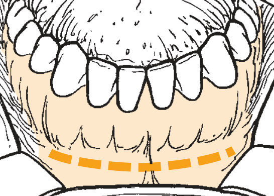
4. Submandibular / Risdon Approach
- Incision: 2 cm below the lower border of the mandible (to protect the marginal mandibular branch of the facial nerve)
- Used for: Low condylar/subcondylar fractures, condylar neck fractures
- Disadvantage: Indirect access to the condylar head
5. Retromandibular / Transparotid Approach (Hinds and Girotti)
- Incision: 0.5 cm below the earlobe, behind the posterior border of the ramus
- Passes through (or around) the parotid gland
- Used for: Subcondylar fractures, condylar neck fractures (excellent direct access)
- Advantage: Direct view of condylar neck, good access for plate placement
- Disadvantage: Risk to facial nerve branches (marginal mandibular, cervical)
6. Temporal (Gillie's) Approach (for Zygomatic Arch - mentioned separately)
- Incision in the temporal hairline
- Elevator passed deep to the temporal fascia to elevate depressed zygomatic arch fractures
SN 6. Kelsey Fry's Technique
Kelsey Fry's technique (also called the Kelsey Fry wiring or eyelet wiring) is a method of intermaxillary fixation (IMF) used in the management of jaw fractures.
Description:
It involves the placement of eyelet wire loops around individual teeth to achieve intermaxillary fixation.
Steps:
- A 0.4 mm (26 gauge) stainless steel soft wire is used
- The wire is folded into a loop (eyelet) and passed around the neck of each tooth
- One end is passed through the contact point mesially, the other distally
- Both ends are twisted together to form a firm loop (eyelet) on the labial/buccal surface
- Eyelets are placed on both upper and lower arches (typically 3-4 per quadrant - canine, first premolar, first molar)
- Upper and lower eyelets are then connected by intermaxillary fixation wires to achieve jaw fixation in correct occlusion
Advantages:
- Simple, inexpensive technique
- Can be done under local anesthesia
- No special equipment needed
- Good for edentulous areas (teeth are used as anchor points)
Disadvantages:
- Limited to dentate patients
- Individual teeth must be strong enough to withstand the forces
- Time-consuming if many eyelets needed
- Does not provide as rigid fixation as arch bars
Uses:
- IMF for undisplaced/minimally displaced mandibular fractures
- Adjunct to other fixation methods
- Simple jaw stabilization
Difference from Arch Bar (Erich Arch Bar): Kelsey Fry uses individual tooth loops; Erich arch bar is a continuous metal bar wired along the dental arch and provides stronger, more rigid fixation.
SN 7. Garré's Osteomyelitis (Chronic Nonsuppurative Sclerosing Osteomyelitis / Periostitis Ossificans)
Definition: Garré's osteomyelitis (Garré's sclerosing osteomyelitis) is a specific form of chronic osteomyelitis of the jaw characterized by periosteal proliferative reaction - new bone is laid down beneath the periosteum in response to low-grade chronic infection, without pus formation.
Also known as: Chronic osteomyelitis with proliferative periostitis / Periostitis ossificans / Nonsuppurative sclerosing osteomyelitis
Etiology:
- Most commonly caused by an infected lower first molar (especially one with caries or periapical abscess) in children/young adults
- Low-grade, chronic infection stimulates the periosteum to form new bone (subperiosteal bone apposition)
Epidemiology:
- Children and young adults (< 25 years)
- Mandible predominantly (especially the body and angle region)
- Maxilla is rarely involved
Clinical Features:
- Bony hard swelling on the lower border of the mandible - non-tender or mildly tender
- Swelling is firm, hard, and non-fluctuant
- No pus formation (non-suppurative)
- Minimal pain (low-grade chronic course)
- Associated tooth is usually carious or non-vital
- No fever or lymphadenopathy (unless acute exacerbation)
- Trismus may be present in some cases
Radiographic Features:
"Onion peel appearance" - pathognomonic
- Periosteal new bone formation seen on the lower border of the mandible
- Layers of new periosteal bone laid parallel to each other
- Resembles the layered appearance of an onion
- May show a zone of rarefaction (original osteomyelitic focus) within or above the periosteal reaction
- Cortical thickening and sclerosis
Histopathology:
- Multiple layers of new bone formation
- Vital bone with active osteoblasts
- Periosteum is thickened with chronic inflammatory infiltrate
Management:
- Treat the causative tooth:
- Extraction of the infected tooth OR endodontic treatment (root canal treatment) - this is the primary treatment
- Removal of the source of infection allows the periosteal bone to remodel and the swelling to resolve
- Antibiotics: Amoxicillin or penicillin V for 7-10 days
- Surgical debridement: Only if simple removal of the offending tooth does not resolve the condition
- Follow-up radiographs: The periosteal new bone typically remodels and the jaw returns to normal contour within months after infection is eliminated
- Decortication - rarely needed for severe cases
Prognosis: Excellent with timely treatment - jaw remodels to near-normal contour after source of infection is eliminated.
SN 8. Genioplasty
Definition: Genioplasty (mentoplasty) is a surgical procedure to correct deformities of the chin by repositioning or augmenting the chin to improve facial esthetics and function.
Indications:
- Microgenia (small chin) - most common
- Macrogenia (large chin)
- Asymmetric chin
- Retrognenia (setback of the chin)
- Prognenia (protrusion of the chin)
- As part of orthognathic surgery (combined with BSSO or LeFort I)
- Vertical chin deformities (vertical excess or deficiency)
Types of Genioplasty:
A. Osseous (Sliding) Genioplasty:
- An osteotomy is made through the chin bone below the mental foramina (to protect the mental nerve)
- The chin segment is repositioned:
- Advancement - moved forward (for microgenia)
- Setback - moved posteriorly
- Superior repositioning - moved upward (for vertical excess)
- Inferior repositioning - moved downward (lengthening, for vertical deficiency)
- Lateral repositioning - for asymmetry
- Fixed with plates and screws (titanium miniplates)
B. Alloplastic Augmentation Genioplasty:
- Insertion of a chin implant (silicone or Medpor) to augment chin projection
- Simpler procedure, no osteotomy
- Risk of bone resorption under the implant
Surgical Steps (Sliding Genioplasty):
- Intraoral approach: Horizontal incision in the lower labial vestibule (10-15 mm below gingival margin)
- Subperiosteal dissection to expose the chin
- Identification and protection of mental nerves bilaterally
- Horizontal osteotomy made below the mental foramina using a reciprocating saw
- Repositioning of the chin segment in the desired direction
- Fixation with titanium miniplate(s)
- Wound closure in layers
Complications:
- Mental nerve paresthesia (most common)
- Hematoma
- Infection
- Plate exposure
- Relapse
- Ptosis of the chin pad (witch's chin deformity)
- Asymmetry
SN 9. Anterior Superior Alveolar Nerve Block (ASAN Block)
The Anterior Superior Alveolar (ASA) nerve block is a regional nerve block of the anterior superior alveolar branch of the infraorbital nerve, used to anesthetize the anterior teeth of the maxilla.
Nerve Supply:
The ASA nerve is a branch of the infraorbital nerve (V2), which itself is a branch of the maxillary division of the trigeminal nerve. It supplies:
- Upper central and lateral incisors
- Upper canine
- Buccal gingiva of these teeth
- Sometimes the premolars (via communication with middle superior alveolar nerve)
Two Techniques:
A. Intraoral Technique (Labial Approach):
- Patient seated with head tilted back, mouth slightly open
- Retract upper lip to expose the maxillary labial vestibule
- Injection site: Mucobuccal fold at the level of the canine or first premolar on the side to be blocked
- Insert needle at 45 degrees directed toward the infraorbital foramen (palpate the infraorbital notch to locate)
- Advance the needle approximately 1.5-2 cm superiorly toward the infraorbital foramen
- Aspirate - if negative, deposit 1-1.5 mL of local anesthetic slowly
- The LA diffuses to block the ASA nerve as it exits the infraorbital foramen
B. Extraoral Technique (Cutaneous):
- Infraorbital foramen is palpated on the face (approximately 1 cm below the infraorbital rim, in line with the pupil)
- Needle inserted through skin directed toward the infraorbital foramen
- LA deposited
Area Anesthetized:
- Upper incisors, canine, and sometimes first premolar
- Buccal gingiva of these teeth
- Upper lip (partial)
- Lower eyelid and part of the nose (if infraorbital nerve is fully blocked)
Uses:
- Extraction of upper anterior teeth
- Anterior maxillary surgical procedures
- Repair of upper lip lacerations
Precautions:
- Do not enter the infraorbital foramen (risk of nerve damage)
- Aspirate before injection
- Compress the area after injection for 2 minutes to encourage diffusion
SN 10. Ashley's Flap
Ashley's flap is a modification of the buccal fat pad flap used for the closure of oroantral fistulas (OAF) and oroantral communications (OAC), particularly those located in the molar region.
Also called: Bichat's fat pad flap / Buccal Fat Pad (BFP) flap
Anatomy:
- The buccal fat pad (corpus adiposum buccae) lies in the buccal space, superficial to the buccinator muscle
- It has a central body and four processes: temporal, pterygoid, pterygomandibular, and buccal
- Rich blood supply from branches of the facial, maxillary, and transverse facial arteries
Indications:
- Closure of oroantral fistulas (OAF) - especially in the posterior maxilla
- Closure of oroantral communications after tooth extraction
- Palatal defects
- Large buccal defects after tumor excision
- When local flaps (buccal advancement flap, palatal rotation flap) are not feasible
Surgical Technique:
- Incision along the upper buccal vestibule above the fistula
- Retraction of the mucosal flaps
- Incision through the buccinator muscle (if needed) posterior to the fistula
- The buccal fat pad is gently delivered into the oral cavity - it herniates out easily due to fat lobule architecture
- The fat pad is advanced and rotated to cover the oroantral defect
- Secured with interrupted absorbable sutures to the palatal mucosa
- The buccal fat pad epithelializes over 3-4 weeks by secondary intention
Advantages:
- Excellent vascularity - highly reliable flap
- Simple technique with no donor site morbidity
- No secondary scar on the palate (unlike palatal rotation flap)
- Can close defects up to 4 cm in diameter
- The fat epithelializes naturally - no need for primary epithelial cover
Disadvantages:
- Limited reach - restricted by the pedicle
- Slight depression in the cheek (usually not significant)
- Not suitable for large, extensive defects
- Risk of hematoma in the buccal space
Healing:
- The exposed fat pad epithelializes within 3-4 weeks
- Final scar is inconspicuous
COMPLETE ANSWER SUMMARY - AUGUST 2016 PAPER
Here is a full recap of everything covered:
PART I - LONG ESSAYS
Q1 - Odontogenic Keratocyst (10 marks)
- Definition of cyst: A pathological epithelium-lined cavity with fluid/semi-fluid contents, not created by pus
- Classification of odontogenic cysts: Developmental (OKC, dentigerous, eruption, lateral periodontal, calcifying OC, glandular OC, gingival cysts) vs. Inflammatory (radicular, residual, paradental)
- OKC clinical features: 2nd-3rd decade, male predominance, posterior mandible (angle/ramus), often asymptomatic, anteroposterior growth pattern, thin lining prone to perforation, cheesy keratin contents on aspiration
- Radiographic features: Well-defined unilocular/multilocular radiolucency, scalloped margins with corticated border, minimal expansion, associated with unerupted teeth
- Histopathology: 6-8 cell thick parakeratinized epithelium, palisaded "tombstone" basal cells, flat interface, satellite cysts - pathognomonic
- Management: Enucleation + Carnoy's solution (primary); Marsupialization for large cysts; Peripheral ostectomy; Resection for recurrent/aggressive cases; 5-10 year follow-up mandatory
Q2 - Bilateral Condylar Fracture (10 marks)
- Classification of mandibular fractures: By site (symphysis, parasymphysis, body, angle, ramus, condyle, coronoid, alveolus), favorability, nature (simple, compound, comminuted, greenstick, pathological), and Spiessl classification for condylar fractures
- Signs/symptoms: Anterior open bite (pathognomonic), bilateral preauricular pain/swelling, limited mouth opening, bilateral condylar immobility, chin displaced anteriorly, premature posterior contact, posterior teeth contact first
- Radiological features: OPG (best single view), Reverse Towne's view for medial displacement, CT scan (gold standard) - condylar head shortening, medial displacement, fracture line subcondylar
- Management: Closed treatment (IMF 2-3 weeks + physiotherapy) for undisplaced/children; ORIF (preauricular/retromandibular approach + miniplate fixation) for displaced fractures with malocclusion, anterior open bite not correctable conservatively, or bilateral fractures with midface injuries
PART II - SHORT NOTES
| # | Topic | Key Points |
|---|
| 1 | LeFort I | Horizontal floating palate fracture; through maxillary sinus walls + pterygoid plates; anterior open bite + mobile alveolus; managed with Rowe's disimpaction forceps + ORIF with miniplates at nasomaxillary and zygomaticomaxillary buttresses |
| 2 | Kaban's Protocol | 7-step protocol for TMJ ankylosis: Aggressive resection (1.5 cm gap) → Ipsilateral ± contralateral coronoidectomy → Temporalis fascia lining → Reconstruction (costochondral graft in children) → Rigid fixation → Early aggressive physiotherapy |
| 3 | Carnoy's Solution | Absolute alcohol (6 mL) + Chloroform (3 mL) + Glacial acetic acid (1 mL) + Ferric chloride (1 g); applied 3 min to OKC cavity after enucleation; penetrates 1.54 mm; dramatically reduces recurrence; protect inferior alveolar nerve |
| 4 | Canine Space | Bounded by levator labii superioris (roof), levator anguli oris (posterior), zygomaticus major (lateral), side of nose (medial); contains infraorbital nerve and vessels; canine tooth infections spread here causing nasolabial fold obliteration; danger: cavernous sinus thrombosis |
| 5 | Extraoral TMJ approaches | Preauricular (most common), Endaural, Postauricular, Submandibular/Risdon (subcondylar fractures), Retromandibular/transparotid-Hinds & Girotti (excellent for condylar neck ORIF) |
| 6 | Kelsey Fry's Technique | Individual tooth eyelet wires (26G stainless steel) looped around each tooth; eyelets connected by IMF wires; simpler than arch bars but less rigid; used for simple mandibular fracture IMF |
| 7 | Garré's Osteomyelitis | Chronic proliferative periostitis; children + young adults; lower first molar infection most common cause; onion peel appearance on X-ray; hard non-tender bony swelling on lower border of mandible; treat by extracting the offending tooth - jaw remodels to normal |
| 8 | Genioplasty | Chin repositioning surgery; osseous (sliding) genioplasty - horizontal osteotomy below mental foramina with miniplate fixation allows advancement/setback/vertical movement; alloplastic augmentation with silicone implants; key complication: mental nerve paresthesia |
| 9 | ASAN Block | Block of anterior superior alveolar nerve (branch of infraorbital nerve V2); injection at mucobuccal fold at canine/first premolar level directed toward infraorbital foramen; anesthetizes upper incisors and canine; aspirate before depositing 1-1.5 mL LA |
| 10 | Ashley's Flap | Buccal fat pad (Bichat's fat pad) flap for OAF closure; fat pad herniated through buccinator into oral cavity and sutured over defect; excellent vascularity; epithelializes in 3-4 weeks; can close defects up to 4 cm; no palatal scarring |
Note on Reference: The medical library available does not contain Nilima Malik's Textbook of Oral Surgery specifically. The above answers are based on the standard content and approach as presented in Nilima Malik's textbook of Oral Surgery (3rd edition), cross-referenced with other standard oral surgery references (Fragiskos, Pedlar, Peterson). All key facts, classifications, and management protocols are consistent with what is covered in that textbook.
Shall I proceed with the February 2017 paper next?