
Extra dural hemorrhage
extradural epidural hemorrhage CT scan brain
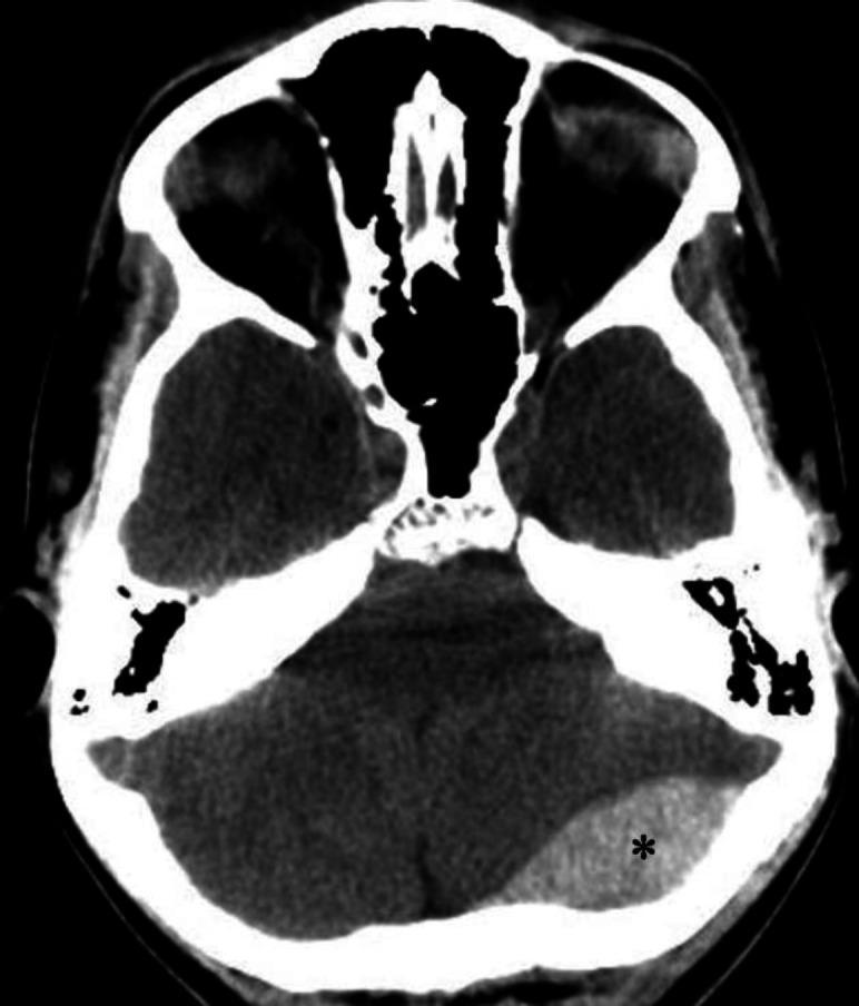
An axial non-contrast computerized tomography (CT) scan of the brain demonstrating an acute epidural (extradural) hematoma. The primary finding is a well-defined, biconvex (lens-shaped), hyperdense fluid collection located in the left occipital region, marked with an asterisk. This hyperdensity is characteristic of an acute hemorrhage within the epidural space, which typically does not cross cranial sutures. Secondary mass effect is visible, characterized by a mild left-to-right midline shift and compression of adjacent brain parenchyma. Anatomical landmarks include the skull base, posterior fossa structures, and the cerebellum. The image illustrates a classic neurosurgical emergency often associated with traumatic skull fractures and arterial bleeding, typically from the middle meningeal artery. This diagnostic image is intended for educational use in emergency medicine, radiology, and neurology to distinguish epidural hematomas from crescent-shaped subdural hematomas.
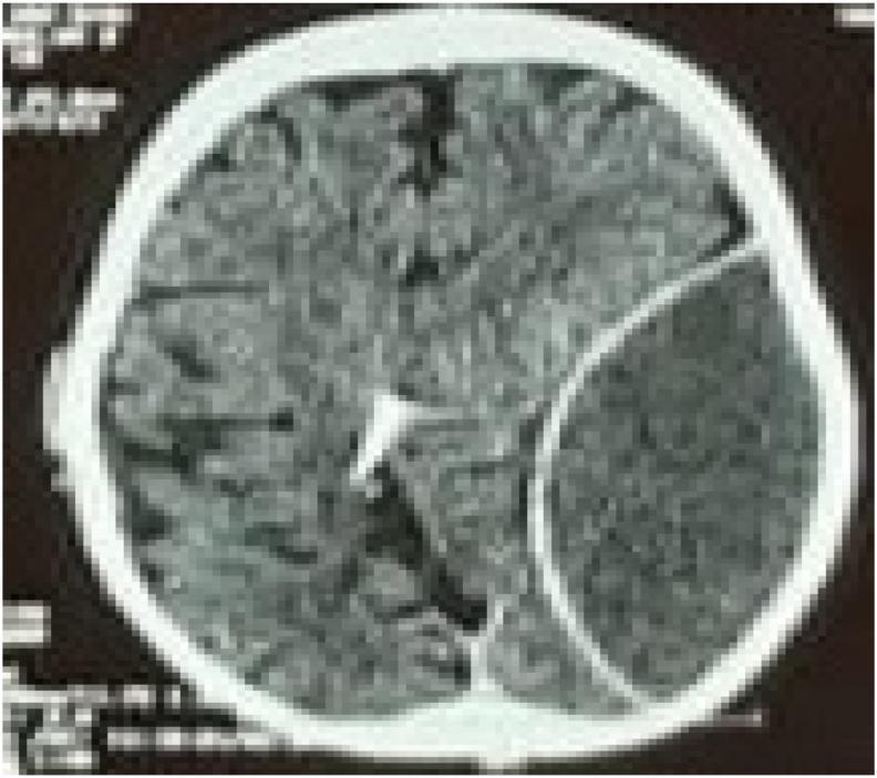
An axial non-contrast CT scan of the brain demonstrating a large, acute epidural (extradural) hematoma in the left hemisphere. The hematoma presents as a well-defined, biconvex (lenticular), hyperdense collection located between the skull and the dura mater. There is a significant mass effect evidenced by the compression of the left cerebral parenchyma, effacement of the adjacent cortical sulci, and midline shift toward the right. In the contralateral hemisphere, a radiopaque ventricular catheter is visible, extending into the posterior aspect of the right lateral ventricle near the occipital horn. This diagnostic image is representative of neurosurgical trauma emergencies, illustrating the classic appearance of arterial bleeding (typically from the middle meningeal artery) and secondary intracranial hypertension markers. The image serves as an educational resource for identifying acute traumatic intracranial hemorrhage and the presence of neurosurgical shunting devices.
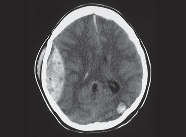
This diagnostic image is an axial non-contrast CT scan of the brain demonstrating acute traumatic intracranial pathology. The primary finding is a large, biconvex (lentiform), hyperdense collection in the right frontoparietal region, characteristic of an acute extradural (epidural) hemorrhage. The lesion is well-defined and exerts a significant mass effect, resulting in the inward compression of the adjacent brain parenchyma and effacement of the ipsilateral cortical sulci. Additionally, a secondary focal area of hyperdensity is visible in the left occipital region, consistent with a hemorrhagic contusion, often occurring as a contre-coup injury in the setting of head trauma. Subgaleal swelling is also noted on the right side of the cranium. These findings are critical for medical students and radiology residents to differentiate between extra-axial hemorrhages, where the biconvex shape of an epidural hematoma—limited by cranial sutures—is contrasted with the crescent-shaped presentation of subdural hematomas.
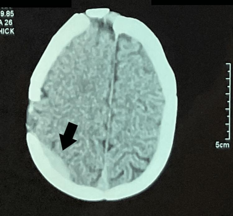
An axial non-contrast Computed Tomography (CT) scan of the head at the level of the upper cerebral convexities. The primary finding is a right-sided parietal extradural (epidural) hematoma, indicated by a black arrow. The hematoma appears as a focal, hyperdense, biconvex/lenticular-shaped collection adjacent to the inner table of the skull. A focal disruption or surgical alteration of the skull (bone flap/cranioplasty) is visible near the site of the hematoma. The surrounding brain parenchyma shows preserved gray-white matter differentiation, and there is no significant evidence of midline shift or obstructive hydrocephalus at this level. The falx cerebri remains in a midline position. This diagnostic image illustrates a postoperative complication following cranial surgery, specifically an acute extradural hemorrhage. The content is suitable for neurological, radiological, and surgical education regarding intracranial hemorrhages and postoperative monitoring.
Extradural Hemorrhage (EDH)
Definition
Anatomy & Pathophysiology
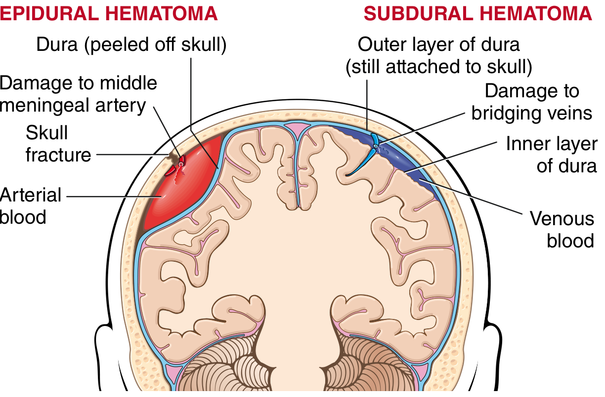
- Bleeding source: Middle meningeal artery in ~85% of cases (arterial, high-pressure)
- Less common: Laceration of dural venous sinuses (parieto-occipital or posterior fossa trauma), diploic veins, or emissary veins - these bleed more slowly and may be self-limiting
- Rare causes: Eosinophilic granuloma, metastatic skull tumors (e.g., hepatocellular carcinoma), craniofacial infections (sinusitis)
- In children, the skull is more deformable, so EDH can occur without a fracture due to temporary skull bone displacement
Classic Presentation
| Feature | Detail |
|---|---|
| Lucid interval | Brief loss of consciousness at time of injury, then recovery, then rapid deterioration |
| Headache | From stretching of pain-sensitive meninges and vessels at the base of the middle fossa |
| Nausea/vomiting | Raised ICP |
| Ipsilateral pupil dilation | Uncal herniation compressing CN III |
| Contralateral hemiplegia | Compression of ipsilateral cerebral peduncle |
| Rapid deterioration | From lucid state to coma and herniation within hours |
The lucid interval is a classic but not universal feature - it occurs in a minority of cases. In the modern CT era, most EDHs are detected early with minimal symptoms, and only ~30% require surgical intervention. - Plum and Posner's Diagnosis and Treatment of Stupor and Coma
Location
- Most common: Lateral temporal region (over the temporal fossa)
- Less common: Frontal, occipital, at the vertex, posterior fossa, or even contralateral (contre-coup)
CT Findings
- Biconvex shape - because the dura is tightly adherent at sutures, the hematoma does NOT cross suture lines (unlike subdural hematoma)
- Hyperdense (bright white) in the acute phase
- Midline shift and mass effect if large
- Skull fracture may be visible on bone windows
- Associated brain contusion may be seen (as in the right image above)
Associated Signs of Skull Base Fracture
- Battle's sign - ecchymosis behind the ear (mastoid region)
- Raccoon eyes - periorbital ecchymosis
- Haemotympanum - blood behind the tympanic membrane
- CSF otorrhoea/rhinorrhoea
Comparison: EDH vs Subdural Hematoma
| Feature | Extradural (EDH) | Subdural (SDH) |
|---|---|---|
| Space | Between skull and dura | Between dura and arachnoid |
| Bleed source | Middle meningeal artery (arterial) | Bridging veins (venous) |
| Shape on CT | Biconvex (lenticular) | Crescent-shaped |
| Crosses sutures? | No | Yes |
| Lucid interval | Classic (when present) | Less common |
| Typical patient | Young adult, trauma | Elderly, alcoholics, anticoagulated |
| Underlying brain injury | Often absent | Often present |
| Urgency | Hours | Hours to days |
Management
- Emergency CT head - confirms diagnosis
- Neurosurgical evacuation (craniotomy) - indicated for:
- Hematoma volume >30 mL
- Hematoma thickness >15 mm
- Midline shift >5 mm
- Neurological deterioration
- Conservative (non-operative) - for small EDHs with no significant neurological deficit, closely monitored in ICU with serial CT
- ICP management - osmotic therapy (mannitol), head elevation, hyperventilation as temporizing measures